

 PDF
PDF ePub
ePub Citation
 Print
Print


The drug development process is typically divided into three major steps: discovery, preclinical development, and clinical trials. The preclinical development of a new drug generally includes studies of efficacy, pharmacology, and experimental toxicology to define the dose, route, and frequency to be investigated in subsequent studies. Efficacy studies demonstrate that treatment with drug candidates has the desired therapeutic effect. Efficacy studies also help to identify the best drug candidates for further development [1].
The regulatory requirements of international guidelines generally demand toxicological and pharmacological studies in at least two laboratory animal species, a rodent and a nonrodent [1]. Beagle dogs are frequently used as model non-rodents for studying the efficacy and safety of newly developed drugs and chemicals, because of similarities in their anatomical morphology and physiology compared to humans [2]. In biomedical research, it is well recognized that individual animal variability can affect study results. To minimize animal variability, researchers should be provided with normal, healthy, well-adapted animals that represent a physiological model characterized by a narrow range of physical, behavioral, and clinical parameters [3-5]. Basic data of the test animal's blood, urine, and blood chemical values have detected many differences depending on sex and age, and the conditions where animals are maintained [7-9]. Thus, animals may be genetically identical but environmental effects during development cannot be ignored and these differences mean that is essential to establish basic data [10]. The establishment of standard basic data is important in the biosciences, because it can reduce and refine the use of laboratory animals [11].
Baseline parameters for hematology, urine, and blood chemical values play an important role in estimating target organ effects when it is difficult to observe the changes in tissues or when they are likely to be ignored during histopathological examinations [3-5]. These basic data can provide a foundation for interpreting certain test results more accurately [10,12]. Thus, it is necessary to acquire different types of basic data for laboratory animals to facilitate the reliable interpretation of a broad range of research studies using model animals. There have been some studies to acquire basic data for laboratory animals [10,12,13] but the volume of data is not great, so many tests are conducted based on foreign basic data [13,14].
The current study aimed to provide basic data on physiological and hematological characteristics, and organ weights for beagle dogs. This study analyzed whether there were differences in physiological and hematological parameters, and the organ weights of male and female beagle dogs. Thus, basic data was generated that should prove valuable to research in fields relevant to basic medical sciences where basic data are required to evaluate tests on dogs.
Materials and Methods
Animal husbandry and maintenance
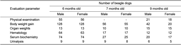
Male and female beagle dogs (5 months old) were obtained from Samtako (Beijing Marshall Biotechnology, Beijing, China) and housed in stainless net cages (80W×90L×65H cm) or a fenced room (130W×372L×125H cm) (1 dog per cage). Quarantine and acclimatization periods were 10 days. The animals were housed in a room maintained at a temperature (23±3℃) and a relative humidity (55±10%). The light and dark cycle was 12:12 h (light from 08:00 to 20:00). The temperature and humidity of the housing rooms was measured every hour and no changes were detected that could be considered to have any effect on the tests. The study evaluated 237 beagles including 148 (male 74, female 74) aged 6 months, 52 (male 27, female 25) aged 7 months, and 37 (male 20, female 17) aged 9 months. The 7-month-old and 9-month-old beagles were used as a control group in the toxicity test. The number of beagle dogs in each evaluation class is shown in Table 1.
Food and water intakes
Dry food for dogs (Oriental Yeast, Tokyo, Japan) was provided at a rate of 300 g per day, so no food-related factors could have affected the study. Water was sterilized and disinfected using a membrane filter with an UV ray flowing-water sterilizer, and provided ad libitum. Water examination was conducted by the Research Institute of Public Health and Environment (Daejeon, Korea) twice each year. No pollutants were detected in the water that could have influenced the evaluations conducted in this study.
Blood collection
Dogs were fasted overnight prior to blood collection. Blood samples were collected from the cephalic veins using a syringe with a 21- or 22-gauge needle. The blood samples were collected in CBC bottles containing EDTA-2K (Green Cross Medical Industry, Yongin, Korea) and used for hematological examination. For the serum biochemical evaluation, 3 mL blood samples were centrifuged at 3,000 rpm for 10 min within 1 h of collection. Sera were stored at -80℃ in a freezer prior to analysis. All processes used in the animal testing, including blood-gathering and collection, were reviewed by the Institutional Animal Care and Use Committees before they were conducted. Blood collection was conducted after the animals adapted to their feeding conditions.
Hematology
Blood samples were analyzed within 20 min using automatic blood analyzers (T-540 Coulter Counter, Coulter Electronics, Hialeah, FL, USA; ADVIA120 Hematology System, Bayer Healthcare LLC, Tarrytown, NY, USA). The following parameters were determined: white blood cell (WBC) count, differential leukocyte count, red blood cell (RBC) count, hemoglobin concentration, hematocrit, platelet count, mean corpuscular volume, mean corpuscular hemoglobin, and mean corpuscular hemoglobin concentration.
Serum biochemistry
The following serum biochemical parameters were evaluated using an automatic analyzer (Shimadzu CL-7200, Shimadzu, Kyoto, Japan): alkaline phosphatase (ALP), alanine aminotransferase (ALT), aspartate aminotransferase (AST), total bilirubin (TB), total cholesterol (TC), triglycerides (TG), phospholipids, total protein (TP), albumin, albumin/globulin (A/G) ratio, glucose, blood urea nitrogen (BUN), creatinine, creatine phosphokinase (CPK), calcium, and inorganic phosphorus. Sodium, potassium, and chloride were measured using an automatic electrolyte analyzer (Ciba-Corning 644 Na/K/Cl Analyzer, Ciba-Corning, Medfield, MA, USA).
Statistical analysis
Data processing and statistical analysis were performed using SAS version 9.2 (SAS Institute Inc., Cary, NC, USA). Values determined for different sexes and ages were compared using two-way ANOVA (SAS Version 9.2). A one-way ANOVA test (SAS Version 9.2) was applied at each time interval to compare the reproductive organ weight within each sex. Duncan's multiple-comparison test was used to assess the statistical significance of differences among the study groups. A level of P<0.05 was accepted as statistically significant.
Results
Physiological values
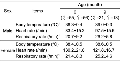
A significant increase in the body temperature was observed in male dogs (P<0.001). The mean body temperature in 6-month-old male dogs was 38.3±0.4℃, whereas that in 9-month-old male dogs was 39.0±0.3℃. In both sexes, the respiratory rate increased significantly with age (P<0.001), while the heart rate of females was slightly higher than that of males (Table 2).
Body weight gain and organ weights
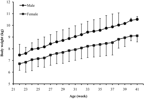
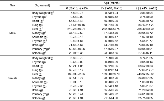
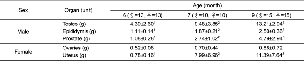
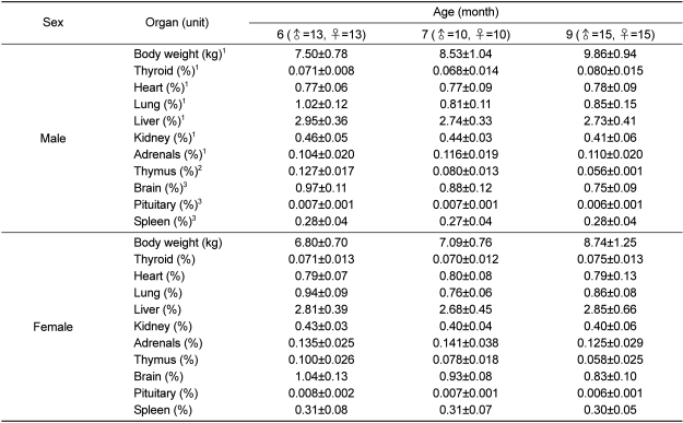
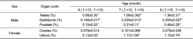
The body weight of male and females increased rapidly up to 33 weeks (0.18±0.07 kg/week for male dogs, 0.14±0.06 kg/week for female dogs), before increasing gradually up to 41 weeks (0.14±0.06 kg/week for males, 0.11±0.08 kg/week for females). The body weight of males was significantly higher than that of females (Figure 1, P<0.001). The absolute and relative weights of the thyroid, heart, lung, liver, kidney, and adrenals increased significantly as both sexes grew older, whereas male organs weighed significantly more than those of females (Table 3, P<0.001). The thymus gradually decreased in weight as both sexes grew older (P<0.001). Brain, pituitary, and spleen weights increased significantly with age (Tables 3 and 5, P<0.001). In absolute and relative terms, the reproductive organ weight of males and females, including the epididymis, prostate, and uterus, increased significantly as they grew older (P<0.05), whereas the weight of the ovaries did not increase significantly with age (Table 4). The absolute weight of testes increased significantly from 6 to 9 months, while their relative weight increased significantly from 6 to 7 months (P<0.05).
Hematological parameters
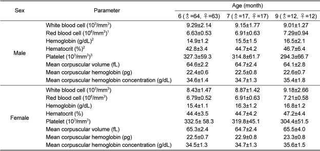
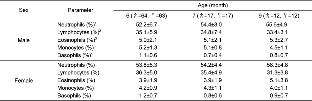
The results of hematological examination are shown in Tables 7 and 8. The platelet count of female dogs was slightly higher than that of males. The RBC, hemoglobin, and hematocrit of both sexes increased non-significantly with age. In the leukocyte differential count, the neutrophils and eosinophils of both sexes tended to increase with age, whereas basophils, lymphocytes, and monocytes decreased.
Biochemical parameters
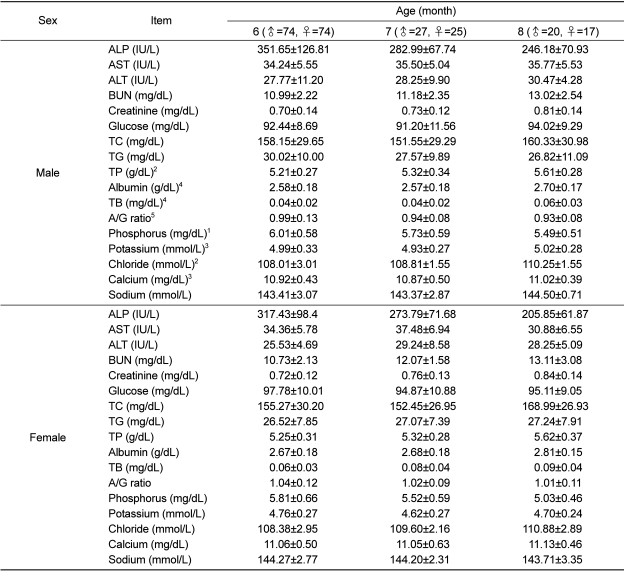
The results of the serum biochemical profiles are shown in Table 9. The ALP of male beagles was slightly higher than that of females, while the TC in female dogs aged 9 months was higher than that of males at the same age. The remaining biochemical parameters, including, ALT, BUN, creatinine, TG, and TP, increased non-significantly with age in both sexes.
Urological parameters
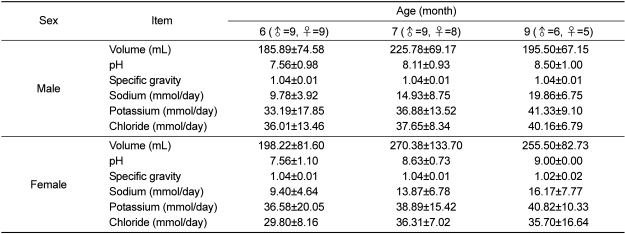
The amount of urine and sodium ions tended to increase as dogs grew older. However, there were large individual differences (Table 10).
Discussion
Beagle dogs are the most frequently used nonrodent animals in biomedical research, because of similarities in their anatomical morphology and physiology to humans [2]. In biomedical research, it is well recognized that individual animal variability can affect study results. Therefore, highly defined dog models should be used in biomedical studies [3]. In clinical or non-clinical tests, the normal range of blood constituent is a standard means of diagnosis when identifying diseases or abnormalities. Thus, they are very important values, although they can vary significantly depending on the individual environment (e.g., species, sex, age, and conditions) or genetic characteristics [13,15-17]. This means it is essential to reduce errors by determining the normal range as accurately as possible to facilitate the exact diagnosis of diseases or confirming abnormalities. The current study was conducted to determine basic physiological data on the body weight, organ weight, body temperature, heart rate, and hematological, biochemical and urological parameters for male and female beagle dogs.
In the physical examination of male and female beagle dogs aged 6 and 9 months, the body temperature and respiratory rate did not differ significantly between the sexes, although the respiratory rate of both sexes tended to increase as they grew older, while the heart rate of females was slightly higher than that of males (Table 2). Ferasin et al. [18] reported that dogs aged less than 1 year appeared to have a significantly higher heart rate than older dogs, while the heart rate of healthy dogs undergoing routine clinical examination was related to their age.
The body weight of male and female dogs increased rapidly up to 33 weeks, followed by a gradual increase to 41 weeks. The body weight was significantly different between the sexes (Figure 1). Their body weight increased by 0.18±0.07 kg/week in male dogs and by 0.14±0.06 kg/week in female dogs. Nunamaker et al. [19] reported that the body weight increased by 19.7 g/day in a control group of dogs. The relative organ weights were similar to those reported for a control group in a previous report [20]. The relative reproductive organ weights of both sexes increased rapidly up to 7 months (Table 6). These results indicate that puberty may be reached at 7 months of age.
Age-related differences were found in terms of RBC count, hemoglobin, hematocrit, neutrophils, eosinophils, basophils, lymphocytes, monocytes, ALT, BUN, creatinine, TG, and TP. Sex-related differences were found for ALP and TC (Tables 7, 8, and 9). However, these parameters were not significantly related to age or sex. These results were slightly different to those reported by Jeong et al. [12]. The number of eosinophils was higher than that reported by Jeong et al [12], but similar to that reported by Wolford et al. [21]. The number of lymphocyte was also higher than that reported by Jeong et al. [12] and Wolford et al. [21]. This was considered to be a consequence of transportation stress, but it may also be related to a vaccination before their final export. ALP is an enzyme found in the canalicular membrane or sinusoidal membrane, and it can be used to measure the degree of damages to these parts. However, ALP is also secreted by the bones, intestines, and placenta [22,23]. Thus, it can be increased with motion and during periods of growth and pregnancy [14]. We found a progressive reduction in serum ALP concentration as both sexes grew older. This suggests that the observed change in the ALP level is related to the turnover of bone. Uchiyama et al. [23] and Ikeuchi et al. [14] also observed a reduction in plasma ALP concentration with age in dogs. However, this biochemical blood parameter did not exceed the normal range and there was no difference between males and females. Hematological examinations, biochemical examinations of blood, and urine examinations indicate considerable differences depending on the species, source of supply, maintenance conditions of animals, and age, as well asthe analytical equipment used to conduct tests [25-27].
Therefore, it is important that each research institute maintains standard data appropriate for their facility to facilitate the accurate interpretation of test results. Test results are also influenced by various factors, so it is necessary to minimize any deviations due to the external environment by formulating an SOP covering conditions such as the period of specimen collection, environmental conditions, animal stress status, and animal feeding activity.
 XML Download