Materials and Methods
Direct administration of inorganic metals into yolk sac
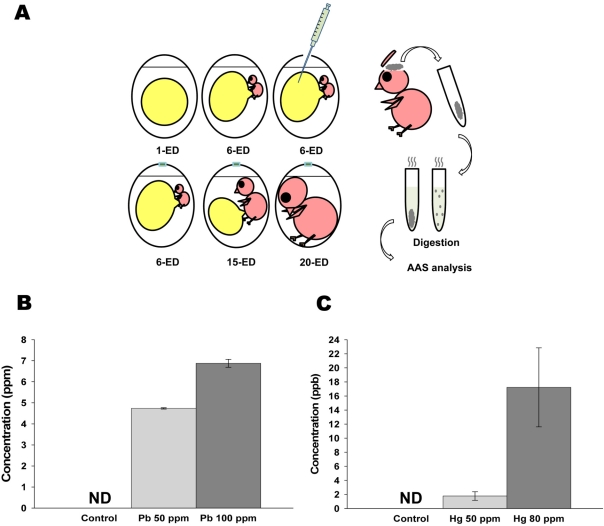
 | Figure 1Administered lead (Pb) and mercury (Hg) in early developmental stage of chick embryo were still detectable in the brain of adult stage. (A) Schematic diagram of experimental design for AAS analysis. The prepared solution [0, 50 (25 µg/500 µL) and 100 ppm (50 µg/500 µL) of Pb; and 0, 50 (25 µg/500 µL) and 80 ppm (40 µg/500 µL) of Hg] was injected into each yolk sac of 6-ED chick embryos. On 20-ED, the collected chick embryo brains were digested for Atomic Absorption Spectrometry analysis. (B-C) Accumulated Pb (B) and Hg (C) in neonatal brain. Indicated concentration of inorganic Pb or Hg were injected into yolk sac on 6-ED. On day 20, the accumulated concentration of Pb (B) and Hg (C) in neonatal brain were determined by F-AAS as described in Materials and Methods. All data represent three independent experiments, each carried out in triplicate, and significance was tested using ANOVA with a Newman-Keuls post-hoc test, where ** represents P<0.05 vs control. ND represents non-detectable. |
Atomic absorption spectrophotometry (AAS)
The 3-(4,5-dimethylthiazol-2-yl)-2,5-diphenyltetrazolium bromide (MTT) assay
Western blot analysis
Neuronal cell culture (astrocytes and neuronal cells primary culture)
Immunocytochemistry
Statistical analysis
Results
Direct administration of inorganic lead and mercury in chick embryo resulted in the accumulation of inorganics in neonatal brain
Lead and mercury treatment resulted in the cytotoxicity of primary neuronal cells
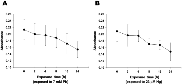
 | Figure 2Lead and mercury induces cytotoxicity of neuronal cells. A-B. Decreased cell viability with the treatment of Pb (A) and Hg (B). Neuronal cells were treated with Pb (7 mM) and Hg (23 µM) for overnight, and MTT colorimetric assay was performed as described in Materials and Methods. All data represent three independent experiments, each carried out in triplicate, and significance was tested using ANOVA with a Newman-Keuls post-hoc test, where ** represents P<0.05 vs control. |
Lead and mercury treatment resulted in the cytoskeletal reorganization of neonatal brain
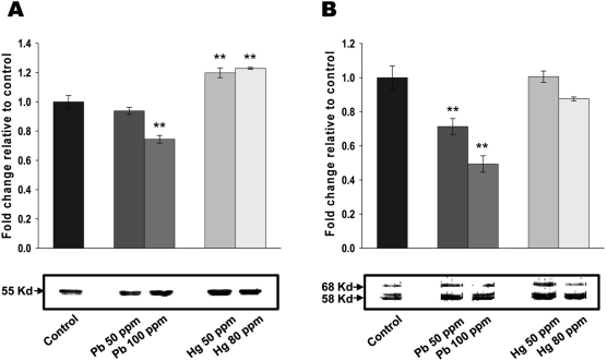
 | Figure 3Lead and mercury modulate cytoskeletal reorganization in neonatal brain. (A-B) Effect of lead and mercury on tubulin (A) and tau (B) expression. Indicated concentration of inorganic lead or mercury were injected into yolk sac on 6-ED. On day 20, total cellular extracts were isolated from neonatal brain and Western blot analyses performed with antibodies against tubulin or tau. Western blots were quantified using densitometric analysis, and values expressed relative to control. Western blots are representative of n=3, and significance was tested using ANOVA with a Newman-Keuls post-hoc test, where ** represents P<0.05 vs control. |
Lead and mercury treatment resulted in the reorganization of cytoskeleton in primary neuronal cells
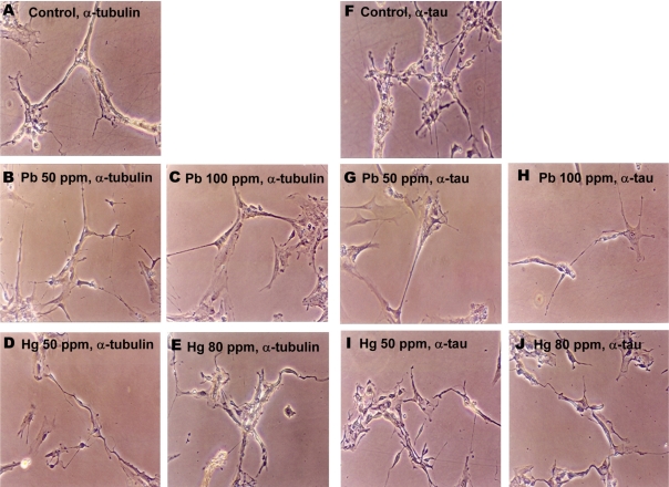
 | Figure 4Lead and mercury modulate cytoskeletal reorganization in neuronal cells. Neuronal cells were treated with Pb (4.8 and 7 mM for 50 and 100 ppm groups, respectively) and Hg (2.4 and 23 µM for 50 and 80 ppm groups, respectively) for overnight, and immunocytochemical staining was performed using antibody against tubulin (A-E) or tau (F-J). |


 PDF
PDF ePub
ePub Citation
 Print
Print


 XML Download