This article has been
cited by other articles in ScienceCentral.
Abstract
A male one year-old beagle dog with unilateral cryptorchism was presented for investigation of reduced appetite. Abdominal sonography and radiography demonstrated abnormal enlargement of the left testicle in the abdominal cavity. Both the retroperitoneal cryptorchid testicle and the other contralateral testicle were removed surgically. The retroperitoneal cryptorchid testicle was an enlarged, firm and bulging sphere mass. The cut surface revealed a homogeneous white color. The contralateral testicle in the scrotum showed an almost normal appearance. Histopathologically, the retroperitoneal cryptorchid testicle was diagnosed as a Sertoli cell tumor. This report describes a case of Sertoli cell tumor with cryptorchism in a beagle dog.
Go to :

Keywords: Sertoli cell tumor, cryptorchism, testis, beagle, dog
Testicular neoplasia and Leydig cell hyperplasia are common findings in aged dogs, unlike in other domestic animals and in men [
1]. The prevalence varies from 0.068 to 4.6% in male dogs, and up to 60% of the aged animals appeared to have testicular tumors in studies that included old dogs [
2,
3]. The three main types of testicular tumors in dogs are Sertoli cell tumors, seminomas and Leydig cell tumors, occurring at about equal frequencies [
4]. Cryptorchism is an important risk factor for the development of testicular tumors, causing a 26-fold increase in the risk for Sertoli cell tumors and a 15-fold increase for seminomas [
5]. Here, we describe the characteristics of spontaneous Sertoli cell tumor occurrence with the cryptorchism in a beagle dog.
A one year-old male beagle dog was obtained from the Animal Facilities of Center for Animal Resources Development, Wonkwang University, Korea. In the previous 3 days, the dog had shown appetite reduction, and the dog was given a health examination. Abdominal sonography and radiography demonstrated abnormal enlargement of the left testicle in the abdominal cavity. Both the retroperitoneal cryptorchid testicle and the other contralateral testicle were removed surgically. The orchidectomized testicles were submitted to gross examination and trimmed. The trimmed tissues was fixed in 10% neutral buffered formalin and embedded in paraffin. Four µm sections were made and stained with hematoxylin and eosin for histopathological examination.
The retroperitoneal cryptorchid testicle was an enlarged, firm and bulging sphere mass (
Figure 1A). The cut surface revealed a homogeneous white color (
Figure 1B). The contralateral testicle in the scrotum showed an almost normal appearance. Histopathologically, the tumor cells had abundant eosinophilic cytoplasm, round or oval nuclei, and punctuate nucleoli (
Figure 2). Malignant behavior was rare. As the results of histopathological analysis, the retroperitoneal cryptorchid testicle was diagnosed as a Sertoli cell tumor.
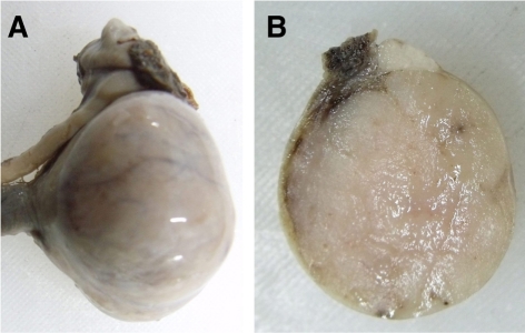 | Figure 1Gross findings of retroperitoneal cryptorchid testicle. A) The testicle was an enlarged, firm and bulging sphere mass. B) The cut surface revealed a homogeneous white color. 
|
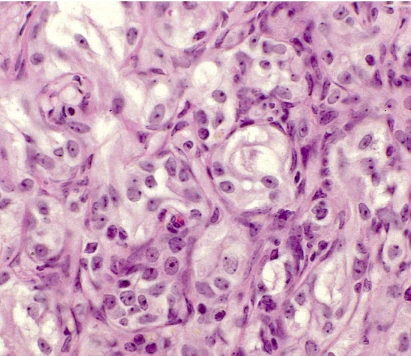 | Figure 2Histopathological findings of retroperitoneal cryptorchid testicle. The tumor cells had abundant eosinophilic cytoplasm, round or oval nuclei, and punctuate nucleoli. Malignant behavior was rare. Hematoxylin-eosin stain. ×400.
|
Testicular neoplasms are frequent in aged dogs [
6], with seminoma, Sertoli cell tumors or sertolioma, and Leydig cell tumors being the most prevalent types of testicular neoplasia [
6], although cases of mixed germ cell tumors [
7] and mesenchymal tumors [
8] have been described. Older dogs are commonly affected by testicular enlargement [
9], especially in cryptorchid testes, where the incidence of neoplasia is higher compared with scrotal testes [
10]. It is common to observe two or more types of neoplasia in the same testicle [
4]. A Sertoli cell tumor in the dog is the most prone to produce metastases, occurring in 10 to 14% of the cases. In contrast, seminoma produces metastasis in 6 to 11%, and metastasis from a Leydig cell tumor is extremely rare [
4,
10]. In this case, metastases were not detected. The more frequent occurrence of these tumors in the right testicle can be accounted for by cryptorchism, which is more common on the right side [
4]. The right testicle arises more cranially than the left and has a longer distance to travel to reach the scrotum [
4,
10]. In this study, the dog revealed unusual cryptorchism in the left testicle. This report describes a case of a Sertoli cell tumor with cryptorchism in a beagle dog.
Acknowledgments
This research was supported by the Basic Science Research Program through the National Research Foundation of Korea (NRF) funded by the Ministry of Education, Science, and Technology (2010-0021940).
Go to :
References
1. Peters MA, de Jong FH, Teerds KJ, de Rooij DG, Dieleman SJ, van Sluijs FJ. Ageing, testicular tumours and the pituitary-testis axis in dogs. J Endocrinol. 2000; 166(1):153–161. PMID:
10856894.

2. Weller RE, Dagle GE, Buschbom RL, Park JF. Examination of testicular tumours in the beagle dog exposed to inhaled plutonium. Int J Radiat Biol. 1995; 68(1):63–70. PMID:
7629439.
3. Peters MA, Teerds KJ, van der Gaag I, de Rooij DG, van Sluijs FJ. Use of antibodies against LH receptor, 3β-hydoxysteroid dehydrogenase and vimentin to characterize different types of testicular tumour in dogs. Reproduction. 2001; 121(2):287–296. PMID:
11226053.
4. Nielsen SW, Kennedy PC. Moulton JE, editor. Tumors of the genital system. Tumors in Domestic Animals. 1990. 3rd ed. Berkeley: University of California Press;p. 479–513.
5. Hayes HM Jr, Wilson GP, Pendergrass TW, Cox VS. Canine cryptorchism and subsequent testicular neoplasia: case-control study with epidemiologic update. Teratology. 1985; 32(1):51–56. PMID:
2863879.
6. Hayes HM Jr, Pendergrass TW. Canine testicular tumors: epidemiologic features of 410 dogs. Int J Cancer. 1976; 18(4):482–487. PMID:
977190.
7. Patnaik AK, Mostofi FK. A clinicopathologic, histologic and immunohistochemical study of mixed germ cell-stromal tumors of the testis in 16 dogs. Vet Pathol. 1993; 30(3):287–295. PMID:
8392765.
8. Herron MA. Tumors of the canine genital system. J Am Anim Hosp Assoc. 1983; 19(6):981–994.
9. Reif JS, Brodey RS. The relationship between cryptorchidism and canine testicular neoplasia. J Am Vet Med Assoc. 1969; 155(12):2005–2010. PMID:
4391618.
10. Brearley MJ. White RAS, editor. The urogenital system. Manual of Small Animal Oncology. 1991. Gloucester: British Small Animal Veterinary Association;p. 306–307.
Go to :


 PDF
PDF ePub
ePub Citation
 Print
Print


 XML Download