

 PDF
PDF ePub
ePub Citation
 Print
Print


Abstract
Ultraviolet (UV) irradiation is an environmental factor that causes skin aging, and is also a major factor leading to cumulative alterations of skin structure, function and appearance. To investigate the effects of Selenium (Sel) on UV-induced skin aging, hairless mice were treated for 4 weeks with UV irradiation and topical application of Sel. Then, the effects of Sel were measured in the skin of these mice via histological analysis and Western blotting. According to the results of wrinkle formation analysis, the topical application of Sel induced a reduction in wrinkling formation in the damaged skin of the UV-irradiated mice. Additionally, our histological analysis demonstrated that the skin thickness in the Sel-treated group was less than in the UV-irradiated group. Furthermore, in an effort to investigate the mechanisms underlying the effects of Sel, the expression levels of matrix-metalloproteinase (MMP) and MAPK protein were assessed in both groups. The application of Sel induced a reduction in MMP-1 expression levels to the levels observed in the non-irradiated group. However, the expression level of MMP-9 was increased slightly in the Sel application group as compared with the vehicle application group. Additionally, the levels of ERK phosphorylation were increased by the application of Sel, but the levels of JNK and p38 were not altered by Sel treatment. These results suggest the possibility that Sel should be considered as a skin aging-protective and therapeutic drug candidate, which functions via the regulation of MMP expression levels.
REFERENCES
Azar D.T.., Hahn T.W.., Jain S.., Yeh Y.C.., Stetler-Stevenson W.G.1996. Matrix metalloproteinases are expressed during wound healing after excimer laser keratectomy. Cornea. 15:18–23.

Beck M.A.., Kolbeck P.C.., Shi Q.., Rohr L.H.., Morris V.C.., Levander O.A.1994. Increased virulence of a human enterovirus (coxsackievirus B3) in selenium-deficient mice. J. Infect. Dis. 170(2):351–357.
Birringer M.., Pilawa S.., Flohe L.2002. Trends in selenium biochemistry. Nat. Prod. Rep. 19:693–718.
Bissett D.L.., Chatterjee R.., Hannon D.P.1990. Photoprotective effect of topical anti-inflammatory agents against ultraviolet radiation-induced chronic skin damage in the hairless mouse. Photodermatol. Photoimmunol. Photomed. 7(4):153–158.
Bizot-Foulon V.., Bounchard B.., Hornerbeck W.., Duberter L.., Bertaux B.1995. Uncoordinated expressions of type I and III collagens, collagenase and tissue inhibitor of matrix metalloproteinase 1 along in vitro proliferative life span of human skin fibroblast. Cell Biol. Int. 19:129–135.
Braverman I.M.., Fonferko E.1982. Studies in cutaneous aging. I. The elastic fiber network. J. Invest. Dermatol. 78:434–443.
Brennan M.., Bhatti H.., Nerusu K.C.., Bhagavayhula N.., Kang S.., Fisher G.J.., Varani J.., Voorhees J.J.2003. Matrix metalloproteinase-1 is the major collagenolytic enzyme responsible for collagen damage in UV-irradiated human skin. Photochem. Photobiol. 78:43–48.
Buechner N.., Schroeder P.., Jakob S.., Kunze K.., Maresch T.., Calles C.., Krutmann J.., Haendeler J.2008. Changes of MMP-1 and collagen type Ialpha1 by UVA, UVB and IRA are differentially regulated by Trx-1. Exp. Gerontol. 43(7):633–637.
Carlson B.A.., Novoselov S.V.., Kumaraswamy E.., Lee B.J.., Anver M.R.., Gladyshev V.N.., Hatfield D.L.2004. Specific excision of the selenocysteine tRNA[Ser]Sec (Trsp) gene in mouse liver demonstrates an essetial role of selenoproteins in liver function. J. Biol. Chem. 279:8011–8017.
Csarini J.P.., Michel L.., Maurette J.M.., Adhoute H.., Bjot M.2003. Immediate effects of UV radiation on the skin: modification by an antioxidant complex containing carotenoids. Photodermatol. Photoimmunol. Photomed. 19(4):182–189.
Choi H.K.., Kim D.H.., Kim J.W.., Ngadiran S.., Sarmidi M.R.., Park C.S.2010. Labisia pumila extract protects skin cells from photoaging caused by UVB irradiation. J. Biosci. Bioeng. 109(3):291–296.
Fini M.E.., Girard M.T.., Matsubara M.1992. Collagenolytic/gelatinolytic enzymes in corneal wound healing. Acta Ophthalmol. 70 (Suppl 202). 26–33.
Fisher G.J.., Datta S.C.., Talwar H.S.., Wang Z.Q.., Varani J.., Kang S.., Voorhees J.J.1996. Molecular basis of sun-induced premature skin aging and retinoid antagonism. Nature. 379:335–339.
Fortino V.., Maioli E.., Torricelli C.., Davis P.., Valacchi G.2007. Cutaneous MMPs are differently modulated by environmental stressors in old and young mice. Toxicol. Lett. 173(2):73–79.
Hatfield D.L.., Gladyshev V.N.2002. How selenium has altered our understanding of the genetic code. Mol. Cell. Biol. 22:3565–3576.
Jenkins G.2002. Molecular mechanisms of skin aging. Mech. Aging Dev. 123:801–810.
Kang T.H.., Park H.M.., Kim Y.B.., Kim H.., Kim N.., Do J.H.., Kang C.., Cho Y.., Kim S.Y.2009. Effects of red ginseng extract on UVB irradiation-induced skin aging in hairless mice. J. Ethnopharmacol. 123(3):446–451.
Karunasinghe N.., Ryan J.., Tuckey J.., Masters J.., Jamieson M.., Clarke L.C.., Marshall J.R.., Ferguson L.R.2004. DNA stability and serum selenium levels in a high-risk group for prostate cancer. Cancer Epidemiol. Biomarkers Prev. 13(3):391–397.
Kashanian S.., Gholivand M.B.., Ahmadi F.., Ravan H.2008. Interaction of diazinon with DNA and the protective role of selenium in DNA damage. DNA Cell Biol. 27(6):325–332.
Kim H.H.., Cho S.., Lee S.., Kim K.H.., Cho K.H.., Eun H.C.., Chung J.H.2006. Photoprotective and anti-skin-aging effects of eicosapentaenoic acid in human skin in vivo. J.
Kiremidjian-Schumacher L.., Roy M.., Glickman R.., Schneider K.., Rothstein S.., Cooper J.., Hochster H.., Kim M.., Newman R.2000. Selenium and immunocompetence in patients with head and neck cancer. Biol. Trace Elem. Res. 73(2):97–111.
Kohrle J.., Brigelius-Flohe R.., Bock A.., Gartner R.., Meyer O.., Flohe L.2000. Selenium in biology: facts and medical perspectives. Biol. Chem. 381:849–864.
Kowalska E.., Narod S.A.., Huzarski T.., Zajaczek S.., Huzarska J.., Gorski B.., Lubinski J.2005. Increased rates of chromosome breakage in BRCA1 carriers are normalized by oral selenium supplementation. Cancer Epidemiol. Biomarkers Prev. 14(5):1302–1306.
Lavker R.M.1995. Cutaneous aging: chronologic versus photoaging, Gilchrest, B. ed., pp. 123-135, Blackwell.
Martisian L.M.1990. Metalloproteinase and their inhibitors in matrix remodelling. Trends Genet. 6:121–125.
Park C.H.., Lee M.J.., Kim J.P.., Yoo I.D.., Chung J.H.2006. Prevention of UV radiation-induced premature skin aging in hairless mice by the novel compound Melanocin A. Photochem. Photobiol. 82(2):574–578.
Rayman M.P.2000. The importance of selenium to human health. Lancent. 356:233–241.
Rittie L.., Fisher G.J.2002. UV-light-induced signal cascades and skin aging. Aging Res. Rev. 1:705–720.
Ropke C.D.., da Silva V.V.., Kera C.Z.., Miranda D.V.., de Almeida R.L.., Sawada T.C.., Barros S.B.2006. In vitro and in vivo inhibition of skin matrix metalloproteinases by Pothomorphe umbellata root extract. Photochem. Photobiol. 82(2):439–442.
Schwarz K.., Foltz C.M.1957. Selenium as an integral part of factor 3 against dietary necrotic liver degeneration Nutrition. J. Am. Chem. Soc. 15:255–264.
Sevanian A.., Hodis H.1997. Antioxidants and atherosclerosis: an overview. Biofactors. 6(4):385–390.
Smith M.L.., Lancia J.K.., Mercer T.I.., Ip C.2004. ).Selenium compounds regulate p53 by common and distinctive mechanisms. Anticancer Res. 24(3a):1401–1408.
Stadman T.C.1996. Selenocysteine. Annu. Rev. Biochem. 65:83–100.
Steinberg D.1997. Low density lipoprotein oxidation and its pathobiological significance. J. Biol. Chem. 272(34):20963–20966.
Stetler-Stevenson W.G.., Karutzsch H.C.., Wacher M.P.1989. The activation of human Type IV collagenase. J. Biol. Chem. 264:1353–1356.
Tsukahara K.., Moriwaki S.., Fujimura T.., Takema Y.2001. Inhibitory effect of an extract of Sanguisorba officinalis L. on ultraviolet-B-induced photodamage of rat skin. Biol. Pharm. Bull. 24(9):998–1003.
Uitto J.1986. Connective tissue biochemistry of the aging dermis. Dermatol. Clin. 4:433–446.
van Rij A.M.., Thomson C.D.., McKenzie J.M.., Robinson M.F.1979. Selenium deficiency in total parenteral nutrition. Am. J. Clin. Nutr. 32:2085–2086.
Varani J.., Spearman D.., Perone P.., Fligiel S.E.G.., Datta S.., Wang Z.Q.., Shao Y.., Kang S.., Fisher G.J.., Voorhees J.J.2001. Inhibition of type I procollagen synthesis by damaged collagen in photoaged skin and by collagenase-degraded collagen in vitro. Am. J. Pathol. 158:931–942.
Visse R.., Nagase H.2003. Matrix metalloproteinase and tissue inhibitors of metalloproteinase: structure, function and biochemistry. Circ. Res. 92:827–839.
Wilber C.G.1980. Toxicology of selenium: a review. Clin. Toxicol. 17:171–230.
Wlaschek M.., Heinen G.., Poswig A.., Schwarz A.., Krieg T.., Scharffetter-Kochanek K.1994. UVA-induced autocrine stimulation of fibroblast-derived collagenase/MMP-1 by interrelated loops of interleukin-1 and interleukin-6. Photochem. Photobiol. 59:550–556.
Figure 1.
Schematic diagram of the irradiation site in the dorsal skin surface of hairless mice (A) and the irradiation schedule lead to induce skin aging (B). The UV dose was increased weekly by 1 MED up to 4 MED. Vehicle or Selenium was applied to the backs of hairless mice after UV irradiation.
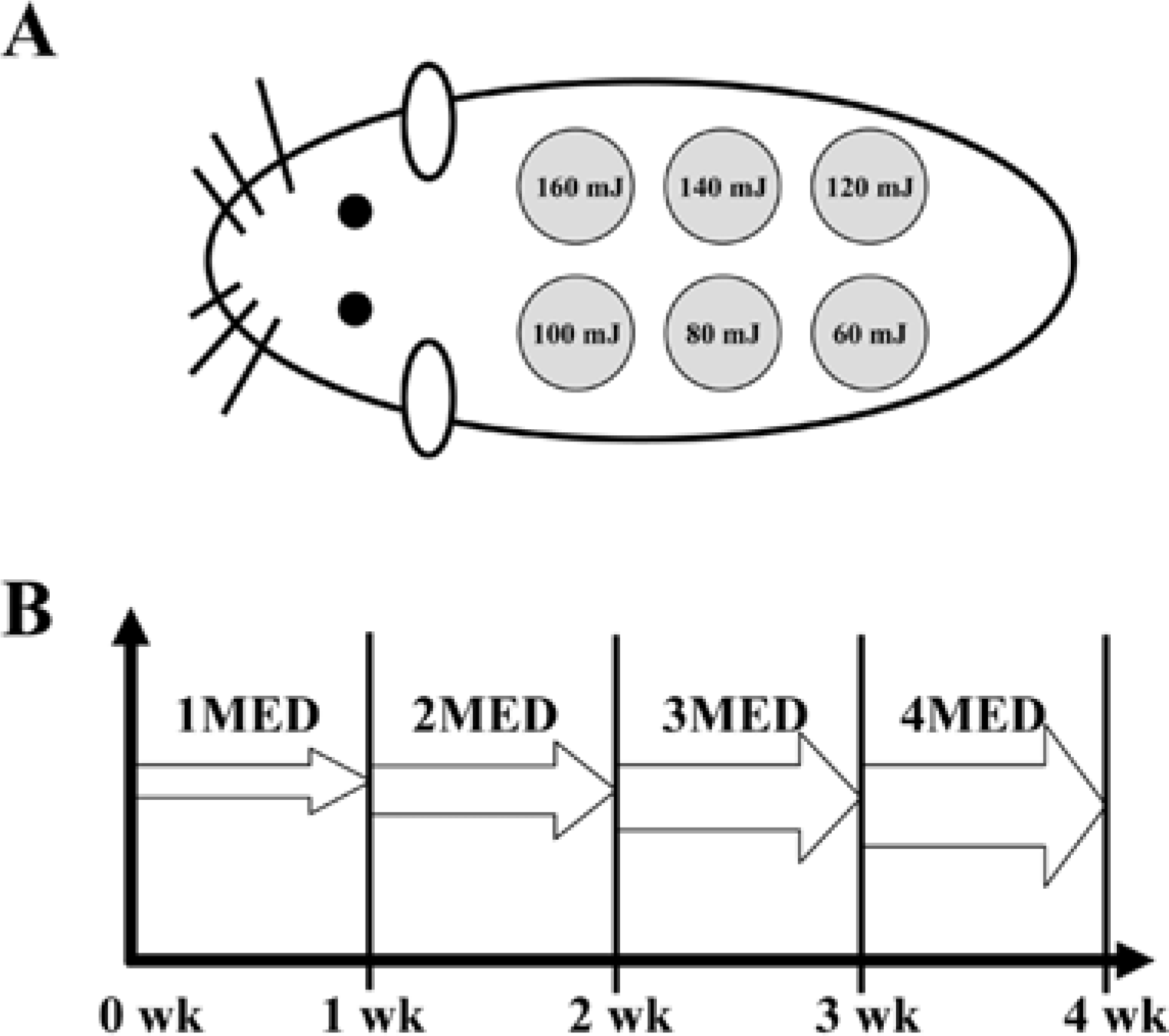
Figure 2.
Inhibition of UV-induced wrinkle formation by Sel in the hairless mice. Wrinkle formation was measured by replica grading at final week. The level of wrinkle were assessed according to the scoring system suggested by Bissette et al. (1990) (grad 0: no wrinkles, grade 1: a few shallow wrinkles, grade 2: some wrinkles, grade 3: several deep wrinkles). The values of data represented mean±SD of three experiments. ∗P<0.05 is the significance level compared to the control (No irradiation) group, P<0.05 is the significance level compared to the vehicle applicated group.
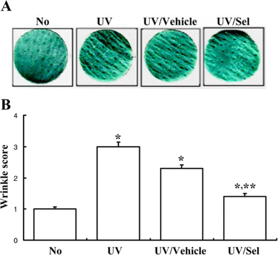
Figure 3.
Inhibition of UV-induced skin thickness by Sel in hairless mice. The dorsal skin of hairless mice was prepared into histological slide and skin thickness were measured the wide from epidermis to dermis. Cellular morphology was viewed at 20x magnification and the values of data represented mean±SD of three experiments. ∗P<0.05 is the significance level compared to the control (No irradiation) group, P<0.05 is the significance level compared to the vehicle applicated group.
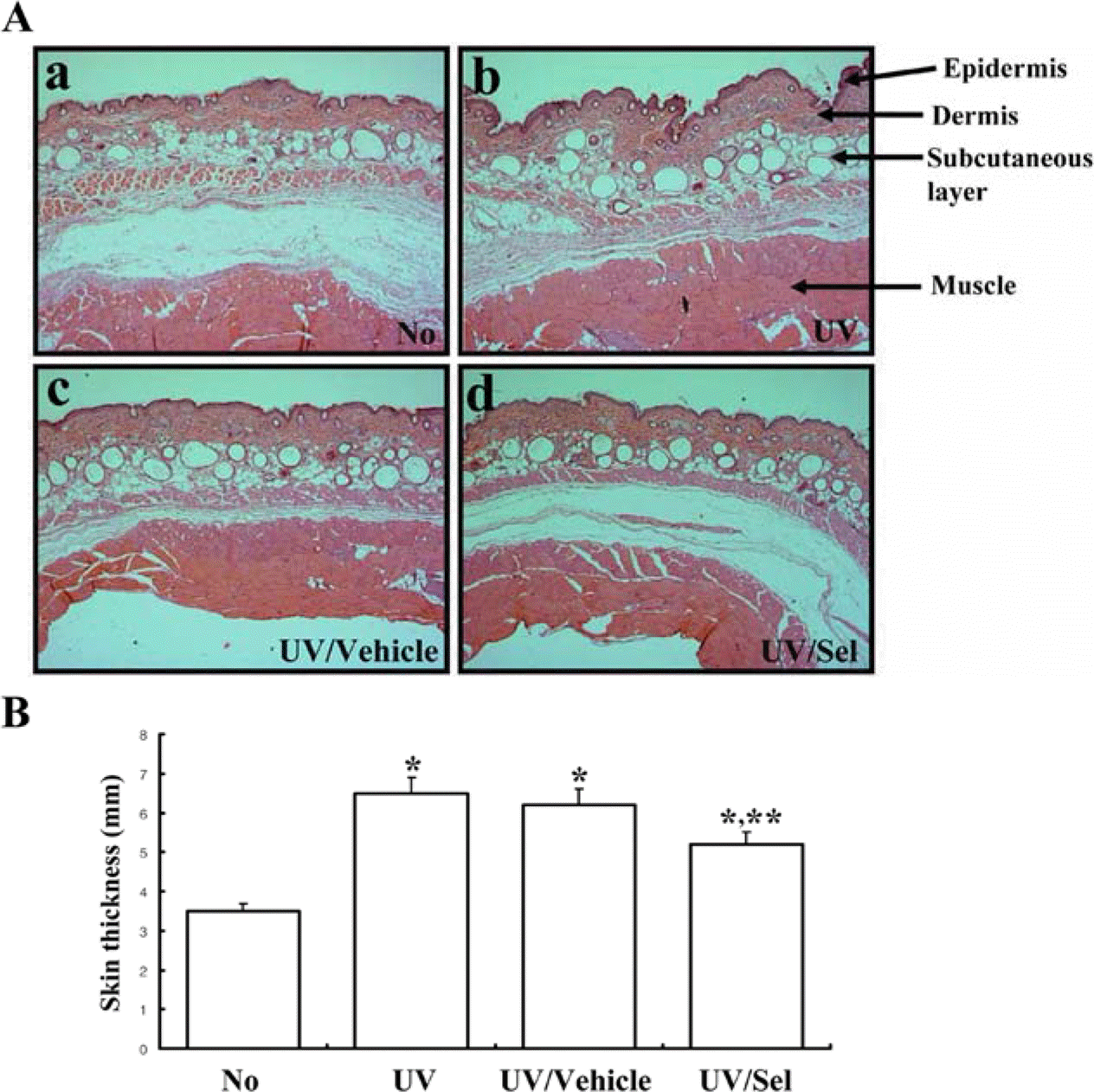
Figure 4.
Effect of Selenium (Sel) on UV-induced MMP expression using Western blot (A) and immunohistochemical staining (B). Total cell lysate were prepared from skin tissues of control, UV-irradiation, UV/Vehicle and UV/Sel group, as described in Materials and Methods. Fifty micrograms of protein per sample were immunoblotted with antibodies for each protein. Three samples were assayed in triplicate using Western blotting. The values are the mean±SD. ∗P<0.05 is the significance level compared to the control (No irradiation) group, ∗∗P<0.05 is the significance level compared to the vehicle applicated group.
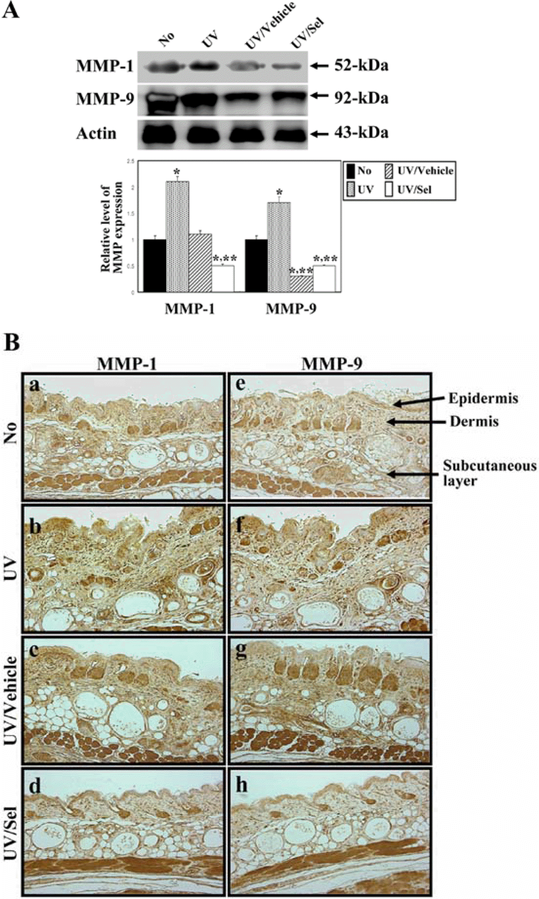
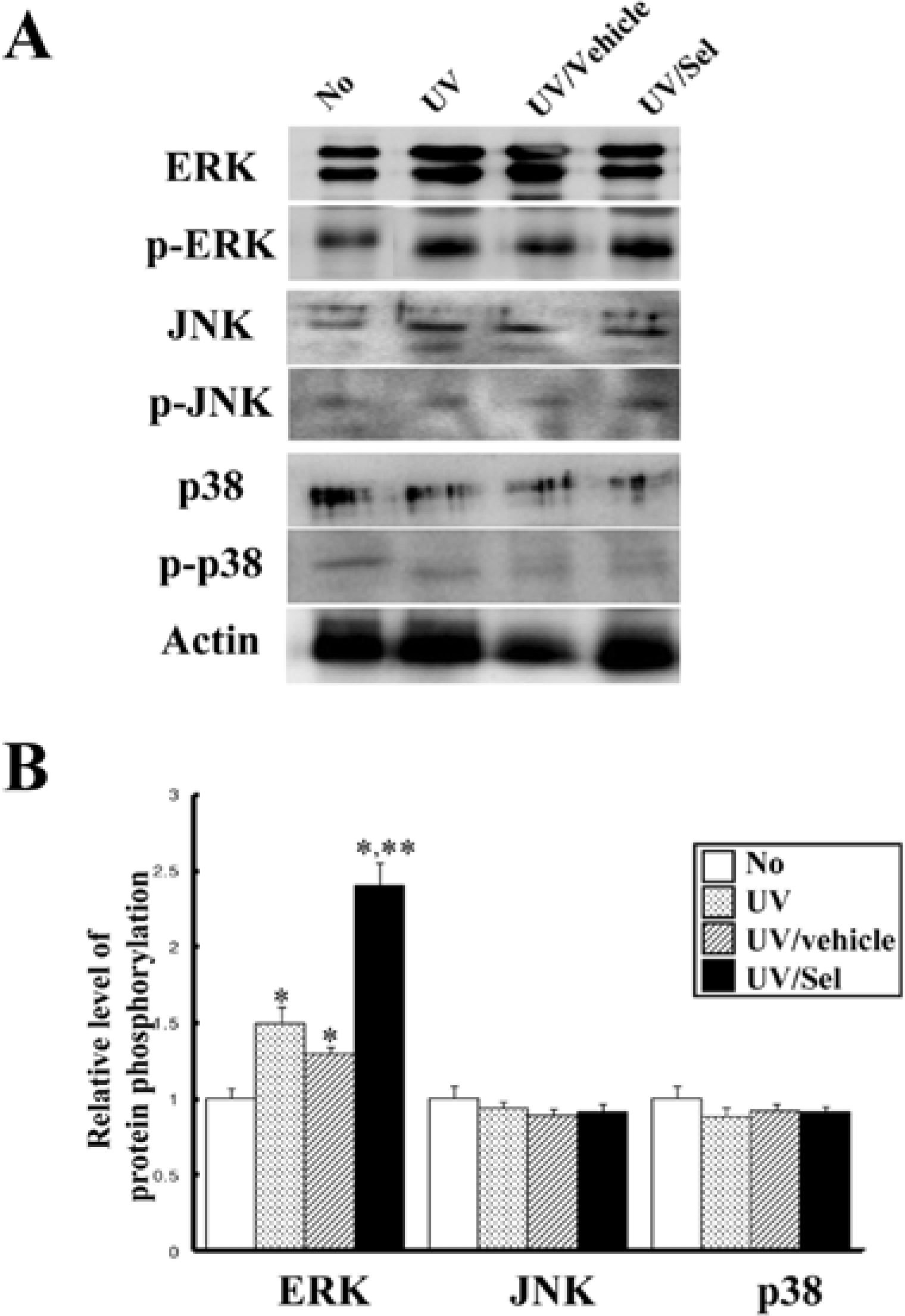
Figure 5.
Effect of selenium (Sel) on UV-induced MAPK protein expression using Western blot. Total cell lysate were prepared from skin tissues of control, UV-irradiation, UV/Vehicle and UV/ Sel group, as described in Materials and Methods. Fifty micrograms of protein per sample were immunoblotted with antibodies for each protein. Three samples were assayed in triplicate using Western blotting. The values are the mean±SD. ∗P<0.05 is the significance level compared to the control (No irradiation) group, ∗∗P<0.05 is the significance level compared to the vehicle applicated group.
 XML Download