

 PDF
PDF ePub
ePub Citation
 Print
Print


Abstract
We describe a case of tumoral calcinosis in a 13-month-old female Beagle dog presenting for surgical removal of a 4×3×3 cm mass in the soft tissues of the medial right shoulder joint. On radiologic examination, the mass showed increased radiopacity. Blood analysis data indicated chronic renal failure with hyperphosphatemia and hypercalcemia. Grossly, the mass was irregular, round and multilocular, with hard consistency. Histologically, there were many variable-sized loculi surrounded by capsule and interstitial connective tissues were generated among the loculi. Inflammatory cells, fibroblasts, fibrocytes and multinucleated giant cells were present at the margins of the loculi. Most of the loculi were filled with calcium or chalky material. To our knowledge, this is the first case of tumoral calcinosis in Korea.
REFERENCES
Balachandran S.., Abbud Y.., Prince M.J.., Chausmer A.B.1980. Tumoral calcinosis: scintigraphic studies of an affected family. Br. J. Radiol. 53(634):960–964.

Black J.R.., Sladek G.D.1983. Tumoral calcinosis in the foot and hand. A case report. J. Am. Pediatry Assoc. 73(3):153–155.
Chew D.J.., Nagode L.A.1992. Calcitriol in the treatment of chronic renal failure. In Current Veterinary Therapy XI Small Animal Practice (Kirk, R.W. and Bonagura, J.D. ed.), 11th ed., pp. 857-860, WB Saunders, Philadelphia.
Chew D.J.., DiBartola S.P.., Nagode L.A.., Starkery R.J.1992. Phosphorus restriction in the treatment of chronic renal failure. In Current Veterinary Therapy XI Small Animal Practice (Kirk, R.W. and Bonagura, J.D.ed.), 11th ed., pp. 853-857, WB Saunders, Philadelphia.
Cofan F.., Gracia S.., Combalia A.., Campistol J.M.., Oppenheimer F.., Ramon R.1999. Uremic tumoral calcinosis in patients receiving long term hemodialysis therapy. J. Rheumatol. 26(2):379–385.
Flo G.L.., Tvedten H.1975. Cervical calcinosis circumscripta in three related Great Dane dogs. J. Am. Vet. Med. Assoc. 11:507–510.
Garcia S.., Cofan F.., Fernandez de R.P.., Sala P.., Oppenheimer F.2002. Uremic tumoral calcinosis of the foot mimicking infection. Foot Ankle Int. 23(3):260–263.
Krawiec D.R.1987. Renal failure in immature dogs. J. Am. Anim. Hosp. Assoc. 23:101–107.
Legendre A.M.., Dade A.W.1974. Calcinosis circumscripta in a dog. J. Am. Vet. Med. Assoc. 164:1192–1194.
Malik M.., Acharya S.1993. Tumoral calcinosis of the fingers. A report of two cases. Int. Orthop. 17(5):279–281.
Mitnick P.D.., Goldfarb S.., Slatopolsky E.., Lemann J. Jr.., Gray R.W.., Agus Z.S.1980. Calcium and phosphate metabolism in tumoral calcinosis. Ann. Intern. Med. 92(4):482–487.
Nagode L.A.., Chew D.J.., Steinmeyer C.L.., Carothers M.A.1993. Renal secondary hyperparathyroidism: Toxic aspects, mechanism of development, and control by oral calcitriol treatment. Proc. 11th Annu. Vet. Meet. Coll. Vet. Intern. Med.154–157.
Prahinski J.R.., Schaefer R.A.2001. Tumoral calcinosis of the foot. Foot Ankle Int. 22(11):911–913.
Prince M.J.., Schaeffer P.C.., Goldsmith R.S.., Chausmer A.B.1982. Hyperphosphatemic tumoral calcinosis: association with elevation of serum 1,25-dihydroxycholecalciferol concentrations. Ann. Intern. Med. 96(5):586–591.
Roudebush P.., Maslin W.R.., Cooper R.C.1988. Canine tumoral calcinosis. Compend. Contin. Educ. Pract. Vet. 10:1162–1164.
Scott D.W.., Buerger R.G.1988. Idiopathic calcinosis circumscripta in the dog: A retrospective analysis of 130 cases. J. Am. Anim. Hosp. Assoc. 24:651–658.
Seawright A.A.., Grono L.R.1961. Calcinosis circumscripta in dogs. Aust. Vet. J. 37:421–425.
Smack D.., Norton S.A.., Fitzpatrick J.E.1996. Proposal for a pathogenesis-based classification of tumoral calcinosis. Int. J. Dermatol. 35(4):265–271.
Vaughan L.C.1962. The radiographic features of calcinosis circumscripta (Kalkgichy, calcium goat) in the dog. Vet. Rec. 74:988–989.
Viegas S.F.., Evans E.B.., Calhoun J.., Goodwiller S.E.1985. Tumoral calcinosis: a case report and review of the literature. J. Hand Surg. Am. 10(5):744–748.
Figure 1.
Radiographic appearance. Ventrodorsal view of right shoulder joint. Note the 3.7×2.7 cm mass with increased radiopacity in soft tissue of medial right shoulder joint. The mass shows round shape with irregular margin.
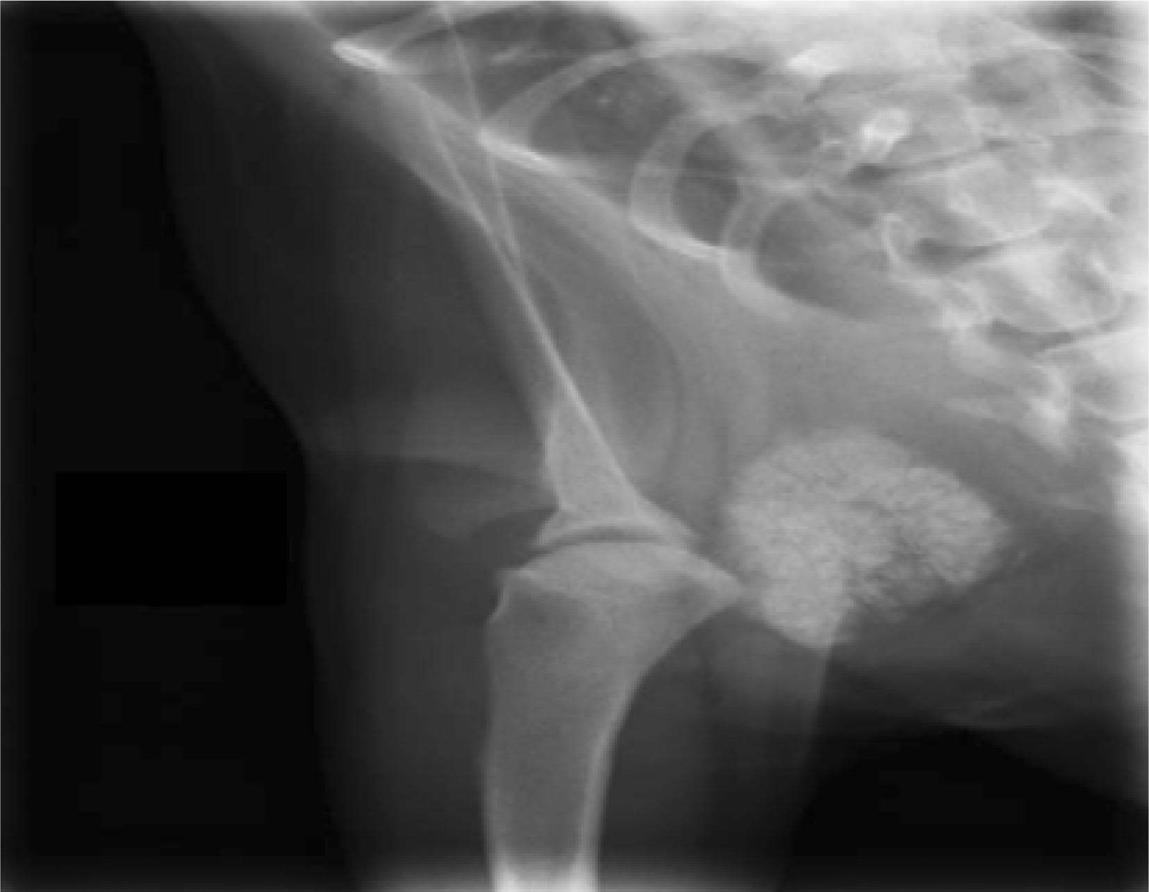
Figure 2.
Gross lesion of the mass. The mass of 4×4×3 cm was irregular and hard. The mass has multiple lobes with variable sizes.
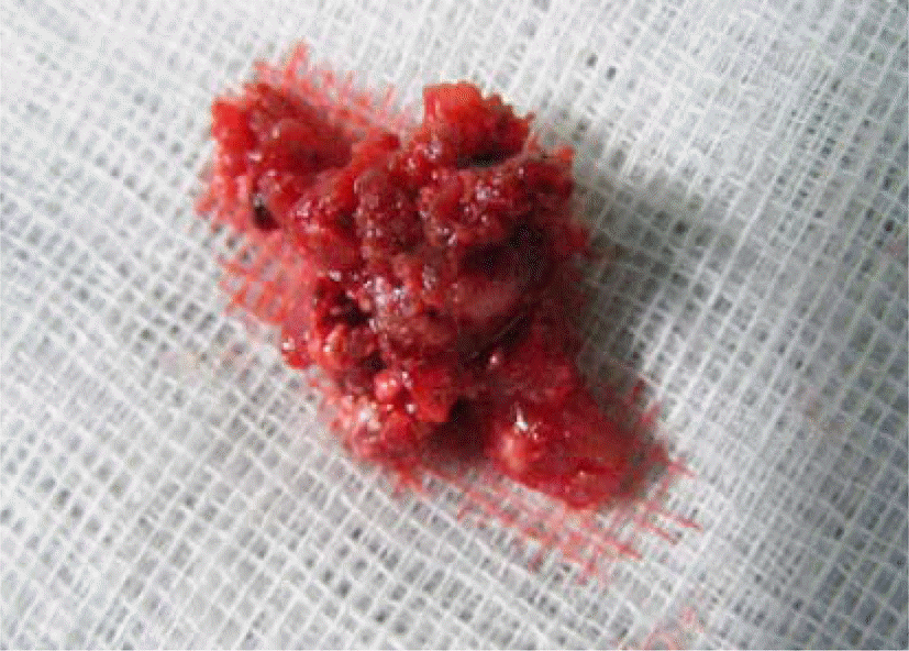
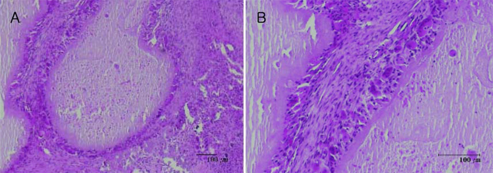
Figure 3.
Histologic lesion of tumoral calcinosis. The loculi were surrounded by the interstitial tissues. Note the calcium or chalky material in the lumen of the locule (A). Multinucleated giant cells, histiocytes, and fibroblasts form a cellular palisading along the margins of the locule (B). H & E stain.
 XML Download