

 PDF
PDF ePub
ePub Citation
 Print
Print


Abstract
A 12-year-old female Maltese dog brought to a local veterinary clinic with two nodules in the subcutis of left shoulder. The nodules were grown gradually from 2 years ago. The nodules were removed surgically under anesthesia and examined histopathologically. They were 15×26 mm in diameter (oval form) and 12×15 mm in diameter (round form), respectively. Histopathologically, the lesions consisted of multilayered basaloid cells in the peripheral of the mass and ghost cells in the central region. Typical findings of these two nodules were gradually keratinized basaloid cell toward central area result in forming anuclear ghost cells. These microscopic features of cutaneous nodules were diagnosed as pilomatrixoma and the identified findings were similar to fully developed stage of human pilomatrixoma. This report may assist in the categorizing of canine pilomatrixoma using histopathological features.
REFERENCES
Danny W.S.., Wayne I.A.1991. Canine hair follicle neoplasms: a retrospective analysis of 80 cases (1986-1987). Veterinary Dermatology. 2:143–150.
Du Vivier A.., McKee P.1993. Benign tumours of the skin. In Atlas of Clinical Dermatology (Du Bivier, A. ed.), pp.8.11-8.12, Gower Medical Publishing, London.
Forbis R.., Helwig E.B.1961. Pilomatrixoma (calcifying epithelioma). Arch. Dermatol. 83:606–618.

Goldshmidt M.H.., Shofer F.S.1992. Skin Tumours of the Dog and Cat. 125–130. Butterworth-Heinemann, Oxford.
Grabczynska S.A.., Budny P.., Calonjet E.., Ratnavel R.2002. Case 3: Multiple familial pilomatrixoma. Clin. Exp. Dermatol. 27:343–344.
Gross T.L.., Ihrke P.J.., Walder E.J.1992. Veterinary Dermatopathology, pp. 365–367. Mosby Year Book, St. Louis.
Hashimoto K.., Lever W.F.1999. Tumor of skin appendages. In Dermatology in General Medicine (Fitzpatrick, T.B. ed.), 5th ed., pp.890–914. McGraw-Hill, New York.
Kaddu S.., Soyer H.P.., Hodl S.., Kerl H.1996. Morphological stages of pilomatricoma. Am. J. Dermatopathol. 18:333–8.
Kaddu S.., Soyer H.P.., Wolf I.H.., Kerl H.1997. Proliferating pilomatricoma. A histopathologic simulator of matrical carcinoma. J. Cutan. Pathol. 24:228–234.
MacKie R.M.1998. Tumours of the skin appendages. In Textbook of Dermatology (Champion, R.H. ed.), 6th ed., pp.1685–1715. McGraw-Hill, New York.
McKee P.1999. Essential Skin Pathology. p. 160. Mosby International Ltd;London:
Moehlenbeck F.W.1973. Pilomatrixoma (calcifying epithelioma). A statistical study. Arch. dermatol. 108:532–534.
Theilen G.H.., Medewell B.R.1987. Veterinary Cancer Medicine. pp.257, Lea and Febiger, Philadelphia.
Toma S.., Noli C.2005. Isotretinoin in the treatment of multiple benign pilomatrixomas in a mixed-breed dog. Veterinary Dermatology. 16:346–350.
Walsh J.S.., Fairley J.A.1999. Cutaneous mineralization and ossification. In Dermatology in General Medicine (Fitzpatrick, T.B. ed.), 5th ed., pp.1829–1835. McGraw-Hill, New York.
Wang J.., Cobb C.J.., Martin S.E.., Venegas R.., Wu N.., Greaves T.S.2002. Pilomatrixoma: Clinicopathologic study of 51 cases with emphasis on cytologic features. Diagn. Cytopathol. 27:167–172.
Figure 1.
A: Dome-shaped surface with mild pigmentation and alopecia. B: Creamy-white to yellowish color, firm, well-lobulated and solitary appearance on the cut sections.
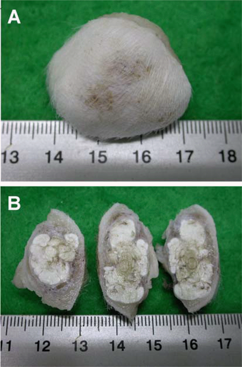
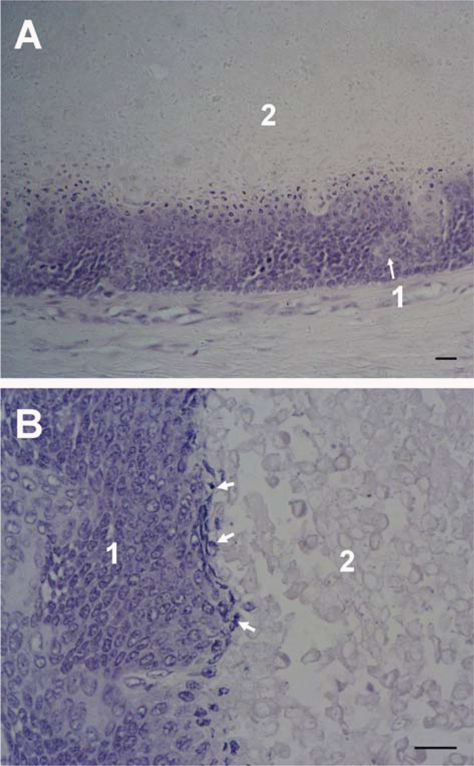
Figure 2.
A: The subcutaneous cyst showed enlarged lumen and irregularly shaped and densely packed basaloid cells (1) in the rim. Gradually degenerated cells, ghost cells (2), were in central lesion. B: In the larger magnification of A, basophilic basaloid cells (1) showed nuclear mitosis (arrow) in the junction of degenerated cells. Ghost cell (shadow cells) (2) were disappeared nuclei and poorly stained but maintaining their outline. H&E stain. Scale bar=150 µm.
 XML Download