Abstract
Chronic spontaneous urticaria (CSU) is a complex idiopathic disease of the skin with various cellular infiltrations. Although mast cells are key effector cells in the pathogenesis of CSU, CD4+ T helper 2 cells also have particular roles in the development and maintenance of CSU. Periostin is known as a downstream molecule of interleukin (IL)-4 and IL-13, key cytokines of type 2 immune responses. In this study, we examined periostin and IL-13 levels in the sera of patients with CSU (n=84) and healthy normal controls (NCs, n=43). Periostin levels were significantly lower in the CSU group than in NCs (71.4±21.8 vs 85.1±22.4 ng/mL, P=0.04). Periostin levels were also lower in the severe CSU group than those in mild CSU (59.7±18.0 vs 73.4±22.0 ng/mL, P=0.04). However, IL-13 levels were significantly higher in patients with CSU than in NCs (508.5±51.2 vs 200.7±13.3 pg/mL, P=0.001). In conclusion, periostin and IL-13 may be independently related to the pathogenesis of CSU.
Chronic spontaneous urticaria (CSU) is defined as urticaria that persists for at least 6 weeks.12 Apart from mast cells which are the major effector cell type in urticaria, other cells of the skin tissue are infiltrated in CSU patients, including CD4+ T lymphocytes (especially the Th2 subclass), eosinophils, and neutrophils.34 A few studies demonstrated that Th2 cytokines, such as interleukin (IL)-4 and IL-13, are increased in the sera of CSU patients or their production is increased in peripheral mononuclear cells from CSU patients.56
Periostin, an extracellular matrix protein, plays an important role in the pathogenesis of chronic allergic inflammatory diseases, such as bronchial asthma and atopic dermatitis.789101112 IL-4 and IL-13, key cytokines of the Th2-type immune response, have considerable influences on the synthesis and amplification of periostin by stimulating fibroblasts, the major source of periostin.7 It has been demonstrated that serum periostin levels are the most significant single predictor of eosinophilic airway inflammation in asthmatics, rather than fraction of exhaled nitric oxide (FENO), blood eosinophil count, or serum IgE levels, suggesting that periostin is a useful biomarker of eosinophilic airway inflammation in asthmatics and Th2-targeted asthma therapy.8 Furthermore, Periostin levels has been shown to be well correlated with disease severity and chronicity in atopic dermatitis.12 In this regard, periostin might be involved in the pathogenesis of CSU, for which a Th2 immune response has been implicated, and periostin levels could be a biomarker of disease severity or treatment response in CSU.
Thus, we measured serum concentrations of periostin and IL-13 in patients with CSU to evaluate the significance of periostin and IL-13 in the pathogenesis of CSU and possible application as a biological marker of disease severity.
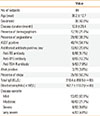
Eighty-four patients with CSU and 43 normal healthy controls (NCs) were included in this study. CSU was diagnosed as daily appearance of wheals and associated itching sensation lasting at least 6 weeks when any known causes of chronic urticaria were ruled out by history, and physical and laboratory evaluations. We excluded patients with clinical evidence of urticaria vasculitis and physical urticaria, such as dermographism, cholinergic urticaria, and cold urticaria. NCs had no history of allergic diseases with a mean age of 30.4±9.3 years, and 20.9% were men. The characteristics of the patients (n=84) are shown in Table. The study was approved by the Institutional Review Board of Hallym University Dongtan Sacred Heart Hospital, and each subject gave informed consent.
In most cases, serum total IgE (normal <100 IU/mL), antinuclear antibody and antithyroid antibodies, including antithyroid peroxidase (anti-TPO) antibodies (normal <60 U/mL), antithyroglobulin (anti-TG) antibodies (normal <60 U/mL), and anti-thyroid stimulating hormone receptor (anti-TSH-R) antibodies (normal <1.75 IU/L), were evaluated. Total IgE and antithyroid antibody levels were measured using chemiluminescence immunoassay (Cobas e 411, Roche, Germany for total IgE; ADVIA Centaur® XP, Siemens, Germany for anti-TPO and anti-TG antibody and anti-TSH-R antibody). Atopy was defined as 1 or more positive reactions to 12 common inhalant allergens by allergy skin prick tests or simultaneous multiple-allergen tests (inhalant panel, AdvanSure AlloScreen®, LG, Seoul, Korea): Dermatophagoides (D.) pteronyssinus, D. farinae, birch, oak, grass mix, ragweed, mugwort, Japanese hop, Alternaria, Aspergillus, and dog and cat epithelia.
CSU patients were divided into 4 groups according to disease severity, which was decided by treatment levels modified from the guidelines for the treatment of chronic urticaria1:mild, well controlled with 1 or 2 second-generation antihistamine(s); moderate, well controlled with 3 or more second-generation antihistamines; severe, well controlled with addition of leukotriene receptor antagonists and/or H2-antagonists; very severe, not adequately controlled with level 3 treatment and added cyclosporine or omalizumab.
An autologus serum skin test (ASST) was performed following the method recommended by EAACI/GA2LEN at least 3 days after stopping all the medications for urticaria, including antihistamines and corticosteroids,13 and the remaining serum was preserved at -80℃ until periostin and IL-13 were measured. Wheal with a diameter of at least 1.5 mm greater than that of a control wheal induced by saline was defined as positive.
Periostin levels were measured with a proprietary sandwich enzyme-linked immunosorbent assay (ELISA; Shino-test, Kanagawa, Japan) as previously reported using anti-periostin antibody (clones SS18A and SS17B).12 IL-13 levels were also measured using an ELISA kit (Millipore, Billerica, MA, USA) according to the manufacturer's instructions.
Data are expressed as mean±SD. Comparisons between CSU patients and NCs were performed using Chi-square and Fisher's exact tests for categorical variables and using Student's t test for continuous variables. Comparisons of periostin and IL-13 levels between CSU patients and NCs were performed using analysis of covariance for control of age and sex. Correlations between periostin and IL-13, and between periostin/IL-13 and clinical parameters were examined using Pearson's correlation coefficients. A P value of less than 0.05 was considered significant. Statistical analyses were performed using SPSS statistics 21 (IBM SPSS Inc., Chicago, IL, USA).
Periostin levels were significantly lower in CSU patients than those in NCs (ng/mL, mean±SD, 71.4±21.8 vs 85.1±22.4, P=0.04; Fig. 1A). There were no significant differences in periostin levels according to the presence of angioedema, dermographism, atopy, antinuclear antibody, thyroid autoantibody, or the positivity of ASST. Periostin levels were not significantly correlated with IL-13 levels, total IgE levels, or blood eosinophil count. Interestingly, Severe to very severe group of CSU patients had lower periostin levels compared to the mild to moderate group of CSU patients with borderline significance (59.7±18.0 vs 73.4±22.0 ng/mL, P=0.04, Fig. 2).
On the contrary, IL-13 levels were higher in CSU patients compared to the NCs (508.5±51.2 vs 200.7±13.3 pg/mL, P=0.001, Fig. 1B). However, IL-13 levels were not significantly different between the presence and the absence of angioedema, dermographism, atopy, antinuclear antibody, thyroid autoantibody, or the positivity of ASST. IL-13 levels were not significantly correlated with serum total IgE levels or blood eosinophil count. When we analyzed an association of IL-13 levels with treatment level of CSU patients, there were no significant differences in IL-13 levels between treatment levels.
In this study, we demonstrated that IL-13 levels in serum were significantly increased in CSU patients compared to NCs (P=0.001), while serum periostin levels were decreased in CSU patients compared to NCs with borderline significance (P=0.04). Furthermore, serum periostin levels were significantly lower in the severe to very severe group than in the mild to moderate group of CSU patients (P=0.04).
Although CSU is a complex idiopathic disease that may be triggered by various etiologic factors, it has been indicated that cutaneous mast cells are major effector cells in most cases. However, mast cell degranulation is followed by many cellular infiltrations that are responsible for changes in vasopermeability, upregulation of adhesion molecules on endothelial cells, and rolling and attachment of blood leukocytes.5 CD4+ T-lymphocytes of the Th2 subclass particularly affect late-phase responses, such as tender, deep, erythematous, or poorly demarcated areas of swelling that persist for up to 24 hours.6 Elevation in the Th2-initiating cytokines, such as IL-33, IL-25, and thymic stromal lymphopoietin (TSLP), was recently demonstrated in the lesional skin of CSU patients.14
Periostin is an intrinsic mediator of the amplification and chronicity of allergic skin inflammation.9 A positive correlation between periostin and key Th2 cytokines, such as IL-4 and IL-13, has been well established in bronchial asthma and atopic dermatitis.789 Caproni et al.6 showed that serum IL-13 levels were significantly higher in CSU patients than healthy controls and even higher in the ASST-positive group than in the ASST-negative group. They also found that CSU patients with higher IL-13 levels have more frequent and diffuse exacerbations and severe itch, suggesting that IL-13 might be a serologic marker of disease activity. However, we could not find any association between IL-13 levels and clinical manifestation. Serum IL-4 levels did not show any consistency among studies.56 Moreover, periostin has not yet been investigated in chronic urticaria.
It is noteworthy that the two Th2 markers behaved independently in CSU patients. Although periostin expression may be upregulated by Th2 cytokines, other unknown factors that suppress or downregulate periostin expression could be present in CSU patients. Unlike in atopic dermatitis or asthma, more complicated cellular infiltrates, followed by the release of various inflammatory cytokines, are involved in CSU pathogenesis, although Th2 immune responses might be critical in the development and maintenance of CSU. As it is well established that fibroblasts are major producers of periostin and that periostin contributes to the development of fibrosis of the airways and skin, the activity of dermal fibroblast may be decreased in chronic urticaria. Further studies are needed to elucidate fibroblast activity in CSU patients to support our findings.
Recently, Yang et al.15 reported that histamine was found to directly induce periostin expression whereas the expression levels of IL-4 and IL-13 were not altered by histamine stimulation. Lower periostin levels in CSU patients in this study might be influenced by antihistamine treatment. This might be another explanation for the discrepancy between periostin and IL-13 levels in CSU patients in this study. In addition, they also demonstrated that periostin induction was inhibited by H1 antagonist treatment in vitro and was attenuated in histamine receptor 1 knock-out mice. It could be the reason periostin levels were further decreased in severe CSU patients taking more antihistamines.
Our study has two limitations. One is the relatively small number of study subjects. In addition, the number of the severe to very severe group patients (n=12) was much smaller than the mild to moderate group patients (n=71) during data analyses according to the disease severity. The comparison between these 2 groups does not seem appropriate. The other is that the urticaria activity score was not assessed. The severity and long-term prognosis of the disease can also be estimated based on the treatment level for disease control. Therefore, the association between the serum biomarker and treatment level could have clinical implications as a prognostic factor of the disease. In the present study, periostin levels were significantly lower in the severe to very severe group than in the mild to moderate group. However, caution is needed before concluding that periostin levels may be a prognostic indicator or a severity marker of CSU and further studies with a larger sample size are required to confirm our results.
Figures and Tables
 | Fig. 1(A) Serum periostin and (B) IL-13 levels in CSU patients and NCs. CSU, chronic spontaneous urticaria; NCs, normal controls. |
Table
Clinical characteristics of patients with chronic spontaneous urticaria

Values are presented as means±SD. In case of missing data, the number of subjects for whom data are available is indicated next to the value.
ASST, autologous serum skin test; ANA, antinuclear antibody; M, male; TG, thyroglobulin; TPO, thyroid peroxidase; TSH-R, thyroid stimulating hormone receptor.
ACKNOWLEDGMENTS
This study was supported by a grant from the Basic Science Research Program through the National Research Foundation of Korea (NRF), as funded by the Ministry of Education, Science and Technology (2012R1A1A1012349).
References
1. Zuberbier T, Aberer W, Asero R, Bindslev-Jensen C, Brzoza Z, Canonica GW, et al. Methods report on the development of the 2013 revision and update of the EAACI/GA2 LEN/EDF/WAO guideline for the definition, classification, diagnosis, and management of urticaria. Allergy. 2014; 69:e1–e29.
2. Losol P, Yoo HS, Park HS. Molecular genetic mechanisms of chronic urticaria. Allergy Asthma Immunol Res. 2014; 6:13–21.
3. Ying S, Kikuchi Y, Meng Q, Kay AB, Kaplan AP. TH1/TH2 cytokines and inflammatory cells in skin biopsy specimens from patients with chronic idiopathic urticaria: comparison with the allergen-induced late-phase cutaneous reaction. J Allergy Clin Immunol. 2002; 109:694–700.
4. Zweiman B, Haralabatos IC, Pham NC, David M, von Allmen C. Sequential patterns of inflammatory events during developing and expressed skin late-phase reactions. J Allergy Clin Immunol. 2000; 105:776–781.
5. Ferrer M, Luquin E, Sanchez-Ibarrola A, Moreno C, Sanz ML, Kaplan AP. Secretion of cytokines, histamine and leukotrienes in chronic urticaria. Int Arch Allergy Immunol. 2002; 129:254–260.
6. Caproni M, Cardinali C, Giomi B, Antiga E, D'Agata A, Walter S, et al. Serological detection of eotaxin, IL-4, IL13, INF-γ, MIP-1α, TARC and IP-10 in chronic autoimmune urticaria and chronic idiopathic urticaria. J Dermatol Sci. 2004; 36:57–59.
7. Takayama G, Arima K, Kanaji T, Toda S, Tanaka H, Shoji S, et al. Periostin: a novel component of subepithelial fibrosis of bronchial asthma downstream of IL-4 and IL-13 signals. J Allergy Clin Immunol. 2006; 118:98–104.
8. Jia G, Erickson RW, Choy DF, Mosesova S, Wu LC, Solberg OD, et al. Periostin is a systemic biomarker of eosinophilic airway inflammation in asthmatic patients. J Allergy Clin Immunol. 2012; 130:647–654.e10.
9. Masuoka M, Shiraishi H, Ohta S, Suzuki S, Arima K, Aoki S, et al. Periostin promotes chronic allergic inflammation in response to Th2 cytokines. J Clin Invest. 2012; 122:2590–2600.
10. Wenzel SE. Asthma phenotypes: the evolution from clinical to molecular approaches. Nat Med. 2012; 18:716–725.
11. Izuhara K, Arima K, Ohta S, Suzuki S, Inamitsu M, Yamamoto K. Periostin in allergic inflammation. Allergol Int. 2014; 63:143–151.
12. Kou K, Okawa T, Yamaguchi Y, Ono J, Inoue Y, Kohno M, et al. Periostin levels correlate with disease severity and chronicity in patients with atopic dermatitis. Br J Dermatol. 2014; 171:283–291.
13. Konstantinou GN, Asero R, Maurer M, Sabroe RA, Schmid-Grendelmeier P, Grattan CE. EAACI/GA(2)LEN task force consensus report: the autologous serum skin test in urticaria. Allergy. 2009; 64:1256–1268.
14. Kay AB, Clark P, Maurer M, Ying S. Elevations in Th2-initiating cytokines (IL-33, IL-25, TSLP) in lesional skin from chronic spontaneous ("idiopathic") urticaria. Br J Dermatol. 2014; 172:1294–1302.
15. Yang L, Murota H, Serada S, Fujimoto M, Kudo A, Naka T, et al. Histamine contributes to tissue remodeling via periostin expression. J Invest Dermatol. 2014; 134:2105–2113.


 PDF
PDF ePub
ePub Citation
 Print
Print


 XML Download