

 PDF
PDF ePub
ePub Citation
 Print
Print


INTRODUCTION
Allergic diseases, including asthma, allergic rhinitis, and atopic dermatitis, are mediated by allergen-triggered hyperreactivity and have been prevalent worldwide in recent decades.123 It is clear that the allergic diseases result from chronic inflammation mediated by repeated exposure to allergens.4 Th2-polarized immune responses mainly contribute to the progress and persistence of allergic diseases and are orchestrated by various immune cells and integral mediators such as cytokines.56 Based on the prevailing view so far, it is evident that cytokine network plays an important role in Th2-mediated allergic responses through controlling the balance between Th1 and Th2 cells.7 However, distinguishing allergic phenotypes on the basis of the type 1 and type 2 cytokine profiles has a limitation due to pleiotropic effects of cytokines.6 To better understand redundant functions of cytokine cascades, we need more information about the balance of Th1 and Th2 responses as well as the allergy-related cytokine profiles in human on normal or allergic conditions.
Allergic diseases have a unique characteristic that symptoms of each allergic disease manifest in a predictable sequence by age, termed the 'allergic (or atopic) march.'89 Early childhood which has immature immune systems and is the onset of allergic sensitization, is thought to be critical to the development and progress of allergic disease. Barcharier et al.10 have recently reported that origins of asthma are mainly in pre-schooled children and that early interventions for children with risk factors are effective in preventing ensuing asthma. However, the patterns and differences of allergy-related parameters on an immunological basis during childhood, as critical period of allergic disease, have not yet been established.
This study was conducted to determine Th1/Th2 ratios and cytokines, including chemokines and growth factors, across a wide range of ages in non-atopic healthy individuals. We also examined whether there is significant differences immunological parameters between healthy and allergic individuals during childhood.
MATERIALS AND METHODS
Study subjects and design
In total, 137 of normal children and adolescents under the age of 15 years were recruited together with 119 of adults over the age of 20 years. All of these participants were defined as healthy by physician's diagnosis. All of the healthy participants experienced no allergic symptoms, such as wheezing, and were defined as non-atopic by an allergen-specific IgE test or a skin prick test (SPT). Seven allergens were used for the specific IgE test (ThermoFisher, Scientific, Uppsala, Sweden): Dermatophagoides farinae (Der f), cat dander, dog dander, cockroach, Alternaria, mugwort, and alder. Thirteen allergens were applied for the SPT: Der f, Dermatophagoides pteronyssinus (Der p), cat dander, dog dander, cockroach, grass, alder, birch, oak, ragweed, mugwort, Alternaria, and Aspergillus. The individuals have experienced neither wheezing nor other allergic symptoms during at least the preceding 6 weeks.
Eighty-seven allergic children and adolescents under the age of 17 years who had a positive reaction to more than 1 allergen were recruited and compared to the healthy participants. All of the allergic patients suffered from more than 1 of the 4 allergic diseases (asthma, allergic rhinitis, atopic dermatitis, and food allergy), and blood was withdrawn when they visited the hospital.
Peripheral blood mononuclear cells (PBMCs) and plasma were obtained from all of the participants and examined by flow cytometric analysis and the Multiplex assay. This study was approved by the Institutional Review Board (2012-11EXP-02-3C, 2011-04-EXP-02-R, 2010-02CON-14-P), and informed consent was obtained from each individual prior to participation.
Intracellular cytokine staining and flow cytometric analysis
Cell Preparation Tube (BD Vacutainer® CPT™; BD Biosciences, San Jose, CA, USA) which contains citrate anticoagulant was used to obtain whole blood for the rapid isolation of fresh mononuclear cells. PBMCs were isolated by centrifuging the tube according to the manufacturer's instructions (1,500 g, 20 min, at 20℃) within 24 hours of the blood being withdrawn and then incubated for 4 hours in RPMI1640 media (Gibco, Grand Island, NY, USA) with 10% fetal bovine serum (FBS; Gemini Bio-Product, West Sacramento, CA, USA) containing phorbolmyristate acetate (50 ng/mL, Sigma-Aldrich, St. Louis, MO, USA), ionomycin (1 µg/mL, Sigma-Aldrich), and Golgi-Plug (BD Biosciences). After stimulation, the cells were stained with anti-CD3-fluorescein isothiocyanate (BD Biosciences) and anti-CD4-energy-coupled dye (Beckman-Coulter, Brea, CA, USA) monoclonal antibodies, and then fixed and permeabilized using the Cytofix/Cytoperm Plus kit (BD Biosciences) according to the manufacturers' protocols. Th1 and Th2 cells were regarded as IFN-γ- or IL-4-expressing helper T cells, respectively. For intracellular cytokine staining, permeabilized cells were stained with anti-IFN-γ-allophycocyanin and anti-IL-4-phycoerythrin monoclonal antibodies (both BD Biosciences). Finally, the cells were washed and resuspended in PBS containing 10% FBS and analyzed using an FC-500 cytometer (Beckman-Coulter). After gating on helper T cells (CD3+ CD4+ cells) in the PBMCs, IFN-γ- or IL-4-expressing cells were analyzed simultaneously.
Analysis of soluble factors by multiplex assay
Plasma chosen randomly from participants was used for the analysis: 16 healthy children and 19 allergic children, 15 healthy adolescents and 11 allergic adolescents. The levels of 22 soluble factors in plasma were measured in duplicate using a customized Procarta® cytokine assay kit (Affymetrix/Panomics, Santa Clara, CA, USA) according to the manufacturer's instructions. Briefly, antibody beads that bound to each targeted cytokine or chemokine were first added on pre-wet filter plate. After washing, samples and standard were added with assay buffer, followed by incubation with 1-hour shaking. Washing the plate again, premixed detection antibodies were added, and the plate was incubated for 30 minutes with shaking. Finally, the plate was incubated for 30 minutes again with streptavidin-PE, followed by analysis using a Luminex instruments (Bio-Plex® 200 systems; Bio-rad, Hercules, CA, USA). The following soluble factors were measured: interleukin (IL)-2, IL-4, IL-5, IL-6, IL-10, IL-12p70, IL-13, IL-15, IL-17, Interferon (IFN)-γ, Tumor Necrosis Factor (TNF)-α, Transforming Growth Factor (TGF)-β, Chemokine C-C motif Ligand 2 (CCL2)/Monocyte Chemotactic Protein (MCP)-1, CCL7/MCP-3, CCL11/Eotaxin, Chemokine C-X-C motif Ligand 5 (CXCL5)/Epthelial cell-derived Neutrophil Activating peptide-78 (ENA-78), CXCL10/Interferon gamma-induced Protein 10 (IP-10), Vascular Endothelial Growth Factor (VEGF), Nerve Growth Factor (NGF), Platelet-Derived Growth Factor BB (PDGFBB), Leptin, and IL-1 receptor antagonist (IL-1ra).
RESULTS
Changes in Th1/Th2 ratios in healthy individuals with age
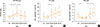
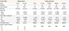
In total, 256 non-atopic healthy and 87 allergic individuals were participated in this study, and their characteristics are summarized in Table. Peripheral blood mononuclear cells (PBMCs) were isolated from the non-atopic healthy participants to evaluate age-related changes in Th1/Th2 ratios. As shown in Fig. 1A, Th1/Th2 ratios were significantly increased from the 3-9 to 20-39 age groups, were decreased in the 40-59 age group, and remained stable thereafter. We also found that the percentages of Th1 cells were gradually increased between the ≥10 age groups and significantly decreased in the 20-39 age group. In contrast, the percentages of Th2 cells in the 10-19 and 20-39 age group was significantly lower than in other age groups and increased again in the 40-59 age group (Fig. 1B and C). Therefore, these results demonstrated that the Th1/Th2 ratio in non-atopic healthy individuals changed with age and that the peak value of Th1/Th2 ratios in young adults was due to the decrease in the percentage of Th2 cells as well as the difference in increased points between Th1 and Th2 cell percentages.
Changes in the levels of cytokines in healthy individuals with age
A total of 22 cytokines, including 5 chemokines and 3 growth factors (Supplemental Table 1), were assessed simultaneously by using the Multiplex assay to investigate whether profiles of cytokines could be associated with changes in Th1/Th2 ratios and cell percentages according to increasing age in healthy individuals. It was found that plasma cytokine levels were increased 2-4 times from the 3-9 age group to the 10-19 age group, and most of them were stabilized or peaked in adults (20-39 and 40-59 age groups), and decreased in the 60-79 age group (Supplemental Table 2). Although there were no unique patterns in the levels of 22 cytokines, this result may support the importance of the growth period (3 to 19 years of age) for the establishment of the immune system.
Comparison of Th1/Th2 ratios between healthy and allergic individuals
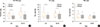
The sharp increase in cytokines during the growth period led us to identify the differences in immunological parameters between allergic and healthy children or adolescents. We thus investigated the ratio of Th1/Th2 in allergic and healthy individuals during childhood and adolescence to find out the effect of allergic diseases on the ratio. The ratio of Th1/Th2 cells was significantly lower in allergic patients than in healthy controls during both childhood and adolescence (Fig. 2A). The percentage of Th1 cells was consistently lower in allergic patients than in healthy controls during both childhood and adolescence, whereas that of Th2 cells did not differ between allergic and healthy individuals (Fig. 2B and C). These results suggest that the difference in Th1/Th2 ratios between allergic and healthy individuals was due to the decline in the percentage of Th1 cells in the blood, but not due to the increase in the percentage of Th2 cells.
Increases in allergy-related soluble factors in allergic children
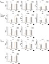
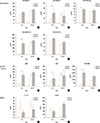
We finally investigated the plasma levels of 22 cytokines in healthy and allergic participants to find out their effect on allergic responses. As a result, the plasma levels of 9 cytokines (IL-4, IL-5, IL-12p70, IFN-γ, IL-17, TNF-α, IL-2, IL-10, and TGF-β) were dramatically increased in allergic children (3 to 9 years of age) compared to healthy controls (Fig. 3). Particularly, the majority of cytokines that were increased in allergic children reached levels similar to those observed in healthy adolescents that had higher levels. The plasma levels of 2 chemokines (CCL7/MCP-3 and CXCL5/ENA-78) and all the growth factors (VEGF, NGF, and PDGFBB) were also increased in allergic children compared to healthy controls (Fig. 4). Overall, the levels of the majority of soluble factors associated with allergy were significantly higher in allergic children than in healthy controls, and these differences disappeared during adolescence.
DISCUSSION
We demonstrated that the Th1/Th2 ratio and allergy-related cytokines changed with age in non-atopic healthy individuals. Previous studies have reported aging effects on the immune response. The production of naïve lymphocytes declines with age,11 and CD4 and CD8 T cells especially CD8 T cells undergo phenotypic changes with age.12 Some research on underlying mechanisms of atopic march have indicated that the nature of allergic responses changes with age.913 It has been also reported that various serum cytokines of healthy individuals show up and down patterns with age.14 However, investigations of multiple immune factors in the same participants are required to monitor immunological changes according to age and to establish reference values of immune factors related to allergic responses. We thus examined the Th1/Th2 ratio and the plasma levels of 22 cytokines in normal healthy participants from all the age groups. In this study, it was found that the Th1/Th2 ratio in healthy participants was gradually increased from early childhood, peaked in the 20-39 age group, and then decreased and stabilized in the ≥40 age groups. The peak was due to the decrease in Th2-cell portions and the difference in increased points between Th1-and Th2-cell percentages. It was also found that allergy-related cytokines had diverse trends according to age, but that most of them were sharply increased from childhood to adolescence. Although the Th1/Th2 balance hypothesis remains controversial,15 the Th1/Th2 balance could be one of the key determinants in the susceptibility and development of various immune-related diseases, such as allergy. Therefore, those results could help understand the relationship between aging and the immune system and provide more information about immunological changes in normal individuals.
We characterized differences in Th1/Th2 ratios and plasma levels of allergy-related cytokines between allergic and healthy individuals during childhood and adolescence. Allergic children and adolescents had a lower Th1/Th2 ratio than healthy controls, and the low ratio of Th1/Th2 was due to the decrease in Th1-cell portions. A previous study has reported that patients with allergic bronchopulmonary aspergillosis (ABPA) have decreased IFN-γ producing CD3+ cells and unchanged IL-4 producing CD3+ cells compared to healthy controls,16 which is consistent with the result of our study. Further studies are needed to determine the cause of the decrease in Th1 cells and its role in allergic diseases.
It is supposed that allergy-related cytokines would be differently regulated and expressed according to their function in allergic responses. However, our results showed that most of the cytokines were expressed at a significantly higher level in allergic children than in healthy controls during childhood regardless of their functions, whereas the levels of the immunological factors did not differ between healthy and allergic individuals during adolescence. In other words, healthy cases showed the lowest level of cytokines during childhood, followed by a sharp increase during adolescence, but allergic cases had a high level of cytokines during childhood.
The levels of 14 cytokines were markedly increased in children with allergic diseases, but not in healthy children. The 14 cytokines included Th2 cytokines (IL-4 and IL-5), Th1 and proinflammatory cytokines (IL-12p70, IL-17, IFN-γ, and TNF-α) chemokines (CCL7/MCP-3 and CXCL5/ENA-78), and others (IL-2, IL-10, TGF-β, VEGF, PDGFBB, and NGF). Several studies have shown that asthmatic patients have increased serum levels of Th2 cytokines during both adulthood17 and childhood,18 and that serum IL-17 levels correlate significantly with the severity of allergic rhinitis.19 It has been demonstrated that the plasma concentrations of IL-10, IL-12, and IL-13 are higher in asthmatic patients compared to healthy controls20 and that baseline levels of serum TNF-α are higher in asthmatic children than in healthy controls.21 The results of previous studies are consistent with ours. However, it is difficult to interpret the overall increase in various allergy-related cytokines because their patterns are not consistent with their functions. More investigation should be performed to determine mechanisms for increases in various cytokines during childhood and their definite roles in allergic responses.
Interestingly, PDGFBB was increased remarkably in allergic children compared to healthy individuals. PDGFBB is a homodimeric isoform of PDGF modulating fibroblastic activity and airway remodeling.22 Several studies have reported that PDGFBB augments the expression of procollagen I in airway fibroblasts of patients with severe asthma,22 so that it is a pro-wheezing biomarker in preterm children.23 However, the function of PDGFBB in allergic diseases remains unclear. This study suggests that the elevated level of PDGFBB could be a marker of allergic diseases in children, and we plan to investigate further age-related changes in PDGFBB levels in healthy and allergic adults to clearly reveal their functions in allergic responses.
We demonstrated the significance of childhood for the onset of allergic disease in terms of higher levels of cytokines in children with allergic disease compared to healthy controls. It has already been suggested that atopic diseases appear in the first decade of life,24 and Stern et al.25 have also reported that atopic symptoms, such as wheezing and bronchial hyperresponsiveness, during early childhood could be predictors of adult asthma. In other words, childhood is a critical period for the development of allergic diseases, and it is thought that some immunological markers can predict risk of allergy during childhood.
Although more studies with samples derived from specific tissues or organs are needed to examine local allergic responses, our findings may have clinical implications in reference plasma values of multiple cytokines for healthy individuals and comparison of their levels between patients and normal subjects. Further studies in terms of sampling time, results of clinical tests, and symptoms of participants are required to the plasma levels of cytokines to give more meaningful information.
In summary, our results indicated that Th1/Th2 ratios showed age-related changes in normal healthy individuals, increased in the growth period, and decreased with age. The Th1/Th2 ratio and the plasma levels of various cytokines showed different patterns between allergic and healthy children. Allergic children had a lower Th1/Th2 ratio and a higher level of cytokines compared to healthy controls. The results of this study may be helpful as reference values in both monitoring immunological changes according to aging in healthy individuals and in distinguishing difference in the levels of immune cells and soluble factors between normal and allergic subject.
 XML Download