

 PDF
PDF ePub
ePub Citation
 Print
Print


INTRODUCTION
Asthma, chronic obstructive pulmonary disease (COPD), and lung cancer are major diseases that impose high burden and mortality worldwide.12 Biological contaminants in indoor air can induce immune dysfunction and inflammation, resulting in inflammatory pulmonary disorders, such as asthma and COPD.34 Mild and moderate asthma are related to eosinophilic inflammation, whereas severe asthma and COPD are associated with neutrophilic inflammation.56 Cluster analysis to identify asthma subtypes has shown that neutrophilic severe asthma is characterized by fixed airway obstruction, whereas the eosinophilic subtype is reversible.78 COPD and lung cancer have been suggested to share a common cause, namely, cigarette smoking. However, some patients with COPD or lung cancer have no history of exposure to cigarette smoking. In such cases, COPD or lung cancer may be caused by agents other than cigarette smoke, such as occupational exposure to gas or dust, indoor exposure to biomass fuel combustion, or exposure to an unknown etiological agent.9
During recent several decades, the prevalence of asthma and COPD has been increasing, and this increase may be related with the change of housing styles, which results in increased biological contaminants in indoor environments.101112 Although as yet poorly researched, possible etiological agents may be biological ones in indoor dust. Indoor dust contains extracellular vesicles (EVs) that originate from microorganisms. Bacteria-derived EVs are spherical-shaped, nanometer-sized, lipid-bilayered nanoparticles, which are produced by gram-negative and some gram-positive bacteria.1314 Animal experiments have shown that EVs from indoor dust can induce neutrophilic pulmonary inflammation15 and that airway exposure to bacteria-derived EVs, which are present in indoor dust, can also induce neutrophilic inflammation and subsequently emphysema in the lung.1617 Asthma and COPD (with or without emphysema) are characterized by chronic inflammation in the airways.18 Cancer has also been associated with chronic inflammation.19 This evidence led us to the notion that exposure to EVs in indoor dust (dust EVs) may increase the risk of asthma, COPD, and lung cancer. We performed a cross-sectional association study to determine whether serum antibodies against dust EVs associate with the increased risk of asthma, COPD, and lung cancer.
MATERIALS AND METHODS
Ethics approval
The study was approved by the Research Ethics Committee of Asan Medical Center (approval number, IRB 2014-0360) and of Dankook University Hospital (approval number, IRG 2012-04-0140). Each study participant provided informed consent.
Study subjects
The study subjects were divided into 4 groups. The first group consisted of 294 patients who visited Asan Medical Center (Seoul) and were diagnosed with asthma by physicians on the basis of age (>40 years) and reversible airway obstruction (post-bronchodilator or post-treatment FEV1 increase >15% of baseline value), irrespective of cigarette smoking history. The second group consisted of 242 patients who visited Asan Medical Center and were diagnosed with COPD by physicians on the basis of age (>40 years) and irreversible airway obstruction (post-bronchodilator FEV1/FVC <0.7), regardless of cigarette smoking history. The third group consisted of 325 patients with lung cancer who were enrolled at a referral hospital (Dankook University Hospital) in Cheonan, on the basis of the histologic confirmation of lung cancer. The fourth group consisted of 90 healthy control subjects with no respiratory disease on the basis of medical checkup at Asan Health Screening and Promotion Center in Seoul.
Clinical phenotyping
To evaluate atopy, skin prick testing was performed with 55 common inhalant allergens (Allergopharma, Reinbeck, Germany) as previously described.20 Atopy was defined as a positive skin prick test response (allergen/histamine ratio >1 and mean wheal size >4 mm) to 1 or more allergens. To evaluate reversible airway obstruction, FEV1 was measured by spirometry. A positive reversible airway obstruction was defined as an FEV1 increase of >15% after bronchodilator or anti-asthma treatment. COPD severity was evaluated by the FFV1 value; the mean FEV1 of the 242 COPD patients enrolled was 50% of predicted value (standard deviation, 16%). COPD patients were classified as mild, moderate, or severe according to COPD severity as defined by using the FEV1 tertile values (57% and 41% of predicted value).
Measurement of serum anti-dust EV antibodies
Dust samples, collected from the bed mattress of 2 apartment houses in Seoul, were sieved through a gauze and centrifuged twice at 10,000 g for 15 minutes. Dust EVs were collected from a supernatant fraction using the filtration and ultracentrifugation method as previously described.21 To measure anti-dust EV IgG titers in serum samples, 50 ng of dust EVs were coated in 96-well plates overnight. To quantify anti-dust EV IgG titers, anti-human IgG antibody were coated instead of dust EVs. On the next day, dust EV-coated wells were blocked with PBS containing 5% skim milk and were added by sera samples diluted 1:1,000 with 5% skim milk. Horse radish peroxidase-conjugated anti-IgG (Abcam, Cambridge, UK) served as a detection antibody and were measured by using a microplate reader.
Definition of a high anti-dust EV IgG antibody threshold
IgG sensitization to dust EVs was defined on an arbitrary basis as a high anti-dust EV IgG in serum if it exceeded the 95 percentile value of the control subjects.
Statistical analysis
Group means were compared by using t tests for 2 groups, and 1-way ANOVA for more than 2 groups. The groups were compared in terms of categorical variables by using the chi-square test. Whether a variable had a normal distribution was tested using the Kolmogorov-Smirnov test. To identify variables associated independently with asthma, COPD and lung cancer, multiple logistic regression analysis was performed after adjustment for age, gender, and cigarette smoking history. Results are reported as odds ratios (ORs) with 95% confidence intervals (CIs). A P value of less than 0.05 was considered statistically significant. All analyses were performed by using SPSS version 21.0 (SPSS Inc., Chicago, IL, USA).
RESULTS
General characteristics of the study groups and their high anti-dust EV IgG levels in serum
A total of 90 control subjects, 294 asthmatics, 242 COPD patients, and 325 lung cancer patients were enrolled as shown in Table 1. Compared to the control subjects, COPD and lung cancer patients had a higher mean age (both P<0.001), whereas asthmatic patients had similar ages. COPD and lung cancer patients were more likely to be male (both P<0.001) and to smoke (both P<0.001), whereas asthmatic patients were less likely to be male (P<0.01) and to smoke (P<0.001 vs the control group).
Serum anti-dust EV IgG levels
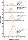
Serum anti-dust EV IgG concentrations of the control group had a normal (Gaussian) distribution (Fig. 1). Subgroup analyses revealed that serum anti-dust EV concentrations did not differ among the age subgroups of the control group This was also true when males and females, or smokers and non-smokers in the control group were compared (Fig. 2). Serum anti-dust EV IgG levels in the asthma, COPD, and lung cancer groups also had a normal distribution (Fig. 1). The positive rate of IgG sensitization to dust EVs was 13.6% in the asthma group, 29.3% in the COPD group, and 54.9% in the lung cancer group, whereas 4.4% in the control group (Table 1 and Fig. 1). Compared to the control subjects, asthmatic, COPD, and lung cancer patients significantly had a high anti-EV IgG antibody in serum (all P<0.001, Table 1 and Fig. 1).
A high serum anti-dust EV IgG level as a potential risk factor for asthma
Multiple logistic regression analysis was performed to evaluate risk factors for asthma after adjustment for age, gender, cigarette smoking history, and dust EV sensitization (Table 2). This evaluation revealed that adjusted ORs for asthma were 1.03 (95% CI 1.01-1.05, P<0.01) for every 1-year increase in age, 0.7 (95% CI 0.3-1.5, P>0.05) for female gender, 0.3 (95% CI 0.1-0.6, P=0.001) for cigarette smoking history, and 3.3 (95% CI 1.1-10.0, P<0.05) for dust EV sensitization. This finding indicates that, in addition to aging, IgG sensitization to dust EVs may be an independent risk factor for asthma, whereas cigarette smoking is a protective factor for asthma.
A high serum anti-dust EV IgG level as a potential risk factor for COPD
Multiple logistic regression analysis was performed to evaluate risk factors for COPD after adjustment for age, gender, cigarette smoking history, and dust EV sensitization (Table 2). This study showed that adjusted ORs for COPD were 1.2 (95% CI 1.1-1.2, P<0.001) for every 1-year increase in age, 1.0 (95% CI 0.3-2.8, P>0.05) for female gender, 3.7 (95% CI 1.4-10.1, P=0.01) for cigarette smoking history and 8.0 (95% CI 2.0-32.5, P=0.003) for dust EV sensitization. This finding indicates that, in addition to aging and cigarette smoking, IgG sensitization to dust EVs is an independent risk factor for COPD.
A high serum anti-dust EV IgG level as a potential risk factor for lung cancer
Multiple logistic regression analysis was performed to evaluate risk factors for lung cancer (Table 2). This study showed that adjusted ORs for lung cancer after adjustment for age, gender, cigarette smoking history, and dust EV sensitization were 1.2 (95% CI 1.1-1.2, P<0.001) for every 1-year increase in age, 0.7 (95% CI 0.3-1.8, P>0.05) for female gender, 2.7 (95% CI 1.1-7.0, P=0.03) for cigarette smoking history, and 38.7 (95% CI 10.4-144.3, P<0.001) for dust EV sensitization. This finding indicates that, in addition to aging and cigarette smoking, IgG sensitization to dust EVs may be an independent major risk factor for lung cancer.
Association between high serum anti-dust EV IgG levels and asthma subtypes
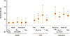
Asthma subtypes were categorized as non-atopic and atopic asthma based on skin prick test responses to common inhalant allergens, and non-eosinophilic and eosinophilic asthma based on the 3% cutoff value of sputum eosinophil percentage. When we compared between non-atopic and atopic asthma using the t test, the mean values of serum anti-dust EV IgG in serum from nonatopic and atopic asthma were 8.86 and 8.87, respectively, and there was no significant difference in anti-dust EV IgG levels between the 2 subtypes (P=0.952) (Table 3). When non-eosinopilic asthma was compared to eosinophilic asthma 1, the mean values of serum anti-dust EV IgG were 8.89 and 8.86 from the 2 subtypes, respectively, and there is no significant difference in anti-dust EV IgG levels between the 2 subtypes (P=0.556) (Table 3). When multiple logistic regression analysis was performed after adjustment for age, gender, and cigarette smoking history in each of the 4 asthma subtypes vs the control group, a high serum anti-dust EV IgG level was found to be an independent risk factor for non-eosinophilic asthma (Fig. 2 and Table 4). Adjusted ORs for the non-atopic, atopic, non-eosinophilic, and eosinophilic asthma subgroups vs the control group were 2.8 (95% CI 0.8-9.6, P=0.1), 2.9 (95% CI 0.9-9.8, P=0.08), 3.7 (95% CI 1.1-12.3, P=0.03), and 3.1 (95% CI 0.8-11.4, P=0.10), respectively.
Association between high serum anti-dust EV IgG levels and COPD severity
The COPD patients enrolled were classified as mild (n=80), moderate (n=82), or severe (n=80) COPD according to COPD severity as defined by using the FEV1 tertile values (57% and 41% of predicted normal value). The mean values of serum anti-dust EV IgG were 8.95, 8.86, and 9.01 in mild, moderate and severe COPD subtypes, respectively, and there were no significant differences in anti-dust EV IgG levels among the 3 subtypes (P=0.065 using the 1-way ANOVA test) (Table 5). When multiple logistic regression analysis was performed after adjustment for age, gender, and cigarette smoking history in each of the 3 COPD tertile subgroups vs the control group, a high anti-dust EV IgG level in serum was found to be an independent risk factor for each of the 3 COPD subgroups (Fig. 2 and Table 6). Adjusted ORs for mild, moderate, and severe COPD subgroups vs the control group were 7.1 (95% CI 1.5-33.4, P=0.01), 12.2 (95% CI 2.4-61.5, P=0.002), and 50.3 (95% CI 1.9-1316.0, P=0.02), respectively.
Association between high anti-dust EV levels and cellular subtypes of lung cancer
The cellular subtypes of 325 lung cancer patients were squamous cell carcinoma (n=94), adenocarcinoma carcinoma (n=90), small cell carcinoma (n=70), adenosquamous carcinoma (n=6), large cell carcinoma (n=4), and non-small cell carcinoma without any specific cellular subtype (n=60). The mean values of serum anti-dust EV IgG were 9.07, 9.07, and 9.15 in the subtypes squamous cell carcinoma, adenocarcinoma, and small cell carcinoma, respectively, but there were no significant differences in anti-dust EV IgG levels among the subtypes (P=0.082, by the 1-way ANOVA test) (Table 7). Multiple logistic regression analysis was performed after adjustment for age, gender, and cigarette smoking history in subjects with squamous cell carcinoma, adenocarcinoma, and small cell lung carcinoma vs the control subjects. This evaluation showed that a high serum anti-dust EV IgG level was an independent risk factor for all the 3 lung cancer subtypes (Fig. 2 and Table 8). Adjusted ORs for the subtypes squamous cell carcinoma, adenocarcinoma, and small carcinoma vs the control group were 81.1 (95% CI 10.4-631.2, P<0.001), 27.2 (95% CI 7.4-99.7, P<0.001), and 61.6 (95% CI 9.0-421.6, P<0.001), respectively.
DISCUSSION
The current study showed that the serum anti-dust EV IgG levels were significantly higher in patients with asthma, COPD, or lung cancer than in the control subjects. Moreover, a high serum anti-dust EV IgG level was found to be an independent risk factor for asthma, especially non-eosinophilic asthma, COPD with mild, moderate, or severe severity, and lung cancer, irrespective of cellular subtypes, after adjustment for age, gender, and cigarette smoking history. Thus, IgG sensitization to dust EVs may increase the risk of non-eosinophilic asthma, COPD, and lung cancer expression and/or development.
The 2014 Global Strategy for Asthma Management and Prevention defines asthma as a heterogeneous disease, usually characterized by chronic airway inflammation. Recent evidence suggests that non-eosinophilic asthma represents a stable phenotype associated with distinct lower airway pathology and structure.22 Specifically, patients are observed to have severe and persistent asthma in the absence of eosinophilic inflammation, and may experience an exacerbation of asthma symptoms without an increase in eosinophilic inflammation.23 In addition, persistent asthma may be associated with the presence of significant neutrophilic inflammation.24 Our previous data also showed that dust EVs induce neutrophilic inflammation in the airways.21 The present data revealed that IgG sensitization to dust EVs is associated with the increased risk of asthma, especially non-eosinophilic asthma. Considering that lower airway inflammation in non-eosinophilic asthma develops in response to etiologic factors acting through immune responses other than inhalant allergens,22 dust EVs might be an important etiological agent of non-eosinophilic asthma.
To the best of our knowledge, there have been few studies directly assessing the relationship between exposure to dust EVs and lung cancer. However, COPD is known to be the risk factor of lung cancer,18 and there is evidence to support that chronic inflammation may promote the development of cancer.1925 Moreover, asthma is also suggested to be a risk factor for lung cancer development in subjects with never-smoking history. Indeed, a recent meta-analysis showed a 1.8-fold increase in lung cancer risk among asthmatics.26 In addition, an epidemiologic study revealed a 1.82-fold risk for lung cancer development in asthmatic patients vs healthy subjects.27 Thus, we postulated that exposure to dust EVs could evoke neutrophilic pulmonary inflammation, which in turn may promote the development of lung cancer. This possibility is supported by the observation that carcinogenesis in organs other than the lung may be promoted by microorganism-induced chronic inflammation.28 For example, it has been proven that Helicobacter pylori causes chronic gastritis and possibly gastric cancer.
A standardized method for evaluating chronic exposure to dust EVs has not yet been established. In our previous study, we used anti-dust EV IgG antibody as a surrogate marker for dust EV exposure. That study showed that children with atopic asthma have higher serum anti-dust EV IgG levels than age-matched atopic children with rhinitis or dermatitis.21 Haneberg et al.29 measured serum antibodies specific for meningococcal EVs and confirmed that vaccination with meningococcal EVs induces an effective immune response. They measured anti-meningococcal EV antibodies by ELISA in serum samples incubated in EV-coated 96-well plates. Their findings support that our present method for measuring anti-dust EV antibodies is valid.
The present study has some limitations. First, we were not able to confirm a causal relationship between exposure to dust EVs and the development of asthma, COPD, or lung cancer because of the cross-sectional design of the present study. To confirm such a causal relationship, a cohort study will be needed. Although an animal study has shown that dust EVs induce neutrophilic inflammation in the lung,30 the degree of exposure to dust EVs would have to be measured in a proposed cohort study to ensure that higher exposure to dust EVs increases the risk for developing asthma, COPD, or lung cancer and that the anti-dust EV IgG level is an appropriate surrogate for dust EV exposure. Second, in the present study, the control subjects were younger than those with COPD or lung cancer; they were also more likely to be female and non-smokers. However, these differences may insignificantly have affected our results because subgroup analysis of the control subjects revealed that serum anti-dust EV IgG levels did not differ between different age, males and females, or smokers and non-smokers. Moreover, our multivariate analysis revealed that a high anti-dust EV IgG level in serum remained an independent risk for COPD and lung cancer, after adjustment for age, gender, and cigarette smoking history. Third, we did not evaluate risk factors that may promote the development of COPD and lung cancer, such as occupational exposure to gas/dust or second-hand exposure to smoke. Fourth, the age- and gender-adjusted ORs of cigarette smoking for lung cancer were lower in the current study than in previous studies. This may reflect the characteristics of recruited subjects in the present study. In particular, the control subjects were on average 15 years younger than the recruited subjects with COPD or lung cancer. Thus, it is possible that, in case 50% of the control subjects were smokers, they may have developed COPD or lung cancer at a later age. In other words, some of the control subjects may have been erroneously included in the control group because they may actually develop COPD or lung cancer in next 15 or more years. The lower OR in the lung cancer group in the current study vs other publications may also be supported by a recent Korean study, which showed that the risk of cigarette smoking for lung cancer may not be as high as previously reported: the adjusted relative risks for lung cancer in subjects who smoked 11-15, 16-20, 21-34, and 35 or more pack-years were 1.99, 3.16, 3.20, and 8.55, respectively.31 In the future, prospectively designed studies will be needed to evaluate the precise role of cigarette smoking in the development of lung cancer, based on the evidence that bacteria-derived EVs in indoor dust induce neutrophilic pulumonary inflammation and emphysema.1721
In conclusion, the present study showed that anti-dust EV IgG antibody titers in serum were significantly higher in patients with non-eosinophilic asthma, COPD, or lung cancer than in healthy control subjects. Multivariable analysis showed that a high serum anti-dust EV IgG concentration was an independent risk factor for non-eosinophilic asthma, COPD, and lung cancer, after adjustment for age, gender, and cigarette smoking. Additional studies of cohort design will be needed to determine whether dust EV exposure has a causal relationship with asthma, COPD, and lung cancer. Nevertheless, the present findings provide an insight into the pathogenesis of non-eosinophilic asthma, COPD, and lung cancer, as well as a clue to developing novel diagnostic and/or therapeutic modalities.


 XML Download