Journal List > Allergy Asthma Immunol Res > v.7(6) > 1052545

Yang, Kim, Lee, Kim, Lee, Yu, Kang, Yu, Lee, Jung, Kim, Seo, Kwon, Song, Jang, Kim, Shim, Lee, Yang, Suh, Hong, Choi, Shin, Ahn, Kim, Kim, Hong, and COCOA Study Group: Prenatal Particulate Matter/Tobacco Smoke Increases Infants' Respiratory Infections: COCOA Study

Abstract

Purpose

To investigate whether prenatal exposure to indoor fine particulate matter (PM2.5) and environmental tobacco smoke (ETS) affects susceptibility to respiratory tract infections (RTIs) in infancy, to compare their effects between prenatal and postnatal exposure, and to determine whether genetic factors modify these environmental effects.

Methods

The study population consisted of 307 birth cohort infants. A diagnosis of RTIs was based on parental report of a physician's diagnosis. Indoor PM2.5 and ETS levels were measured during pregnancy and infancy. TaqMan was used for genotyping of nuclear factor erythroid 2-related factor (Nrf2) (rs6726395), glutathione-S-transferase-pi (GSTP) 1 (rs1695), and glutathione-S-transferase-mu (GSTM) 1. Microarrays were used for genome-wide methylation analysis.

Results

Prenatal exposure to indoor PM2.5 increased the susceptibility of lower RTIs (LRTIs) in infancy (adjusted odds ratio [aOR]=2.11). In terms of combined exposure to both indoor PM2.5 and ETS, prenatal exposure to both pollutants increased susceptibility to LRTIs (aOR=6.56); however, this association was not found for postnatal exposure. The Nrf2 GG (aOR=23.69), GSTM1 null (aOR=8.18), and GSTP1 AG or GG (aOR=7.37) genotypes increased the combined LRTIs-promoting effects of prenatal exposure to the 2 indoor pollutants. Such effects of prenatal indoor PM2.5 and ETS exposure were not found for upper RTIs.

Conclusions

Prenatal exposure to both indoor PM2.5 and ETS may increase susceptibility to LRTIs. This effect can be modified by polymorphisms in reactive oxygen species-related genes.

INTRODUCTION

Environmental tobacco smoke (ETS), which consists of a mixture of gaseous and particulate pollutants, is major indoor air pollution. Indoor particulate matter (PM) is also emitted from cooking, cleaning, and other human activities as well as from smoking.12 Indoor PM and ETS are major indoor air pollutants and may modify the effects of each other.34 Therefore, it is important to understand the combined effect of indoor PM and ETS on health and to elucidate the mechanisms involved. Although there is some epidemiologic evidence on the combined effect of ETS and ambient air pollutants on childhood respiratory outcomes,56789 results on combined exposure to indoor PM and ETS are limited, particularly prenatal exposure.
The prenatal period is critical in terms of the later development of respiratory disorders in childhood because prenatal air pollutant exposure is associated with adverse effects on fetal growth10 and immune responses in early life.11121314 Prenatal and postnatal exposure involve different routes: prenatal air pollutant exposure occurs via transplacental absorption, whereas postnatal exposure occurs via the respiratory route. Therefore, prenatal air pollutant exposure may affect health via a different mechanism from postnatal exposure. Based on these results, we hypothesized that prenatal indoor PM and ETS exposure compared to postnatal exposure would more severely affect the lower respiratory tract than the upper respiratory tract. Since early-childhood respiratory disorders, especially lower-respiratory tract infections (LRTIs), can develop into chronic respiratory impairment later in life,151617 it is important to identify modifiable early life determinants of adverse respiratory outcomes, especially those operating in the prenatal period. However, the impact of prenatal indoor air pollutant exposure, especially the interaction between indoor PM and ETS, on the susceptibility to LRTIs remains poorly understood.
A mechanism through which PM and ETS may lead to respiratory disease is through promotion of reactive oxygen species (ROS).1819 The transcription factor nuclear factor erythroid 2-related factor (Nrf2) is activated by oxidative stress and leads to the transcription of antioxidant genes, such as glutathione Stransferase-pi 1 (GSTP1) and glutathione S-transferase-mu 1 (GSTM1). Therefore, ROS-related genes and polymorphisms may result in different responses to PM and ETS.20 The influence of genetic variation on the association between prenatal exposure to indoor PM and/or ETS and susceptibility to RTIs in infancy remains to be studied.
Epigenetic modifications are one of the mechanisms by which prenatal exposures can affect disease later in life. DNA methylation is a well-characterized epigenetic modification, and there is evidence that it may modulate the lifelong effect of prenatal smoke exposure.212223
To address these issues, a prospective birth cohort study was performed. The effect of prenatal indoor PM and/or ETS exposure on the susceptibility of RTIs in infancy was evaluated. The influence of ROS-related gene polymorphisms on RTI susceptibility in infancy was also assessed. Furthermore, whether prenatal indoor PM and ETS exposure can alter DNA methylation was investigated.

MATERIALS AND METHODS

Study design

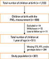
Healthy newborns (n=1,733) were recruited between November 2007 and December 2013. This prospective, general population-based, birth cohort was designated as the COhort for Childhood Origin of Asthma and Allergic Diseases (COCOA); follow-up and further recruitment of this cohort is ongoing. The study methods have been detailed elsewhere.2425 The indoor level of fine particulate matter (PM2.5) has been measured since 2009 for the applicants. In 608 infants, the indoor levels of PM2.5 were evaluated between 26 and 36 weeks of pregnancy. Of these, based on the complete PM2.5, ETS exposure, RTIs, and genotype data, 307 infants were finally included in the study (Fig. 1). Whether the 6- and 12-month-old infants had had any RTIs was determined by parental report of physician-diagnosed RTIs: "Has a doctor diagnosed RTIs in your child during the last 6 months?" Bronchiolitis, tracheobronchitis, and/or pneumonia were considered as LRTIs and common cold, sinusitis, otitis media, and/or croup as upper RTIs (URTIs).

Exposure assessment

Starting in May 2009, indoor PM2.5 samples were collected by specialists during home visit between 26 and 36 weeks of pregnancy. In addition, PM2.5 samples at 6 months after birth were collected in the subgroup (n=75) for the applicants. PM2.5 concentrations were measured 3 times in the parents' bed room by using a particle discriminator (Model GT-331; SIBATA Co., Japan) with a laser light-scattering optical particle counter for 5 minutes. The mean value of 3 measurements was used for evaluation. The indoor PM2.5 values were log-transformed and dichotomized to high or low by using the median value before being entered into the regression models. Mothers were asked the following questions about their ETS exposure at home: "Have you been regularly exposed to passive smoking during your current pregnancy?"
The groups were stratified by exposure time. This led to 4 study population groups receiving the following combination of prenatal/postnatal exposures: prenatal ETS/prenatal PM2.5, prenatal ETS/postnatal PM2.5, postnatal ETS/prenatal PM2.5, and postnatal ETS/postnatal PM2.5. To assess whether the 2 indoor pollutants acted additively to increase RTI susceptibility in infancy, each group was divided into 4 groups according to their ETS exposure and whether the indoor PM2.5 levels were high or low.

Genotyping

Genomic DNA was prepared from heparinized newborn umbilical cord blood by using a G-DEX II kit (Intron, Seoul, Korea). ROS-related genes were analyzed as follows. The Nrf2 (rs6726395) and GSTP1 (rs1695) polymorphisms were genotyped by using a TaqMan assay (ABI, Foster City, CA, USA). The GSTM1 copy number was measured by real-time polymerase chain reaction (PCR). The genotyping method is detailed in the Supplemental Material.

Bisulfite conversion and genome-wide methylation array

Nine subjects were selected from the study population to undergo genome-wide methylation analysis of cord blood genomic DNA. Bisulfite conversion was performed by using the EZ DNA methylation kit (Zymo Research, Irvine, CA, USA) according to the manufacturer's instructions. The bisulfite-converted genomic DNA was analyzed by using the Infinium Human Methylation 450 Beadchip (Illumina, San Diego, CA, USA), with >450,000 probes covering 99% of reference sequence genes, following the Illumina Infinium HD Methylation protocol. The 9 subjects and the methylation array are described in the Supplemental Material.

Statistical analysis

Chi-square and t tests were used to assess the significance of differences between the groups, as appropriate. The associations between prenatal indoor PM2.5 and/or ETS exposure and the incidence of RTIs at 12 months of age were analyzed by using multiple logistic regression. Adjustments were made for potential confounding factors, namely, maternal age at delivery, maternal body mass index, maternal educational degree, gestational age, delivery mode, infant sex, and family history of allergic diseases. The results are expressed as adjusted odds ratios (aORs) and 95% confidence intervals (CIs). All statistical analyses were performed by using SPSS 18.0 software (SPSS Inc., Chicago, IL, USA), with a P value <0.05 considered statistically significant.

Ethics statement

The study was approved by the Institutional Review Board of Asan Medical Center (IRB No. 2008-0616), Samsung Medical Center (IRB No. 2009-02-021), Yonsei University (IRB No. 4-2008-0588), and CHA Medical Center (IRB No. 2010-010).

RESULTS

Study population characteristics

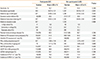
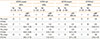
Table 1 summarizes the characteristics of the study population (n=307). The study population consisted of 171 boys and 136 girls. In total, 61.6% had been prenatally exposed to maternal ETS, and the mean indoor PM2.5 level during pregnancy was 6.08 ± 7.64 µg/m3. The frequencies of the Nrf2 (rs6726395) GG, GSTP1 (rs1695) AG or GG, and GSTM1 null genotypes were 40.1%, 36.9%, and 56.7%, respectively. The distribution of the 2 polymorphisms was in Hardy-Weinberg equilibrium. The incidences of LRTIs and URTIs by the age of 12 months were 16.3% and 76.2%, respectively.
Table 1 also shows the characteristics of the infants who were not included for reasons shown in Fig. 1. There were no significant differences between participants and non-participants, except in gestational age and the incidence of URTIs.

Risk factors for RTIs in infancy

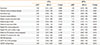
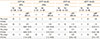
High PM2.5 levels during pregnancy were independent risk factors for LRTIs in infants (aOR=2.11; 95% CI: 1.12, 3.99) (Table 2). However, none of the early-life environmental factors increased the risk of URTIs. When we compared indoor PM2.5 levels according to RTIs and exposure time, prenatal indoor PM2.5 levels were higher in infants with LRTIs than in those without (mean=7.21 vs 5.71, respectively; 95% CI: 4.99, 9.44 vs 4.97, 6.45, respectively; P=0.119, data not shown). These differences were not distinct according to postnatal PM2.5 levels or the presence of URTIs.

Effects of prenatal exposure to both indoor PM2.5 and ETS on RTIs susceptibility in infancy

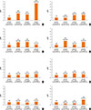
Prenatal high indoor PM2.5 and ETS exposure acted additively to increase the risk of LRTIs (aOR=6.56; 95% CI: 2.02, 21.24) in infants (Fig. 2A and Table S1). Such an additive effect was not replicated when examining the effects of exposure to prenatal PM2.5/postnatal ETS, postnatal PM2.5/prenatal ETS, and postnatal PM2.5/postnatal ETS on any respiratory outcomes in infants. In addition, such additive effects were not observed for URTIs risk, regardless of exposure time (Fig. 2B and Table S1).

Effect of GSTM1, GSTP1, and Nrf2 genotypes on the relationship between prenatal indoor PM2.5/ETS exposure and RTIs in infancy

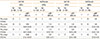
Prenatal exposure to both high indoor PM2.5 and ETS increased LRTIs risk in the GSTM1 null, GSTP1 AG or GG, and Nrf2 GG genotypes (aOR=8.18, 95% CI: 1.53, 43.72; aOR=7.37, 95% CI: 1.12, 48.66; and aOR=23.69, 95% CI: 2.13, 263.07, respectively). Such gene-environment interactions were not observed for URTIs (Tables 3,4,5).

Relationship between DNA methylation patterns and prenatal indoor PM2.5/ETS exposure

Prenatal indoor PM2.5/ETS exposure was associated specifically with 15 CpG sites, 6 of which were located in intergenic regions. Of the remaining 9 CpG sites, 5 were hypomethylated and 4 were hypermethylated by PM2.5/ETS exposure. This analysis is described in the Supplemental Material.

DISCUSSION

The present study showed that indoor PM2.5 and ETS exposure may have an effect on RTIs in infants and revealed that the adverse effect may depend on the timing of the exposure. The ability of indoor PM2.5 and/or ETS to increase susceptibility to LRTIs appeared to be stronger when the exposure occurred during the prenatal period rather than the postnatal period. This study also showed that the genetic polymorphisms GSTM1, GSTP1 (rs1695), and/or Nrf2 (rs6726395) were further associated with the increased susceptibility of LRTIs in indoor PM2.5/ETS-exposed infants. Thus, the susceptibility of LRTIs in infancy may be shaped by gene-environment interactions between ROS-related genes and prenatal indoor PM2.5/ETS exposure. To our knowledge, this is the first study to evaluate the association between combined exposure to indoor PM2.5/ETS and infants' susceptibility to LRTIs that is associated with exposure time and genetic susceptibility.
Most people spend as much as 90% of their time indoors, especially pregnant women and infants. Chronic exposure to indoor pollutants at home or school can increase air pollutant inhalation and significantly impact health.2627 The interaction between PM and ETS, the most important indoor air pollutants, modify their individual harmful effects on respiratory outcomes.78 However, studies about the indoor PM concentration that would have an adverse health outcome and an interaction between PM and ETS, especially prenatal exposure, are scarce. Our study revealed the effect of indoor PM2.5 even in the low concentration and the additive effect of indoor PM2.5/ETS exposure during the prenatal period on the development of LRTIs in infancy.
The fetal period is critical for lung and immune development. Although some epidemiologic studies showed that prenatal PM or ETS exposure increases the risk of wheezing, asthma, and respiratory infections,28293031 studies comparing the effect according to exposure time are limited. A few studies revealed that prenatal exposure has a stronger effect on respiratory outcomes than postnatal exposure.323334 We also found its stronger associations with combined exposures to indoor PM2.5/ETS during the prenatal period than the postnatal period. However, this result must be interpreted with caution because it is difficult to clearly separate exposure periods.
Air pollutants and tobacco smoke exert their harmful effects on health by inducing oxidative stress in exposed cells and tissues.1819 Air pollutants can be directly absorbed to the fetal circulation and produce ROS, ultimately inducing inflammatory and oxidative stress responses in the fetal lung.35 The fetus can also be affected indirectly by the oxidative stress and inflammatory cytokine production induced in the placenta by the pollutants.36 Of particular interest in this regard are several intracellular antioxidant enzymes, including GSTM1 and GSTP1, which defend the airway epithelium from damage caused by oxidants and inflammation. These enzymes are regulated by the transcription factor Nrf2, which translocates to the nucleus after oxidative stress induction.37 These enzymes in respiratory disease pathogenesis after pollutant exposure is provided by results showing that children with the GSTM1 null genotype are more likely to develop asthma and wheezing after prenatal ETS exposure than children with the GSTM1 present genotype.20 Similarly, our study showed that while both prenatal indoor PM2.5 and ETS exposure greatly increased the incidence of LRTIs in infants, this effect was particularly marked in the infants with the GSTM1 null, GSTP1 (rs1695) AG or GG, or Nrf2 (rs6726395) GG genotypes.
ROS reacts with lipids, proteins, and DNA, resulting in cell membrane damage, alteration of gene and protein expression, and even cell death.1837 Secondary mediators generated by oxidant reactions with lipids, proteins, and other biomolecules contribute to the toxic effects of pollutants. Oxidative stress also induces MAP kinase and NF-κB activation, which may ultimately produce a variety of proinflammatory mediators. Proinflammatory mediators from the airway epithelium play a critical role in the pathogenesis of several pulmonary diseases. Previous experimental studies supported the association between air pollutants and oxidative stress by demonstrating that antioxidant pretreatment attenuates oxidative stress and airway epithelial cell injury induced by air pollutants.383940
Prenatal exposure to environmental factors may affect disease susceptibility later in life by inducing epigenetic changes. A cross-sectional study of children under 18 years of age revealed that air pollutants and ETS both associate with significantly increased DNA methylation and decreased transcription of interferon gamma (IFN-γ) in T-effector cells and forkhead box transcription factor 3 (Foxp3) in T-regulatory cells.41 Interestingly, GSTM1 and GSTP1 polymorphisms alter the ability of prenatal tobacco smoke exposure to induce global DNA methylation.21 Although the sample size in our experiment was too small to make firm conclusions, our data suggest that the ability of prenatal indoor PM2.5 and ETS exposure to promote LRTIs in infancy may be due to DNA methylation alterations; the 9 CpG sites whose methylation was significantly altered by PM2.5/ETS exposure were in subjects with LRTIs (Table S2 and Fig. S1). Further studies on this issue are required.
This study has several limitations. First, it was not possible to clearly distinguish between the effects of prenatal and postnatal exposure or exposure that persisted during both the preand postnatal periods. Further studies on the effect of indoor PM and ETS exposure during specific prenatal and postnatal periods may help identify the mechanisms involved. Second, the RTIs and ETS data were derived from questionnaires, and the indoor PM2.5 levels were measured only 1 day between 26 and 36 weeks of pregnancy and at 6 months of age. Therefore, an information bias could not be excluded. Although questionnaires may misclassify ETS exposure, previous studies have shown a fairly good correlation between self-reported ETS exposure and biomarkers of ETS exposure.5424344 Future studies may gain greater sensitivity by using more objective and precise measures of RTIs, and smoke and indoor PM exposure. The third limitation is the relatively small study population, which is because the indoor PM2.5 measurements started later in the COCOA study, and these data were thus only available for about one-third of the whole COCOA cohort. However, it is unlikely that the addition of indoor PM2.5 measurements to the protocol introduced a selection bias because the study participants and non-participants did not differ significantly in terms of their characteristics. An increase in the sample size would be likely to lead to more consistent and significant data. The fourth limitation is that we only selected 1 polymorphism from each gene. However, these polymorphisms have been shown in several studies to contribute to asthma susceptibility.20454647
An important strength of our study is its prospective design: the indoor PM2.5 and ETS exposure data and the data on many potential confounders were collected before the children were born. This is likely to have markedly reduced the study bias. An additional strength is that the PM2.5 measurement was performed at home. This direct measurement of residential indoor PM2.5 probably estimates the actual exposure levels more accurately than other indirect methods. Finally, we investigated genotypic data for GSTM1, GSTP1, and Nrf2 to determine gene-environment interactions between both PM2.5/ETS exposure and LRTIs. These results imply that air pollutant exposure should be reduced, especially in genetically susceptible infants, and support a mechanism for oxidative stress in inducing adverse respiratory outcomes by air pollutants.
It should be noted that there was an important difference between previous studies and ours, namely, that maternal ETS exposure was considered in our study. It was not possible to evaluate the effect of maternal active smoking because the active smoking rate of Korean women is low: only 11.4% of the COCOA cohort mothers had smoked before their pregnancy, of whom only 1 continued to smoke during pregnancy. Thus, maternal ETS exposure was more likely to be an important source of pollutant exposure in our cohort than maternal smoking.

CONCLUSIONS

Indoor PM2.5 and ETS exposure increases susceptibility to LRTIs in infants. This effect was particularly marked when the exposure occurred in the prenatal period. Moreover, the effect was modified by ROS-related gene polymorphisms. Along with studies suggesting that acute LRTIs in early life is associated with a long-standing susceptibility to all forms of lung disease, including asthma,151617 our study highlights the importance of health intervention strategies that focus on the indoor environment in the prenatal period. Additional analyses of genetic and epigenetic variants may help individualize such strategies. Further studies of gene-environment interactions and epigenetic mechanisms that shape the effect of air pollutants on susceptibility to LRTIs and chronic lung diseases are warranted, along with studies assessing the association between LRTIs in infancy and the development of chronic lung diseases, such as asthma.

Figures and Tables

Fig. 1

Flow chart of the study population. Of infants with prenatal indoor PM2.5 measurements (n=608), 14 met the exclusion criteria, 72 were withdrawn from the study, and 11 were lost to follow-up. An additional 204 infants were then excluded because the RTIs, prenatal ETS exposure, or genotype data were missing. ETS, environmental tobacco smoke; PM2.5, fine particulate matter; RTIs, respiratory tract infections.

aair-7-573-g001
Fig. 2

Effect of combined exposure to both PM2.5 and ETS according to exposure time on susceptibility to (A) lower and (B) upper respiratory tract infections in infancy. The groups were stratified by exposure time: (a) prenatal PM2.5/prenatal ETS; (b) prenatal PM2.5/postnatal ETS; (c) postnatal PM2.5/prenatal ETS; and (d) postnatal PM2.5/postnatal ETS. Prenatal high indoor PM2.5 and ETS exposure acted additively to increase the risk of lower respiratory tract infections. *Adjustments for maternal age at delivery, maternal body mass index, maternal educational degree, gestational age, delivery mode, infant sex, and family history of allergy. ETS, environmental tobacco smoke; PM2.5, fine particulate matter.

aair-7-573-g002
Table 1

Characteristics of infants who were included and excluded from the study

aair-7-573-i001
Participants (307) Non-participants (1,426) P value
Number Mean±SD or % Number Mean±SD or %
Sex (male, %) 171 55.7 673 51.8 0.228
Gestational age (week) 303 39.27±1.21 1,257 39.10±1.27 0.042
Maternal age at birth (year) 307 32.69±3.44 1,296 32.56±3.43 0.535
Cesarean section delivery (%) 90/269 33.5 377/1,117 33.8 0.943
Maternal body mass index (kg/m2) 307 20.53±2.24 1,352 20.66±2.63 0.369
Maternal education state 307 1,332 0.999
 ≤High school (%) 21 6.8 90 6.8
 University or college (%) 218 71.0 947 71.1
 Graduate school (%) 68 22.2 295 22.2
Parental history of allergic disease (%) 178/295 60.3 615/1,085 56.7 0.288
Maternal ETS exposure during pregnancy (%) 189/307 61.6 668/1,084 61.6 1.000
Infantile ETS exposure at 1 year (%) 75/241 31.1 196/600 32.7 0.684
PM2.5 during pregnancy (µg/m3) 307 6.08±7.64 307 5.70±4.97 0.461
PM2.5 at 6 months of age (µg/m3) 75 5.11±7.05 245 5.40±5.33 0.699
Nrf2 GG genotype (%) 123/307 40.1 351/913 38.4 0.636
GSTP1 AG or GG genotype (%) 113/306 36.9 295/919 32.1 0.124
GSTM1 null genotype (%) 173/305 56.7 490/911 53.8 0.388
Incidence of LRTIs* at 1 year (%) 49/300 16.3 111/637 17.4 0.917
Incidence of URTIs at 1 year (%) 234/307 76.2 450/668 67.4 0.005

*Lower respiratory tract infections: tracheobronchitis, pneumonia, and bronchiolitis; Upper respiratory tract infections: common cold, sinusitis, otitis media, and croup.

ETS, environmental tobacco smoke; GSTM1, glutathione S-transferase-mu 1; GSTP1, glutathione S-transferase-pi 1; Nrf2, nuclear factor erythroid 2-related factor; PM2.5, fine particulate matter; LRTIs, lower respiratory tract infections; URTIs, upper respiratory tract infections.

Table 2

Risk factors for respiratory tract infections in infancy

aair-7-573-i002
LRTIs URTIs
aOR* 95% CI P value aOR* 95% CI P value
Sex (male) 1.53 (1.01, 2.31) 0.046 1.04 (0.75, 1.45) 0.799
Parental history of allergic disease 0.87 (0.58, 1.30) 0.500 0.97 (0.70, 1.35) 0.863
Gestational age 0.94 (0.80, 1.10) 0.450 0.94 (0.83, 1.08) 0.393
Cesarean section delivery 1.14 (0.74, 1.75) 0.550 0.75 (0.53, 1.06) 0.098
Higher maternal education state 1.08 (0.74, 1.58) 0.683 1.12 (0.82, 1.54) 0.468
Maternal age at birth 0.99 (0.93, 1.05) 0.713 1.06 (1.01, 1.11) 0.019
Maternal body mass index 1.04 (0.96, 1.12) 0.363 0.94 (0.88, 1.01) 0.078
Higher PM2.5 during pregnancy 2.11 (1.12, 3.99) 0.021 1.23 (0.71, 2.15) 0.463
Higher PM2.5 at 6 months of age 0.89 (0.37, 2.18) 0.801 1.00 (0.52, 1.94) 0.999
Maternal ETS exposure during pregnancy 1.33 (0.87, 2.04) 0.193 1.15 (0.82, 1.62) 0.414
Infantile ETS exposure at 1 year 0.87 (0.53, 1.42) 0.576 0.79 (0.53, 1.17) 0.241
Nrf2 GG genotype 1.27 (0.82, 1.97) 0.290 0.55 (0.39, 0.78) 0.001
GSTP1 AG or GG genotype 1.53 (0.98, 2.38) 0.062 0.89 (0.62, 1.28) 0.523
GSTM1 null genotype 1.69 (1.07, 2.67) 0.024 0.91 (0.64, 1.30) 0.612

*Odds ratios were adjusted for maternal age, maternal body mass index, maternal educational state, infant sex, gestational age, delivery mode, and family history of allergy.

ETS, environmental tobacco smoke; GSTM1, glutathione S-transferase-mu 1; GSTP1, glutathione S-transferase-pi 1; Nrf2, nuclear factor erythroid 2-related factor; PM2.5, fine particulate matter; LRTIs, lower respiratory tract infections; URTIs, upper respiratory tract infections.

Table 3

Influence of the glutathione S-transferase-mu 1 (GSTM1) copy number variation on the results of prenatal exposure to both PM2.5 and ETS

aair-7-573-i003
GSTM1 present GSTM1 null GSTM1 present GSTM1 null
LRTIs LRTIs URTIs URTIs
No (n=112) Yes (n=17) No (n=137) Yes (n=32) No (n=36) Yes (n=87) No (n=37) Yes (n=150)
n n aOR* (95% CI) n n aOR* (95% CI) n n aOR* (95% CI) n n aOR* (95% CI)
PM2.5 (Low) 29 2 1.00 34 3 1.00 6 26 1.00 10 27 1.00
ETS (No)
PM2.5 (Low) 37 7 1.68 45 12 4.52 11 33 0.51 12 48 2.02
ETS (Yes) (0.24, 11.79) (0.87, 23.60) (0.14, 1.83) (0.68, 6.01)
PM2.5 (High) 13 2 2.26 28 4 1.65 4 12 0.69 9 25 1.17
ETS (No) (0.26, 19.36) (0.24, 11.29) (0.13, 3.76) (0.38, 3.57)
PM2.5 (High) 33 6 3.75 30 13 8.18 9 32 0.57 11 33 1.86
ETS (Yes) (0.52, 27.10) (1.53, 43.72) (0.14, 2.35) (0.60, 5.75)

*Odds ratios were adjusted for maternal age, maternal body mass index, maternal educational state, infant sex, gestational age, delivery mode, and family history of allergy.

aOR, adjusted odds ratio; CI, confidence interval; ETS, environmental tobacco smoke; PM2.5, fine particulate matter; GSTM1, glutathione S-transferase-mu 1; LRTIs, lower respiratory tract infections; URTIs, upper respiratory tract infections.

Table 4

Influence of the glutathione S-transferase-pi 1 (GSTP1) (rs1695) polymorphism on the results of prenatal exposure to both PM2.5 and ETS

aair-7-573-i004
GSTP1 AA GSTP1 AG+GG GSTP1 AA GSTP1 AG+GG
LRTIs LRTIs URTIs URTIs
No (n=159) Yes (n=29) No (n=91) Yes (n=20) No (n=45) Yes (n=151) No (n=27) Yes (n=86)
n n aOR* (95% CI) n n aOR* (95% CI) n n aOR* (95% CI) n n aOR* (95% CI)
PM2.5 (Low) 38 3 1.00 25 2 1.00 9 33 1.00 7 20 1.00
ETS (No)
PM2.5 (Low) 59 12 3.57 24 7 4.04 15 58 1.14 9 23 1.18
ETS (Yes) (0.66, 19.17) (0.62, 26.30) (0.41, 3.15) (0.29, 4.71)
PM2.5 (High) 26 3 2.82 14 3 1.70 9 23 0.75 3 14 1.43
ETS (No) (0.40, 19.77) (0.19, 15.01) (0.24, 2.34) (0.28, 7.30)
PM2.5 (High) 36 11 6.32 28 8 7.37 12 37 0.83 8 29 1.80
ETS (Yes) (1.18, 33.94) (1.12, 48.66) (0.28, 2.43) (0.41, 7.97)

*Odds ratios were adjusted for maternal age, maternal body mass index, maternal educational state, infant sex, gestational age, delivery mode, and family history of allergy.

aOR, adjusted odds ratio; CI, confidence interval; ETS, environmental tobacco smoke; PM2.5, fine particulate matter; GSTP1, glutathione S-transferase-pi 1, LRTIs, lower respiratory tract infections; URTIs, upper respiratory tract infections.

Table 5

Influence of the nuclear factor erythroid 2-related factor (Nrf2) (rs6726395) on the results of prenatal exposure to both PM2.5 and ETS

aair-7-573-i005
Nrf2 GG Nrf2 GA+AA Nrf2 GG Nrf2 GA+AA
LRTIs LRTIs URTIs URTIs
No (n=98) Yes (n=22) No (n=153) Yes (n=27) No (n=36) Yes (n=87) No (n=37) Yes (n=150)
n n aOR* (95% CI) n n aOR* (95% CI) n n aOR* (95% CI) n n aOR* (95% CI)
PM2.5 (Low) 31 2 1.00 32 3 1.00 9 24 1.00 7 29 1.00
ETS (No)
PM2.5 (Low) 26 10 9.66 57 9 1.19 9 27 1.20 15 54 1.09
ETS (Yes) (0.99, 94.03) (0.27, 5.18) (0.36, 3.94) (0.34, 3.53)
PM2.5 (High) 20 2 4.27 21 4 1.19 9 15 0.66 4 22 1.65
ETS (No) (0.31, 58.04) (0.19, 7.31) (0.19, 2.29) (0.36, 7.52)
PM2.5 (High) 21 8 23.69 43 11 2.82 9 21 0.81 11 45 1.75
ETS (Yes) (2.13, 263.07) (0.65, 12.31) (0.23, 2.94) (0.48, 6.42)

*Odds ratios were adjusted for maternal age, maternal body mass index, maternal educational state, infant sex, gestational age, delivery mode, and family history of allergy.

aOR, adjusted odds ratio; CI, confidence interval; ETS, environmental tobacco smoke; PM2.5, fine particulate matter; Nrf2, nuclear factor erythroid 2-related factor; LRTIs, lower respiratory tract infections; URTIs, upper respiratory tract infections.

ACKNOWLEDGMENTS

The authors thank Kyung-Shin Lee, Jin-Ah Park, and Hee-Suk Kim for organizing the data. We would also like to express our gratitude to Ja-Young Kwon, Suk-Joo Choi, Soo-Young Oh, Kyung-Ju Lee, and Hey-Sung Won for helping collect the obstetric data. We also thank Sung-Ok Kwon, Se-Young Oh, Kyung-Sook Lee, Yee-Jin Shin, Jong-Hwan Lim, Whan-Cheol Kim, and Ho Kim for their participation in this study.

Notes

This research was supported by funds (2008-E33030-00, 2009-E33033-00, 2011-E33021-00, 2012-E33012-00, and 2013-E51011-00) from the Research of Korea Centers for Disease Control and Prevention.

There are no financial or other issues that might lead to conflict of interest.

References

1. Hasheminassab S, Daher N, Shafer MM, Schauer JJ, Delfino RJ, Sioutas C. Chemical characterization and source apportionment of indoor and outdoor fine particulate matter (PM(2.5)) in retirement communities of the Los Angeles Basin. Sci Total Environ. 2014; 490:528–537.
2. Clougherty JE, Houseman EA, Levy JI. Source apportionment of indoor residential fine particulate matter using land use regression and constrained factor analysis. Indoor Air. 2011; 21:53–66.
3. Wallace LA, Mitchell H, O'Connor GT, Neas L, Lippmann M, Kattan M, et al. Particle concentrations in inner-city homes of children with asthma: the effect of smoking, cooking, and outdoor pollution. Environ Health Perspect. 2003; 111:1265–1272.
4. Guarnieri M, Balmes JR. Outdoor air pollution and asthma. Lancet. 2014; 383:1581–1592.
5. Rosa MJ, Jung KH, Perzanowski MS, Kelvin EA, Darling KW, Camann DE, et al. Prenatal exposure to polycyclic aromatic hydrocarbons, environmental tobacco smoke and asthma. Respir Med. 2011; 105:869–876.
6. Miller RL, Garfinkel R, Horton M, Camann D, Perera FP, Whyatt RM, et al. Polycyclic aromatic hydrocarbons, environmental tobacco smoke, and respiratory symptoms in an inner-city birth cohort. Chest. 2004; 126:1071–1078.
7. Sonnenschein-van der Voort AM, de Kluizenaar Y, Jaddoe VW, Gabriele C, Raat H, Moll HA, et al. Air pollution, fetal and infant tobacco smoke exposure, and wheezing in preschool children: a population-based prospective birth cohort. Environ Health. 2012; 11:91.
8. Rabinovitch N, Silveira L, Gelfand EW, Strand M. The response of children with asthma to ambient particulate is modified by tobacco smoke exposure. Am J Respir Crit Care Med. 2011; 184:1350–1357.
9. Nicolai T, Carr D, Weiland SK, Duhme H, von Ehrenstein O, Wagner C, et al. Urban traffic and pollutant exposure related to respiratory outcomes and atopy in a large sample of children. Eur Respir J. 2003; 21:956–963.
10. Jedrychowski W, Bendkowska I, Flak E, Penar A, Jacek R, Kaim I, et al. Estimated risk for altered fetal growth resulting from exposure to fine particles during pregnancy: an epidemiologic prospective cohort study in Poland. Environ Health Perspect. 2004; 112:1398–1402.
11. Hertz-Picciotto I, Park HY, Dostal M, Kocan A, Trnovec T, Sram R. Prenatal exposures to persistent and non-persistent organic compounds and effects on immune system development. Basic Clin Pharmacol Toxicol. 2008; 102:146–154.
12. Herr CE, Ghosh R, Dostal M, Skokanova V, Ashwood P, Lipsett M, et al. Exposure to air pollution in critical prenatal time windows and IgE levels in newborns. Pediatr Allergy Immunol. 2011; 22:75–84.
13. Latzin P, Frey U, Armann J, Kieninger E, Fuchs O, Röösli M, et al. Exposure to moderate air pollution during late pregnancy and cord blood cytokine secretion in healthy neonates. PLoS One. 2011; 6:e23130.
14. Hertz-Picciotto I, Herr CE, Yap PS, Dostál M, Shumway RH, Ashwood P, et al. Air pollution and lymphocyte phenotype proportions in cord blood. Environ Health Perspect. 2005; 113:1391–1398.
15. Gern JE. Viral respiratory infection and the link to asthma. Pediatr Infect Dis J. 2008; 27:S97–S103.
16. Sigurs N, Aljassim F, Kjellman B, Robinson PD, Sigurbergsson F, Bjarnason R, et al. Asthma and allergy patterns over 18 years after severe RSV bronchiolitis in the first year of life. Thorax. 2010; 65:1045–1052.
17. Holt PG, Sly PD. Viral infections and atopy in asthma pathogenesis: new rationales for asthma prevention and treatment. Nat Med. 2012; 18:726–735.
18. Ciencewicki J, Trivedi S, Kleeberger SR. Oxidants and the pathogenesis of lung diseases. J Allergy Clin Immunol. 2008; 122:456–468.
19. Kim BJ, Lee SY, Kim HB, Lee E, Hong SJ. Environmental changes, microbiota, and allergic diseases. Allergy Asthma Immunol Res. 2014; 6:389–400.
20. Gilliland FD, Li YF, Dubeau L, Berhane K, Avol E, McConnell R, et al. Effects of glutathione S-transferase M1, maternal smoking during pregnancy, and environmental tobacco smoke on asthma and wheezing in children. Am J Respir Crit Care Med. 2002; 166:457–463.
21. Breton CV, Byun HM, Wenten M, Pan F, Yang A, Gilliland FD. Prenatal tobacco smoke exposure affects global and gene-specific DNA methylation. Am J Respir Crit Care Med. 2009; 180:462–467.
22. Guerrero-Preston R, Goldman LR, Brebi-Mieville P, Ili-Gangas C, Lebron C, Witter FR, et al. Global DNA hypomethylation is associated with in utero exposure to cotinine and perfluorinated alkyl compounds. Epigenetics. 2010; 5:539–546.
23. Flom JD, Ferris JS, Liao Y, Tehranifar P, Richards CB, Cho YH, et al. Prenatal smoke exposure and genomic DNA methylation in a multiethnic birth cohort. Cancer Epidemiol Biomarkers Prev. 2011; 20:2518–2523.
24. Kim HB, Ahn KM, Kim KW, Shin YH, Yu J, Seo JH, et al. Cord blood cellular proliferative response as a predictive factor for atopic dermatitis at 12 months. J Korean Med Sci. 2012; 27:1320–1326.
25. Yang HJ, Lee SY, Suh DI, Shin YH, Kim BJ, Seo JH, et al. The Cohort for Childhood Origin of Asthma and allergic diseases (COCOA) study: design, rationale and methods. BMC Pulm Med. 2014; 14:109.
26. Pawankar R, Canonica GW, Holgate ST, Lockey RF. World Allergy Organization (WAO) white book on allergy. Milwaukee (WI): World Allergy Organization;2011.
27. Yoon C, Ryu K, Kim J, Lee K, Park D. New approach for particulate exposure monitoring: determination of inhaled particulate mass by 24 h real-time personal exposure monitoring. J Expo Sci Environ Epidemiol. 2012; 22:344–351.
28. Magnusson LL, Olesen AB, Wennborg H, Olsen J. Wheezing, asthma, hayfever, and atopic eczema in childhood following exposure to tobacco smoke in fetal life. Clin Exp Allergy. 2005; 35:1550–1556.
29. Jedrychowski W, Perera FP, Maugeri U, Mrozek-Budzyn D, Mroz E, Flak E, et al. Early wheezing phenotypes and severity of respiratory illness in very early childhood: study on intrauterine exposure to fine particle matter. Environ Int. 2009; 35:877–884.
30. Jedrychowski WA, Perera FP, Maugeri U, Mrozek-Budzyn D, Mroz E, Klimaszewska-Rembiasz M, et al. Intrauterine exposure to polycyclic aromatic hydrocarbons, fine particulate matter and early wheeze. Prospective birth cohort study in 4-year olds. Pediatr Allergy Immunol. 2010; 21:e723–e732.
31. Jedrychowski WA, Perera FP, Spengler JD, Mroz E, Stigter L, Flak E, et al. Intrauterine exposure to fine particulate matter as a risk factor for increased susceptibility to acute broncho-pulmonary infections in early childhood. Int J Hyg Environ Health. 2013; 216:395–401.
32. Raherison C, Pénard-Morand C, Moreau D, Caillaud D, Charpin D, Kopfersmitt C, et al. In utero and childhood exposure to parental tobacco smoke, and allergies in schoolchildren. Respir Med. 2007; 101:107–117.
33. Singh SP, Gundavarapu S, Peña-Philippides JC, Rir-Sima-ah J, Mishra NC, Wilder JA, et al. Prenatal secondhand cigarette smoke promotes Th2 polarization and impairs goblet cell differentiation and airway mucus formation. J Immunol. 2011; 187:4542–4552.
34. Jedrychowski WA, Perera FP, Majewska R, Camman D, Spengler JD, Mroz E, et al. Separate and joint effects of tranplacental and postnatal inhalatory exposure to polycyclic aromatic hydrocarbons: prospective birth cohort study on wheezing events. Pediatr Pulmonol. 2014; 49:162–172.
35. Kelly FJ. Oxidative stress: its role in air pollution and adverse health effects. Occup Environ Med. 2003; 60:612–616.
36. Auten RL, Potts EN, Mason SN, Fischer B, Huang Y, Foster WM. Maternal exposure to particulate matter increases postnatal ozone-induced airway hyperreactivity in juvenile mice. Am J Respir Crit Care Med. 2009; 180:1218–1226.
37. Auerbach A, Hernandez ML. The effect of environmental oxidative stress on airway inflammation. Curr Opin Allergy Clin Immunol. 2012; 12:133–139.
38. Wu W, Peden DB, McConnell R, Fruin S, Diaz-Sanchez D. Glutathione-S-transferase M1 regulation of diesel exhaust particle-induced pro-inflammatory mediator expression in normal human bronchial epithelial cells. Part Fibre Toxicol. 2012; 9:31.
39. Messier EM, Day BJ, Bahmed K, Kleeberger SR, Tuder RM, Bowler RP, et al. N-acetylcysteine protects murine alveolar type II cells from cigarette smoke injury in a nuclear erythroid 2-related factor-2-independent manner. Am J Respir Cell Mol Biol. 2013; 48:559–567.
40. Wu W, Muller R, Berhane K, Fruin S, Liu F, Jaspers I, et al. Inflammatory response of monocytes to ambient particles varies by highway proximity. Am J Respir Cell Mol Biol. 2014; 51:802–809.
41. Kohli A, Garcia MA, Miller RL, Maher C, Humblet O, Hammond SK, et al. Secondhand smoke in combination with ambient air pollution exposure is associated with increasedx CpG methylation and decreased expression of IFN-gamma in T effector cells and Foxp3 in T regulatory cells in children. Clin Epigenetics. 2012; 4:17.
42. Wang IJ, Hsieh WS, Wu KY, Guo YL, Hwang YH, Jee SH, et al. Effect of gestational smoke exposure on atopic dermatitis in the offspring. Pediatr Allergy Immunol. 2008; 19:580–586.
43. Carlsten C, Dimich-Ward H, DyBuncio A, Becker AB, Chan-Yeung M. Cotinine versus questionnaire: early-life environmental tobacco smoke exposure and incident asthma. BMC Pediatr. 2012; 12:187.
44. Yi O, Kwon HJ, Kim H, Ha M, Hong SJ, Hong YC, et al. Effect of environmental tobacco smoke on atopic dermatitis among children in Korea. Environ Res. 2012; 113:40–45.
45. Masuko H, Sakamoto T, Kaneko Y, Iijima H, Naito T, Noguchi E, et al. An interaction between Nrf2 polymorphisms and smoking status affects annual decline in FEV1: a longitudinal retrospective cohort study. BMC Med Genet. 2011; 12:97.
46. Lee E, Chang HY, Lee KS, Suh DI, Yu HS, Kang MJ, et al. The effect of perinatal anxiety on bronchiolitis is influenced by polymorphisms in ROS-related genes. BMC Pulm Med. 2014; 14:154.
47. Kang SH, Jung YH, Kim HY, Seo JH, Lee JY, Kwon JW, et al. Effect of paracetamol use on the modification of the development of asthma by reactive oxygen species genes. Ann Allergy Asthma Immunol. 2013; 110:364–369.e1.
48. Brasch-Andersen C, Christiansen L, Tan Q, Haagerup A, Vestbo J, Kruse TA. Possible gene dosage effect of glutathione-S-transferases on atopic asthma: using real-time PCR for quantification of GSTM1 and GSTT1 gene copy numbers. Hum Mutat. 2004; 24:208–214.

Supplementary Materials

Supplemental Methods

Supplemental Fig. 1

Summary of the genome-wide methylation array data.

Supplemental Table 1

Effect of exposure to both PM2.5 and ETS according to exposure time on susceptibility to respiratory tract infections in infancy

Supplemental Table 2

Different methylation sites depending on prenatal exposure to indoor PM2.5 and ETS
TOOLS
ORCID iDs

Kangmo Ahn
https://orcid.org/http://orcid.org/0000-0001-7751-9829

Similar articles