

 PDF
PDF ePub
ePub Citation
 Print
Print


INTRODUCTION
Chronic urticaria is defined as recurrent eruptions of short-lived swellings due to plasma leakage from small blood vessels into the surrounding connective tissue.1 Several factors are considered causes of the disease, including food allergies, emotional disturbances, intolerance to food additives, and chronic infections. 23 Autoimmunity is also considered an integral part of the pathophysiology of chronic urticaria, as injections of autologous serum have caused wheal and flare reactions, and several autoantibodies, such as anti-IgE and anti-FcεRI, have been identified in patients with chronic urticaria.456 However, these antibodies are only detectable in one-third of chronic urticaria cases, and the autologous serum skin test (ASST) has failed to elicit a wheal-flare reaction in approximately half of chronic urticaria cases.56 Therefore, other factors may be involved in the pathogenesis of chronic urticaria.
Recently, activation of the coagulation pathway, including increased thrombin-antithrombin and D-dimer, has been demonstrated in chronic urticaria.2789 The coagulation pathway is closely associated with the inflammation system. More specifically, inflammation activates coagulation, and coagulation provokes the inflammatory system.10 Tissue factor (TF) has been considered an initiator of coagulation activation.11 For example, the expression of TF in skin specimens from patients with urticaria was considered evidence of coagulation activation in urticarial tissue.12 On the other hand, a contact system that can trigger coagulation activation has been shown to contribute to inflammatory or coagulation processes in vivo.13 There are diverse and widely used clinical laboratory tests for the routine screening and evaluation of the coagulation system, including prothrombin time (PT), activated partial thromboplastin time (aPTT), and thrombin time. However, these assays may fail to detect blood coagulation abnormalities.14 In recent years, a global coagulation assay known as the thrombin generation assay (TGA) has been developed in order to measure coagulation disturbances with greater sensitivity. In the TGA, addition of TF into the test plasma induces dynamical thrombin generation in the plasma, which can then be measured as the endogenous thrombin potential (ETP). The ETP represents combined coagulation effects from all of the coagulation and anticoagulation factors that exist in the plasma.15 Because thrombin is an essential product of coagulation amplification, a low ETP level represents hemorrhagic tendencies or in vivo consumption of coagulation factors, while high ETP levels represent thrombotic tendencies.1617
To elucidate a detailed process of coagulation disturbance in chronic urticaria, it is necessary to assess blood coagulation potential with a keen coagulation test. This study evaluated global coagulation status in patients with chronic urticaria using TGA as well as conventional global assays, such as PT and aPTT. In addition, to clarify the causative contributor(s) of the coagulation disturbance, all coagulation factors were measured.
MATERIALS AND METHODS
Study population
This study was reviewed and approved by the Institutional Review Board of Seoul National University College of Medicine, and all subjects provided signed informed consent. Adult patients diagnosed as having chronic urticaria at the Allergy Department of the Seoul National University Hospital (n=57) were enrolled. Urticaria that persisted for longer than 6 weeks was defined as chronic. We excluded patients who had received anticoagulants or steroids within 7 days of study initiation. Of the total patient population, 38.6% were men and 61.4% were women, and their median age was 40.1 years. Of the 44 patients who underwent ASST as previously described,7 16 (36%) showed positive results. Some of the patients with chronic urticaria exhibited comorbidities, including asthma (n=3), allergic rhinitis (n=7), thyroid cancer (n=1), hyperthyroidism (n=1), hypothyroidism (n=3), systemic lupus erythematosus (n=1), and Sjögren's syndrome (n=1). In addition, 20 healthy controls with a median age of 45.0 years were included.
Specimen collection
Peripheral blood samples were collected in commercial tubes containing 0.109 M sodium citrate (Becton Dickinson, San Jose, CA, USA). Within 1 hour after blood collection, plasma was separated by centrifugation of whole blood at 1,550 g for 15 minutes. The aliquots of plasma were stored at -80℃ and used for the coagulation tests within 6 months.
Coagulation assays
PT and aPTT were measured using HemosIL RecombiPlasTin and SynthASil reagents (Instrumentation Laboratory SpA, Milan, Italy), respectively. Fibrinogen was measured using the HemosIL Fibrinogen-C XL reagent (Instrumentation Laboratory SpA). Coagulation factors were assayed using a PT-based clotting assay (for FII, FV, FVII, and FX) with the HemosIL RecombiPlasTin reagent, and an aPTT-based clotting assay (for FVIII, FXI, FXI, and FXII) with the SynthASil reagent. Antithrombin activity was determined by chromogenic assays (HemosIL liquid Antithrombin; Instrumentation Laboratory SpA). These coagulation tests were performed using an ACL 3000 automated coagulation analyzer (Beckman Coulter, Fullerton, CA, USA). The reference ranges of PT and aPTT were 9.5-12.5 seconds and 26.7-36.6 seconds, respectively.
Thrombin generation assay
TF-triggered thrombin generation in platelet-poor plasma was measured using the calibrated automated thrombogram method (Thrombinoscope BV, Maastricht, The Netherlands) as previously described.18 Briefly, 20 µL of reagent containing TF at a final concentration of 1 pM or 5 pM (PPP Reagent 5 pM or PPP Reagent Low, respectively; Thrombinoscope BV), along with phospholipids or thrombin calibrators, were dispensed into each well of round-bottomed 96-well plates, prior to the addition of 80 µL of test plasma. After the addition of 20 µL of fluorogenic substrate in HEPES buffer containing CaCl2, the fluorescent signal was read in a Fluoroskan Ascent fluorometer (Thermo Labsystems OY, Helsinki, Finland), and thrombin generation curves were calculated using Thrombinoscope software (Thrombinoscope BV). Thrombin generation curves were evaluated using parameters that described initiation, propagation, and termination phases of thrombin generation, namely, lag time, ETP, and peak thrombin height (peak thrombin). The lag time, defined as the time to reach one-sixth of the peak height, is considered a measure of the initiation phase and is equivalent to the clotting time. The peak height was defined as the maximum thrombin concentration produced. ETP, the area under the thrombin generation curve, represented the total amount of the thrombin generated.
Statistical analysis
All statistical analyses were conducted using IBM SPSS Statistics version 21 (IBM Corporation, Armonk, NY, USA). Continuous data comparisons were performed using the Mann-Whitney U and Kruskal-Wallis tests, and the relationships between categorical variables were compared using the chi-square test. The optimal cutoff value was determined with a receiver operating characteristic (ROC) analysis using MedCalc (MedCalc Software, Mariakerke, Belgium). Multivariate logistic regression analysis was conducted in order to estimate the prediction power of urticarial coagulation disturbance. Multiple linear regression was also performed to investigate the influence of clinical and laboratory parameters on ETP values. Two-sided P values <0.05 were considered statistically significant.
RESULTS
Conventional laboratory profiles in patients with chronic urticaria
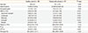
The clinical and laboratory characteristics of the healthy controls and patients with urticaria are shown in Table 1. Although the mean age of the controls was slightly older than that of the patients, there was no difference in gender between the groups. Compared to the controls, patients with urticaria showed significantly higher white blood cell counts. However, there was no difference in hemoglobin or platelet counts between the groups.
Among conventional coagulation tests, the PT of patients was similar to that of controls. Interestingly, the aPTT was slightly prolonged in the patients as compared to the controls. The fibrinogen level was similar between the groups.
Global coagulation status in patients with chronic urticaria
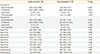
In 1 pM TF-stimulated TGA, peak thrombin and ETP levels were markedly decreased in patients with chronic urticaria (Table 2). However, there were no significant differences in lag time or time-to-peak between controls and patients. Similarly, peak thrombin and ETP were significantly lower in patients than in controls in 5 pM TF-stimulated TGA. The lag time and time-to-peak were correspondingly prolonged in patients as compared to controls.
To investigate detailed causes of the observed hemostatic disturbances, plasma levels of all coagulation factors were measured (Table 2). As expected, intrinsic coagulation factors (VIII, IX, and XII), as well as coagulation factors of the common pathway (II, V, and X), were consistently decreased in patients as compared to controls. Factor XI was also slightly decreased in patients as compared to controls, although this difference did not reach the level of statistical significance. Conversely, D-dimer was significantly increased in the patients as compared to the controls. According to smoking history, there were no significant differences in coagulation results (data not shown). When assessed only in patients, there were no differences according to ASST positivity (data not shown).
Prediction power of global coagulation tests with regard to coagulation disturbances in chronic urticaria
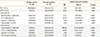
To compare the prediction power of global coagulation tests with regard to coagulation disturbances in chronic urticaria, age- and sex-adjusted logistic regression analyses were performed (Table 3). We divided the study population into 2 groups according to global coagulation test values by using the best cutoff value determined from the ROC analysis. PT did not show any prediction power. However, the aPTT showed a significant power to predict coagulation disturbances in chronic urticaria. In 1 pM TF-stimulated TGA, ETP showed a high odds ratio for predicting coagulation disturbances in chronic urticaria. Notably, the odds ratio of peak thrombin in 5 pM TF-stimulated TGA was the highest among the TGA parameters.
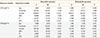
To determine the contributing value of clinical and laboratory parameters to ETP, we performed linear regression analyses in the total population (Table 4). In univariate regression analysis, the presence of urticaria strongly decreased the ETP levels observed in 1 pM TF-stimulated TGA. The aPTT value was also found to be a significant contributing factor to the ETP levels observed in 1 pM TF-stimulated TGA. In a multivariate regression analysis, the presence of urticaria was the only significant independent contributor to the ETP levels observed in 1 pM TF-stimulated TGA. Similar results were also found in the analysis of contributing factor(s) to the ETP levels observed in 5 pM TF-stimulated TGA.
DISCUSSION
Results from the current study demonstrated a coagulation disturbance in patients with chronic urticaria. Although these patients showed prolonged aPTT, differences in PT were not detected between patients and controls. This prolonged aPTT was found to be attributable to deficiencies of intrinsic coagulation factors. TGAs detected the coagulation disturbance associated with chronic urticaria with greater sensitivity than conventional coagulation tests, such as aPTT. Additionally, chronic urticaria was revealed to be an independent contributing factor to the ETP value.
The coagulation cascade has been implicated in the pathogenesis of urticaria.2789 More specifically, study results have revealed elevated plasma markers of thrombin generation, such as factor VIIa, thrombin-antithrombin complex, and D-dimer.9 However, detailed changes in individual coagulation factors have yet to be elucidated. For the first time in chronic urticaria, the current study investigated plasma levels of individual coagulation factors and revealed selective deficiencies of coagulation factors involved in the intrinsic pathway.
In general, the in vivo coagulation process occurs through the extrinsic pathway and is initiated by the binding of factor VII to TF.19 Although factor XII can also act as an initiator of the intrinsic coagulation pathway, it is not thought to mediate the in vivo coagulation process because factor XII deficiency was not associated with bleeding.20 The contact system consists of factor XII, factor XI, prekallikrein, and high-molecular-weight kininogen. Activation of the contact system can initiate the coagulation cascade through factor XII activation. For example, activation of the contact system is a frequent occurrence in angioedema.21 To date, little is known regarding contact factor activation in urticaria; however, it is plausible that the contact factor activation observed in patients with urticaria in the current study was attributable to the selective deficiency of intrinsic coagulation factors, including contact coagulation factor XII, that they exhibited.
The extrinsic pathway is not likely to be involved in the coagulation activation of urticaria because factor VII levels were not decreased in patients with chronic urticaria in the current study. However, reports of TF expression in skin specimens from patients with urticaria1222 may provide indirect evidence for extrinsic coagulation activation. Furthermore, the results of a previous study demonstrated that activated factor VII levels were higher in the acute state of urticaria, but not significantly different in chronic urticaria,23 suggesting that extrinsic coagulation activation may occur in acute urticaria.
Results from the current study also demonstrated high D-dimer levels in patients with urticaria. D-dimer is a sensitive marker of fibrin deposits formed through in vivo activation of the coagulation process. It also provides indirect evidence of thrombin generation, which activates mast cell degranulation, and induces vascular permeability.2425 Therefore, active thrombin generation may aggravate the urticarial process and provide a rationale for anticoagulant treatment.
The results of the current study provide further evidence of the utility of laboratory tests that measure thrombin generation in patients with chronic urticaria. TGA is a sensitive assay used to measure global hemostatic potential in peripheral blood.26 Because the extent of in vivo coagulation activation is determined through combined effects of coagulation and anticoagulation factors, a global coagulation test is more valuable to assess in vivo coagulation activation than measurements of individual coagulation factors. As expected, the ETP and peak thrombin results obtained from TGA sensitively detected the coagulation activation status of patients with chronic urticaria in the current study. Because coagulation activation induces mild consumption of coagulation factors, the amount of residual thrombin generation was low, as exhibited by low levels of ETP and peak thrombin. Correspondingly, the lag time and time-to-peak were prolonged due to decreased coagulation factors, which required more time to form thrombin in vitro. Likewise, the current study demonstrated the influence of urticaria on ETP levels in a linear regression analysis. The presence of urticaria contributed to ETP more strongly than other conventional coagulation parameters, such as PT and aPTT (Table 4).
The current study has several limitations. First, we could not investigate coagulation profiles with regard to disease severity and course. Second, an appropriate biomarker of contact activation remains to be explored in future studies in order to clarify the cause(s) of the observed intrinsic factor deficiencies. Third, the median age of the healthy controls was slightly higher than that of the patients. However, we speculated that the age difference did not influence our results because: (1) the age difference was slight, and (2) the logistic regression analysis was adjusted for age. Last but not least, the clinical meaning of subtle prolongation of aPTT in the urticarial patients was not clarified in this study. Therefore, prospective studies to intervene coagulation activity is needed to evaluate the pathophysiologic role of intrinsic factor deficiencies in chronic urticaria.
In summary, patients with chronic urticaria showed a prolonged aPTT compared to healthy controls. The PT value of patients did not differ from that of healthy controls. We also demonstrated that the intrinsic coagulation factors were mainly decreased in urticaria, suggesting a possible association with contact factor activation. Global coagulation status, as assessed through TGA, keenly demonstrated the in vivo coagulation activation of urticaria by showing more prominent changes in test parameters than those associated with conventional PT and aPTT. In conclusion, chronic urticaria is characterized by in vivo coagulation activation through the intrinsic coagulation pathway, which can be sensitively measured using TGA.
 XML Download