

 PDF
PDF ePub
ePub Citation
 Print
Print


INTRODUCTION
Peroxisome proliferator-activated receptor-γ (PPAR-γ) is a member of the nuclear hormone receptor superfamily regulating gene expression.1 There are thiazolidinedione (TZD) PPAR γ and non-TZD PPAR-γ ligands. TZD PPAR-γ ligands, such as rosiglitazone and pioglitazone, have been studied in terms of not only antidiabetic activity but also anti-inflammatory function. Recently, the effects of pioglitazone, a TZD PPAR-γ ligand, on increased expression of forkhead box P3 (Foxp3) and the population of regulatory T cells have been reported.2 This report suggests that PPAR-γ may affect allergic inflammation as well. However, due to adverse effects of TZD PPAR-γ ligands, such as cardiac hypertrophy, weight gain, edema, and hepatotoxicity, some of the non-TZD PPAR-γ ligands have been introduced and elucidated as having antidiabetic effects and less adipogenic activity.3,4
KR62980 (1-(transmethylimino-N-oxy)-6-(2-morpholinoethyoxy)-3-phenyl-1H-indene-2-carboxylic acid ethyl ester), a non-TZD PPAR-γ ligand, has been proved to have therapeutic effects in an animal model of allergic airway inflammation in vivo by reducing eotaxin-induced eosinophil infiltration and Th2-cell development and by attenuating goblet cell hyperplasia and collagen deposition in the airways.5 This article suggests that KR62980 may have anti-inflammatory effects in allergic rhinitis (AR).
It is well documented that the imbalance between Th1-cytokine (interferon-γ) and Th2-cytokine (IL-4, 13, and IL-5) levels plays a triggering role in the activation of IgE antibody-producing B cells, mast cells, and eosinophils. Accumulating data from mice and humans have identified Th2 cytokine as major contributors to allergy. IL-4, IL-13, and IL-5 are associated with total serum IgE, asthma, and airway sensitivity. Moreover, interferon-γ produced by Th1 cells and IL-4 produced by Th2 cells counter-regulate each other.6,7
IL-17 has been implicated in such situations as a proinflammatory regulator by inducing the expressions of many inflammatory mediators. Also, IL-17 mRNA expression has been shown to be increased in lung cells, bronchoalveolar lavage fluid, and peripheral blood from asthmatics.8,9,10 Moreover, IL-17A-deficient AR mice show a significant decrease in allergic symptoms, serum IgE levels, and eosinophil infiltration into the nasal mucosa compared to wild-type mice.11 IL-10 is also a regulatory cytokine produced by several cell types and exhibits antiallergic inflammatory properties.11
Few studies have been thoroughly conducted on anti-inflammatory effects of KR62980 associated with Th1, Th2, IL-17, and regulatory cytokines. In this study, we investigated effects of KR62980 on nasal symptoms and immunopathological profiles in allergic nasal mucosa of a murine AR model.
MATERIALS AND METHODS
Animals and OVA sensitization
Four-week-old female BALB/c mice were used in all experiments. Each mouse weighed 20 to 30 g. This study followed the principles for laboratory animal research, as outlined in the Animal Welfare Act and Department of Health, Education, and Welfare (National Institutes of Health) guidelines for the experimental use of animals, and the experimental protocol was approved by the Institutional Animal Care Committee of the Clinical Research Institute of Seoul National University Hospital.
Sensitization, KR62980 agent delivery, and allergen challenge
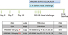
Twenty mice were divided into 4 groups: (1) the negative control group (group A) that was challenged with phosphate-buffered saline (PBS), (2) the AR group (group B) that was challenged with ovalbumin (OVA; Grade V, Sigma, St Louis, MO, USA), (3) the KR62980 intraperitoneal treatment group (group C) that was treated with KR62980 dissolved in dimethyl sulfoxide (DMSO; Sigma) via intraperitoneal injection 3 hours before intranasal OVA challenge, and (4) the KR62980 oral treatment group (group D) that was treated with KR62980 dissolved in DMSO via intragastric administration 3 hours before intranasal OVA challenge. The procedure for allergen sensitization and challenge is summarized in Fig. 1. Briefly, on days 0, 7, and 14, mice were systemically sensitized by intraperitoneal administration of 25 µg of OVA mixed with 1 mg of aluminum hydroxide (Sigma) in 300 µL of phosphate-buffered saline (PBS) or PBS only (group A). From day 22, 2% OVA droplet was administered via the nasal cavity on 7 consecutive days. The treatment groups received intraperitoneal injection (100 mg/kg) (group C) or intragastric administration (100 mg/kg) (group D) of KR62980 (Korea Research Institute of Chemical Technology, Daejeon, Korea) dissolved in DMSO 3 hours before each OVA nasal challenge on days 22, 24, 26, and 28. Twenty-four hours after the final OVA challenge, the mice were sacrificed for analysis. The dosage of KR62980 in this study was referred to reference 6.
Evaluation of allergic responses
Nasal symptom scores
After the final intranasal allergen challenge with 200 µg of OVA, the frequencies of sneezing and nose scratching were counted for 10 minutes to evaluate early allergic responses.
Measurement of OVA-Specific IgE and total IgE
Serum levels of total IgE and OVA-specific IgE were measured by solid-phase enzyme-linked immunosorbent assay (ELISA). Serum samples were harvested 24 hours after the last nasal OVA challenges and plated on 10 µg/mL of OVA-coated microtiter plates. Bound immunoglobulin isotypes were detected with specific secondary antibody (biotin-conjugated rat anti-mouse IgE Ab was purchased from BD Pharmingen, San Jose, CA, USA). Total serum IgE was measured by standard ELISA using anti-mouse IgE capture mAb (BD Pharmingen).
Mesurement of Cytokines in splenocyte culture
The spleen was removed aseptically 24 hours after the last intranasal challenge. A single cell was plated on a 24-well plate at a final concentration of 5×10⁶ cells/well in RPMI-1640 containing 10% fetal bovine serum (FBS) with penicillin-streptomycin. The cell was stimulated with OVA for 72 hours. The supernatant was collected and stored at -70℃ until needed.
The levels of IL-4, 5, 10, 17, and IFN-γ in the splenocyte supernatant were measured using commercially available ELISA kits (R&D Systems, Inc., Minneapolis, MN, USA). After measuring optical density at 450 nm, the concentrations of IL-4, 5, 10, 17, and IFN-γ were determined by interpolation from a standard curve and expressed as pg/mL.
Histological assessment
Experimental mice were sacrificed 24 hours after the final OVA challenge, and nasal tissues were obtained for analysis. Two sections of the nasal septum, 4 µm apart, were made 5 mm posterior to the nasal vestibule and used in histological assessment.
Eosinophil counts
Nose sections were stained with hematoxylin and eosin to assess inflammatory cell infiltration. Eosinophils were defined morphologically by the presence of 2-lobed nucleus and eosinophilic granules in the cytoplasm. Eosinophils were counted under a microscope at ×400 magnification. Four randomly chosen high power fields (HPFs) of each section were examined to yield the mean number of eosinophils per HPF of subepithelial connective tissue. Eosinophils in each section were counted, and the average number of eosinophils per HPF was also calculated. All slides were read by a single pathologist who was blinded to the experimental conditions.
Real-time polymerase chain reaction (PCR)
Total RNA was prepared from the nasal mucosa with TriZolreagent (Invitrogen, Carlsbad, CA, USA). Complementary DNA (cDNA) was synthesized using superscript reverse
transcriptase (Invitrogen) and oligo (dT) primers (Fermentas, Burlington, ON, Canada). For the analysis of IL-4 (Mm00445258_g1), IL-5 (Mm00439646_m1), IL-10 (Mm00439616_m1), IFN-γ (Mm99999071_m1), GAPDH (Mm03302249_g1), PDAR (Pre-Developed AssayReagent) kits of primers, and probes were purchased from Applied Biosystems (Foster City, CA, USA). Amplification of IL-4, IL-5, IL-10, IFN-γ, and GAPDH cDNA was carried out on MicroAmp optical 96-well reaction plates (Applied Biosystems). The reaction was performed using an ABI prism 7000 Sequence Detection System (Applied Biosystems). The average transcript levels of genes were then normalized to GAPDH.
Flow cytometric analysis
For cell-surface staining, splenic mononuclear cells were incubated with fluorescein isothiocyanate-conjugated anti-mouse CD4 antibody (eBioscience, San Diego, CA). For intracellular staining, cells stained with CD4 were incubated with fixation/permeabilization working solution, and Fc receptors were blocked with excess mouse Fc block. Cells were then stained with phycoerythrin (PE)-Cy5-conjugated anti-mouse Foxp3 and allophycocyanin (APC)-CD25 antibody (eBioscience). CD4+CD25+Foxp3+ T cells were analyzed by flow cytometry (FACSCalibur; Becton Dickinson, San Jose, CA).12
RESULTS
Effect of KR62980 on nasal symptoms
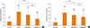
The number of nasal rubbing was more significantly more increased in group B than in group A (7.8±1.3 vs 2.2±1.0). The number of sneezing was also significantly more increased in group B than in group A (7.2±1.3 vs 1.8±1.0). Moreover, nasal rubbing/sneezing scores were significantly more decreased in KR62980-treated groups (groups C and D) than in group B (P=0.018 and P=0.029, respectively) (Fig. 2).
Effects of KR62980 on eosinophilic inflammation
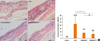
Histological analysis of the nasal mucosa obtained from the OVA group revealed grossly typical pathologic features of AR in the submucosa which was infiltrated with numerous inflammatory cells. There were significant differences in the gross appearance of cross sections between group C/D (KR62980-treated mice) and group B (Fig. 3A).
Eosinophil counts in the nasal mucosa are shown in Fig. 3B. Significant differences in eosinophil counts were found between individual groups (group B vs group C, P=0.023; group B vs group D, P=0.028). The mean eosinophil count in groups B, C, and D were 21.9±11.7, 8.7±3.1, and 6.65±3.8, respectively. The mean eosinophil count was more decreased in group D than in group C, without statistical significance.
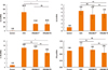
Effecst of KR62980 on the production of OVA-specific IgE and inflammatory cytokines
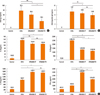
The OVA-treated groups showed significantly higher total IgE and OVA-specific IgE levels than group A. Significant differences were observed in total IgE levels between groups B, C, and D (group B vs group D, P=0.049; group C vs group D, P=0.034) (Fig. 4A). Also, OVA-specific IgE levels between groups B and D were significantly different (group B vs group D, P=0.049) (Fig. 4B). Similarly, the levels of IL-4 and IL-5 in the splenocyte culture were significantly more elevated in the OVA-treated groups (groups B, C, and D) than in group A. KR62980 treatment significantly decreased the levels of IL-4 and IL-5 (P=0.10 for groups B and D, and P=0.01 for group D). In addition, there were significant increments in IL-10 levels in the splenocyte culture supernatant by OVA treatment (P<0.001 for each). KR62980 treatment induced further increments in IL-10 levels in groups C and D (Fig. 4C).
Effects of KR62980 on the Expressions of IL-4, 5, 10, and IFN-r mRNAs
There were increases in the expressions of IL-4, IL-5, IL-10, and IFN-γ mRNAs in the allergic nasal mucosa compared to those in the control group (Fig. 5). However, the expressions of these mRNAs were more reduced in the KR62980-treated groups than in group B.
Effects of KR62980 on the Expressions of IL-17 and IL-17 mRNA
RT-PCR analysis showed that IL-17 mRNA expression in allergic nasal mucosa (group B) was increased, while it was reduced by administration of KR62980 (groups C and D) (Fig. 6A). In addition, the cytokine IL-17 level in the splenocyte culture supernatant seemed to be more decreased in groups C and D compared to group B, though it did not reach statistical significance (Fig. 6B).
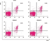
Effects of KR62980 on CD4+CD25+Foxp3 Tregs
We examined the proportion of CD4+ T cells and regulatory T cells from the whole spleen class by flow cytometry. In the AR group, the percentage of regulatory T cells seemed to be increased in the KR62980-treated group D (Fig. 7).
DISCUSSION
PPAR-γ was originally known to regulate adipocyte differentiation and lipid metabolism.13 Since then PPAR-γ has been suggested to be an important immune regulator. PPAR-γ ligands have been proved to have anti-inflammatory activity in the murine models of inflammatory diseases, such as arthritis, inflammatory bowel disease, and asthma.13,14,15,16,17 Recent studies have reported that PPAR-γ activation by administration of PPAR-γ agonists or adenovirus carrying PPAR-γ cDNA decreases allergen-induced airway inflammation, subepithelial fibrosis, eosinophil infiltration, and airway hyperresponsiveness.18 KR62980 has been developed to redeem the limitation of TZD PPAR-γ ligands.19,20,21,22
In this study, we evaluated the effect of KR62980, a novel non-TZD PPAR-γ ligand, on nasal allergic inflammation and examined the role of PPAR-γ in an animal AR model. We found that activation of PPAR-γ by administration of KR62980 also substantially inhibits the expressions of cytokines (IL-4 and IL-5) and eosinophillic inflammation. Suppression of Th2 cytokines has been found in local (nasal mucosa) and systemic (splenocyte culture supernatant) levels, which can explain the reduction in allergic symptoms by KR62980. In case of Th1 cytokines, administration of KR62980 seemed to elevate INF-γ; however, to draw such a conclusion further studies would be needed because there are discrepancies regarding IFN-γ levels between the local nasal tissue and the spleen supernatant. Based on these results, we conclude that, in terms of Th1/Th2 balance, KR62980 can suppress allergic inflammation by regulating Th2 cytokines.
IL-10 is an anti-inflammatory cytokine that down-regulates cellular immunity and allergic inflammation. IL-10 has been shown to modulate allergen-induced airway inflammation, inhibit eosinophil survival and IgE synthesis, and down-regulate the expressions of IL-4 and IL-5 through T-helper type 2 lymphocytes.23 In a previous study regarding IL-10 and PPAR-γ, the IL-10 level was increased in the lung tissue of a murine asthma model after ovalbumin inhalation and was further increased by administration of rosiglitazone and piglitazone.18 However, the definite role of IL-10 has not yet been clearly elucidated in AR. Recently reported data regarding the nasal release of IL-10 and other cytokines or chemokines in AR demonstrated a significant increase in IL-10 at 5 hours after nasal allergen challenge; however, it was significantly lower in AR subjects than in nonallergic subjects during natural pollen exposure, concluding that IL-10 may be related to the alleviation of nasal mucosal allergy.24
Our study with an ovalbumin-induced murine AR model revealed that activation of PPAR-γ by administration of KR62980 reduced eosinophilic inflammation in the nasal mucosal tissue. The elevated IL-10 cytokine levels in the spleen supernatant after ovalbumin challenge were further increased by administration of KR62980. These findings suggest that an anti-inflammatory role of PPAR-γ in the pathogenesis of AR could be partially mediated through an IL-10-dependent mechanism. The elevated IL-10 levels in splenocyte culture may have been attributed to the increased number of T reg cells by administration of KR62980, which was proven by FACS analysis. However, we could not identify further increments in IL-10 by KR62890 in the nasal mucosa. Although it is not clear why the reaction patterns are different in local nasal mucosa versus systemic one, the difference may be due to the different main target of action besides the dissimilarity in protein and mRNA levels as we usually have been noticed. This difference may also be partially due to the sites of interest in the samples and measuring parameters (cytokine levels in the cell supernatant versus mRNA expression in the nasal mucosal tissue).
IL-17 was initially known to be associated with neutrophil-dominant inflammation as a promoter of granulopoiesis, neutrophil accumulation, and neutrophil activation in the lung tissue. Increasing evidence has been published that IL-17 is related to allergic eosinophillic inflammation as well as neutrophillic inflammation. Also, IL-17 is increased in the sera of seasonal AR patients and AR modelmice.10,11 Another study elucidating the relationship between PPAR-γ and IL-17 indicated that administration of PPAR-γ agonists substantially reduced increases IL-17 protein levels and mRNA expression via NF-κB inhibition after ovalbumin inhalation in an animal asthma model.25 Moreover, it has been demonstrated that treatment with rosiglitazone significantly decreases IL-17 expression in an animal model of inflammatory bowel disease. As expected, in our study the expressions of IL-17 cytokine and mRNA were up-regulated in an animal model of OVA-induced AR. Thereafter, our results demonstrated that KR62980 seems to suppress the IL-17 cytokine level in the spleen supernatant and IL-17 mRNA expression in the nasal mucosal tissue. This result is in agreement with that of a previous study conducted with KR62980, in which allergic cytokines in the bronchoalveolar lavage fluid, as assessed by ELISA, were decreased in the KR62980-treated group. Taken together, KR62980, a non-TZD PPAR-γ agonist, like other TZD PPAR-γ agonists, may also play a role in the amelioration of inflammation in a murine AR model.
It has been reported that PPAR-γ enhances regulatory T cells through PPAR-γ-dependent or PPAR-γ-independent mechanisms by increasing retinoic acid synthesis from murine splenic dendtric cells.26,27,28 Recently, the therapeutic effect of pioglitazone on the induction of regulartory T cells in a PPAR-γ-dependent fashion was reported in a murine AR model.29 In this mechanism, pioglitazone can reduce GATA-3 and T bet expression and induce Foxp3+ expression in CD4+ T cells after challenge with OVA. Moreover, it has been reported that regulatory T cells can skew a reduction in Th1 and Th2 subpopulations. In a human study, it has also been demonstrated that the ability of regulatory T cells to suppress Th17 cytokine production is significantly decreased in the AR group.30 In our study, the proportion of CD4+CD25+Foxp3+ T cells in the spleen was slightly increased in the KR62980-treated groups (groups C and D) (Fig. 7). Foxp3+ is an intracellular marker of regulatory T cells. Based on the increasing tendency of IL-10 along with the proportion of CD4+ CD25+Foxp3+ T cells, it is conceivable that the expression of Foxp3+ induced by KR62980 results in an increase in the IL-10 level and, in turn, affects the levels of Th2, Th1, and Th17 cytokines. These findings indicate that KR62980, like other PPAR-γ agonists, may have the potential to regulate induction of specific T-cell subgroups.
Additionally, the anti-inflammatory effect of KR62980 seemed to be more remarkable via oral administration than via intraperitoneal administration in terms of some parameters. The bioavailability after oral administration is reported to be approximately 60.9%, and the metabolic half-life of 0.1-10 µM KR-62980 is reported to be 11.5-15.2 minutes in rat liver microsomes.31 It is reasonable to assume that oral administration of KR62980 would reach a valid concentration more easily. However, further studies are needed to draw a definite conclusion.
In conclusion, our results demonstrate that administration of non-TZD PPAR-γ agonists may modulate allergic symptoms and immunologic features via not only suppression of Th2 cytokines but also regulation of IL-10/IL-17 expression. Hence, non-TZD PPAR-γ agonists may have a therapeutic potential to treat allergic nasal inflammation.

 XML Download