

 PDF
PDF ePub
ePub Citation
 Print
Print


INTRODUCTION
Nasal polyposis (NP) is a common chronic inflammatory disease of the mucous membranes in the nose and paranasal sinuses. NP is characterized by eosinophilic infiltration and tissue remodeling consisting of epithelial proliferation, goblet cell hyperplasia, pseudocyst formation, basement membrane thickening, focal fibrosis and edema.1 NP often accompanies chronic rhinosinusitis (CRS), and symptoms of NP include nasal congestion, loss of smell, and headache caused by secondary infection.2 Although many studies for NP have been reported, its pathogenesis remains poorly understood and the treatment of NP treatment is limited.
Prostaglandin E2 (PGE2), a metabolite of arachidonic acid, has multiple physiological effects.3 These effects are mediated by 4 different E prostanoid (EP) receptors that belong to the G protein-coupled receptor family: EP1 increases Ca2+ channel gating via Gq, EP2 and EP4 increase the levels of cyclic adenosine monophosphate (cAMP) via Gs, whereas EP3 decreases cAMP via Gi.4 In previous studies, low production of PGE2 has been reported in NP and in both fibroblasts and epithelial cells.5 PGE2 suppresses eosinophilia-associated cellular responses induced by staphylococcal enterotoxin, dominantly through an EP2-mediated pathway in NP.6 These observations suggest that PGE2 plays a role as an inflammatory mediator to induce inflammatory diseases, such as NP, allergic rhinitis, and bronchial asthma. However, the effect of PGE2 on the production of interleukin 6 (IL-6) and IL-8 in NP is unknown.
Many kinds of cells such as epithelial cell, T cell, mast and fibroblast, are involved in the pathogenesis of NP. Among these cells, fibroblasts play a key role in the structural modification of the sinonasal mucosa. Fibroblasts differentiate into myofibroblasts and produce large amounts of extracellular matrix molecules such as collagen and fibronectin.7 Recent studies have shown that fibroblasts are not just a structural modifier, but also important modulators of local inflammation.8 Fibroblasts express many receptors for cytokines, growth factors, and hormones.9 Therefore, they play a role for mediator of immune function and have the capacity to release a variety of pro-inflammatory mediators such as eotaxin, IL-6 and IL-8.10,11 Previous studies revealed that IL-6 and IL-8 are increased by PGE2 via the EP4 receptor in different cell types.12,13 However, it is unknown whether PGE2 induces expression of IL-6 and IL-8 in nasal polyp-derived fibroblasts (NPDFs).
The purposes of this study were to determine whether PGE2 has any effect on the increase of IL-6 and IL-8 in NPDFs, and subsequently to investigate the possible mechanism underlying this effect.
MATERIALS AND METHODS
Reagents
PGE2, purchased from Sigma (St. Louis, MO, USA) was dissolved in dimethyl sulfoxide (Sigma) and then diluted with complete medium to concentrations suitable for use in this experiment. The receptor agonists and antagonists, obtained from Cayman Chemical (Ann Arbor, MI) were as follows: EP1 receptor and EP3 receptor agonist (Sulprostone, 10 nM), EP2-receptor agonist (Butaprost, 10 µM), EP4 receptor agonist (CAY10580, 10 µM), EP2 receptor antagonist (AH6809, 10 µM) and the EP4 receptor antagonist (AH23848, 10 µM). Akt inhibitor (LY294002, 10 µM) was purchased from Calbiochem (Billerica, MA, USA). NF-κB inhibitor (BAY-11, 1 µM) was bought from Sigma. NPDFs were previously exposed to PGE2 (20 µM) after pre-treatment for 1 hour with all agonists, antagonists and inhibitors.
Isolation and induction of NPDFs
Fibroblasts were cultured from 8 patients (4 women and 4 men; 32.3±5.2 years of age) who underwent endoscopic sinus surgery for CRS with NP at the Department of Otorhinolaryngology Head and Neck Surgery of the Korea University Medical Center. The study protocol was approved by the Institutional Review Board of the Korea University College of Medicine. NPDFs were isolated from surgical tissues and purified according to our previous study.14 Cells used for the experiments were obtained from the fourth cell passage.
Reverse transcription-polymerase chain reaction (RT-PCR)
NPDFs were stimulated with PGE2 in time (0-24 hours) and dose (0-20 µM, 12 hours) dependent manner. NPDFs were stimulated with PGE2 (20 µM), with or without Sulprostone (10 nM), Butaprost (10 µM), CAY 10580 (10 µM), AH6809 (10 µM), AH23848 (10 µM), LY294002 (10 µM) and BAY-11 (1 µM) for 12 hours. Total RNA was isolated using Trizol reagent (Invitrogen, Carlsbad, CA) and 2 µg of the RNA were reverse-transcribed using MMLV reverse transcriptase (Invitrogen). PCR was performed using the primer pairs targeting specific genes, as follows: IL-6 (sense sequence, 5'-GCCTTCGGTCCAGTTGCC-3'; anti-sense sequence, 5'-GCGCAGAATGAGATGAGTTGTCATG-3'; 566 bp), IL-8 (sense sequence, 5'-ATGACTTCCAAGCTGG CC-3'; anti-sense sequence, 5'-TCTTCAAAAA CTTCTCCACAA CCC-3'; 282 bp), GAPDH (sense sequence, 5'-GTGGATATTGTT GCCATCAATGACC-3'; anti-sense sequence, 5'-GCCCC AGCCT TCTTCATGGTGGT-3'; 271 bp). Amplification reactions were performed as follows: the initial denaturation step was performed at 94℃ for 5 minutes, followed by 30 cycles performed successively at 94℃ for 45 seconds, 55-65℃ for 45 seconds, and 72℃ for 45 seconds. The final extension step was performed at 74℃ for 5 minutes. All these reactions were performed in a volume of 20 µL and the products were electrophoresed on a 1.5% agarose gel and visualized by staining with ethidium bromide. Gel images were acquired using the Molecular Imager ChemiDoc XRS + (Bio-Rad, Hercules, CA, USA).
Enzyme-linked immunosorbent assay (ELISA) of IL-6 and IL-8
NPDFs were stimulated with PGE2 for 48 hours in dose (0-20 µM)-dependent manner. NPDFs were stimulated with PGE2 (20 µM), with or without Sulprostone (10 nM), Butaprost (10 µM), CAY10580 (10 µM), AH6809 (10 µM), AH23848 (10 µM), LY294002 (10 µM) and BAY-11 (1 µM) for 48 hours. IL-6 and IL-8 production in the medium derived from NPDFs was determined by ELISA (R&D Systems, Minneapolis, MN, USA). This assay was performed according to the manufacturer's instructions.
Western blot analysis
NPDFs were stimulated with PGE2 (20 µM), with or without LY294002 (10 µM) or BAY-11 (1 µM) for 1 hour. The fibroblasts were lysed in PRO-PREP™ protein extraction solution (iNtRON Biotechnology, Seongnam, Korea); proteins were separated by 10% sodium dodecyl sulfate-polyacrylamide gel electrophoresis and transferred to polyvinylidene difluoride membranes (Millipore Inc., Billerica, MA, USA). These membranes were incubated with anti-rabbit polyclonal phosphorylated Akt, p50, and GAPDH (Santa Cruz, CA, USA). After incubation, the membranes were washed 3 times (5 minutes per wash) and treated with peroxidase-conjugated anti-rabbit IgG antibody (Vector Laboratories, Burlingame, CA, USA) for 1 hour. After washing, a substrate obtained from an enhanced chemiluminescence reagent kit (Du Pont, Boston, MA, USA) was added to the membranes. The membranes were then exposed to X-ray films.
RESULTS
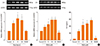
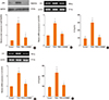
PGE2 induces IL-6 and IL-8 expressions in NPDFs
To determine the effect of PGE2 on IL-6 and IL-8 expressions in NPDFs, NPDFs were stimulated with PGE2 for 12 or 48 hours. PGE2 significantly increased IL-6 and IL-8 mRNA expression levels in time-dependent (Fig. 1A and 2A) and dose-dependent (Fig. 1B and 2B). Also, PGE2 induced production of IL-6 and IL-8 in dose-dependent manner (Fig. 1C and 2C).
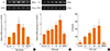
PGE2-induces IL-6 production increases with EP2 and EP4 in NPDFs
To identify the receptors that mediate the effect of PGE2-in duced IL-6 expression, we used agonists and antagonists specific to the corresponding receptors. The specific EP2 agonist (Butaprost, 10 µM) and EP4 agonist (CAY10580, 10 µM) significantly induced IL-6 expression in NPDFs. However, the EP1and EP3 agonist (Sulprostone, 10 nM) did not induce IL-6 expression in NPDFs (Fig. 3A and C). The increased expression of IL-6 was inhibited by both EP4 antagonist (AH23848, 10 µM) and EP2 antagonist (AH6809, 10 µM) (Fig. 3B and D).
PGE2-induces IL-8 production increases with only EP4 in NPDFs
To examine receptors that mediate PGE2-induced IL-8 expression, we treated PGE2 with and without specific agonists and antagonists. The EP4 agonist (CAY10580, 10 µM) significantly induced IL-8 expression in NPDFs. However, EP1 adn EP3 agonist (Sulprostone, 10 nM) and EP2 agonist (Butaprost, 10 µM) did not increase IL-8 expression in NPDFs (Fig. 4A and C). IL-8 expression was inhibited by the EP4 antagonist alone (AH23848, 10 µM) (Fig. 4B and D).
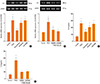
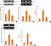
PGE2 increases IL-6 and IL-8 expression via the Akt pathway
To determine the signal pathway for PGE2-induced IL-6 and IL-8 production in NPDFs, we evaluated the stimulation of Akt as a downstream marker of IL-6 and IL-8 signaling by western blot analysis and RT-PCR. In western blot analysis, PGE2-induced stimulation of Akt decreased significantly when an Akt inhibitor (LY294002, 10 µM) was treated (Fig. 5A). Moreover, PGE2-induced expression levels of IL-6 and IL-8 were specifically inhibited by the Akt inhibitor (Fig. 5B-E).
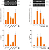
PGE2 increases IL-6 and IL-8 expression via the NF-κB transcription factor
To determine whether the NF-κB transcription factor was involved in PGE2-induced IL-6 and IL-8 production in NPDFs, we treated PGE2 under two conditions: in the presence of the NF-κB inhibitor (BAY-11, 1 µM) and in the absence of the inhibitor. The western blot analysis showed that PGE2 increased the expression of p50 (subunit of NF-κB) and that p50 expression decreased in the inhibitor-treated NPDFs (Fig. 6A). The production of IL-6 and IL-8 was specifically inhibited by treatment with the NF-κB inhibitor (Fig. 6B and C).
DISCUSSION
In this study, we found that PGE2 significantly induced the expression of IL-6 and IL-8 in NPDFs. PGE2 increased IL-6 expression via EP2 and EP4,however, increased IL-8 expression via EP4 alone. PGE2 activated the Akt and NF-κB signal pathways for IL-6 and IL-8 expression.
The roles of cytokines in the development of NP are widely investigated. IL-6 has a chemotactic effect on eosinophils and IL-8 affects both neutrophils and eosinophils, causing their migration to the site of inflammation.15 According to recent studies, increased levels of IL-6 and IL-8 may participate in the primary pathogenesis of CRS and NP as well as in the recurrent episodes.16,17,18 IL-6 is a multifunctional cytokine implicated in various inflammatory conditions including NP pathogenesis. It stimulates fibroblast proliferation, increases collagen deposition, and decreases collagen breakdown. IL-8 releases other inflammatory mediators such as histamine and leukotriene B4.19,20 IL-8 is a critical cytokine in the pathogenesis of CRS, and high levels of IL-8 have been detected within NP.21,22 PGE2-induced IL-6 and IL-8 release is mediated by EP4 receptor in colonic epithelial cells, pulmonary endothelial cells, and astrocytes.12,13,23 In this study, PGE2 induced both IL-6 and IL-8 expression in NPDFs. PGE2-induced IL-6 expression was mediated by EP2 and EP4 receptors. On the other hand, PGE2 induced IL-8 expression only by the EP4 receptor.
PGE2 are mediated via four different G-protein-coupled PGE receptors (e.g. EP1, EP2, EP3, and EP4), which are involved in the activation of phospholipase C (EP1) and activation (EP2, EP4) or inhibition (EP3) of adenyl cyclase.24 Recent studies showed EP4 agonist induced potent relaxations in asthma.25 PGE2 decreases proliferation of bronchial smooth muscle cell via EP2 and EP4 receptors in nonasthmatic eosinophilic bronchitis.26 In our data, activation of EP2 and EP4 receptor induced expression of IL-6 and activation of EP4 receptor stimulated expression of IL-8 in NPDFs. However, sulprostone (EP1/3 agonist) did not induce both IL-6 and IL-8 expression in NPDFs. These data suggest that PGE2 induced expression of IL-6 and IL-8 via EP2 and/or EP4 receptor(s) in NPDFs.
Activation of EP receptors can initiate kinase signaling by phosphatidylinositol 3-kinase (PI3K)/Akt pathways and then affects target gene transcription.27 A previous study has demonstrated that PGE2 induces IL-6 expression in human chondrocytes via the PI3K/Akt-dependent pathway.28 Therefore, we evaluated Akt as a downstream molecule for PGE2-induced IL-6 and IL-8 signaling. In western blot analysis, phosphorylation of Akt significantly increased in PGE2-stimulated NPDFs. Additionally, treatment with the Akt inhibitor (LY294002) specifically inhibited the activation of Akt in PGE2-stimulated NPDFs. In RT-PCR and ELISA data, the expression levels of IL-6 and IL-8 were also inhibited by treatment with LY294002. These findings show that PGE2-induced IL-6 and IL-8 production was mediated by the Akt pathway in NPDFs.
Activated Akt can phosphorylate IκB. The phosphorylated IκB frees from NF-κB, allowing translocation to the NF-κB nucleus. The translocated NF-κB subsequently activates target genes.29,30 Activation of Akt regulates the binding of the NF-κB to the IL-6 promoter and mediates PGE2-induced IL-6 expression.28 We demonstrated that NF-κB is a transcription factor for PGE2-induced IL-6 and IL-8 signaling. PGE2 increased the expression level of p50, a subunit of NF-κB, and p50 was shown to be inhibited by the NF-κB inhibitor (BAY-11) in western blot analysis. PGE2-induced IL-6 and IL-8 production was blocked when treated with the NF-κB inhibitor, as found by ELISA. These findings show that PGE2-induced IL-6 and IL-8 production is mediated by the Akt pathway in NPDFs. Our data reveal that PGE2 enhances IL-6 and IL-8 expression via the activation of NF-κB pathway in NPDFs.
In conclusion, we have shown that PGE2 increases the expression of IL-6 and IL-8 in NPDFs. PGE2-induced expression of IL-6 and IL-8 is mediated by EP2 and/or EP4 receptor(s) and via Akt and /NF-κB downstream pathways in NPDFs. Our findings show the effect of PGE2 on the expression of IL-6 and IL-8 and the underlying pathway in NPDFs. These results suggest that signaling pathway of PGE2 induced-IL-6 and IL-8 expression might provide a therapeutic target for the treatment of NP.
 XML Download