

 PDF
PDF ePub
ePub Citation
 Print
Print


INTRODUCTION
Chronic sinusitis is an inflammatory disease of the nose and sinus mucosa caused by a disturbance of ventilation and nasal cavity drainage.1 In addition, paranasal sinusitis (due to sinomucosal damage and ciliary dysfunction) results in an inflammation that lasts longer than three months.2 Chronic sinusitis is a common upper airway inflammatory condition with a prevalence of 10.1% in the general population in the USA.3 Nasal polyps are smooth, grapelike structures caused by inflamed nasal mucosa. Nasal polyps usually occur in association with chronic sinusitis.4 Upon histopathological examination, chronic sinusitis accompanied by nasal polyp frequently show various degrees of eosinophilic infiltration that are representative of allergic respiratory airway inflammation.5
Chronic sinusitis is associated with lower airway diseases such as asthma, chronic obstructive pulmonary disease (COPD), and bronchiectasis.6,7 It is generally recognized that rhinitis accompanies 90% of asthma cases and that the lifetime occurrence of an asthma event is three times more common in rhinitis patients than the general population.8 In addition to asthma, some studies have reported high prevalence rates for sinonasal symptoms in COPD that range from 75% to 88% with an increased rhinosinusitis prevalence in bronchiectasis patients that range from 58% to 70%.9 Previous studies have suggested the necessity to manage sinusitis in asthma. Patients who did not respond to pre-existing asthma treatment showed improvement after aggressive sinusitis treatment;10,11 therefore, the concept of "united airway disease" or "one linked airway disease" means that upper and lower airway diseases are different manifestations of one pathologic process.8
Studies dealing with associations of the upper and lower respiratory airways have mainly focused on rhinosinusitis patients with lung disease6,12,13; however, the potential effect of upper airway pathology on normal lower airways has not been evaluated. Therefore, this study investigated the impact of sinusitis or nasal polyp on lower airway function in healthy subjects without definite lung disease to prove a "united airway disease" hypothesis in normal subjects.
MATERIALS AND METHODS
Subjects
A retrospective study was performed on patients who chose optional osteomeatal unit computed tomography (OMU CT) in addition to the basic health screening program offered at Seoul National University Hospital, Healthcare System, Gangnam Center in Seoul, South Korea from March 2004 to June 2010. The criteria excluded patients diagnosed with asthma, pulmonary diseases and took regular medication due to lower respiratory symptoms. A chest X-ray of all subjects was reviewed to exclude pulmonary diseases such as bronchiectasis and pulmonary tuberculosis. The Institutional Review Board of Seoul National University Hospital approved this single-institutional retrospective cross-sectional study and waived the requirement for informed consent.
Questionnaire, laboratory tests, and pulmonary function tests
The basic health-screening program consisted of a health questionnaire, laboratory tests, and pulmonary function tests (PFT). The health questionnaire included smoking status, medication history, presence of respiratory symptoms, and comorbid conditions such as hypertension, diabetes, and liver disease. Laboratory tests consisted of complete blood cell counts, blood chemistry, lipid profiles, and C-reactive protein levels. PFT included forced expiratory volume at 1 second (FEV1) and forced vital capacity (FVC).
To identify the atopic status, multiple allergen simultaneous test (MAST)-Immunoblot test (RIDA® Allergy screen kit, R-Biopharm, Darmstadt, Germany) was performed according to the user manual. The applied MAST-Immunoblot test simultaneously measured 39 different specific IgE antibodies to 33 inhalant allergens and 6 food allergens. Class 2 (≥0.7 IU/mL) or higher was defined as positive result.
A methacholine bronchial provocation test assessed bronchial hyperresponsiveness (BHR) using a modified Chai's method as previously described.14 A provocative concentration that caused a 20% fall in FEV1 (PC20) was calculated and a positive BHR was defined as PC20 ≤16 mg/mL.
Assessment of sinuses and nasal polyps
A board-certified radiologist quantified the severity of paranasal sinusitis in OMU CTs based on the CT scoring system reported by Newman et al.15 The CT scoring system ranged from 0-30 points and represented the sum of scores of mucosal thickening in the paranasal sinuses (0 to 3 points for each of the 7 sinus areas) as well as the passage obstruction (0 to 3 points for the nasal passage and each of the 2 osteomeatal complex units). Limited disease was defined as a score less than 12 points and extensive disease defined as a score of 12 points or higher. The presence of a nasal polyp was assessed by the protrusion of a polypoid lesion in the nasal cavity with rhinoscopy.
Statistical analyses
The questionnaire and laboratory test results were compared between groups with and without CT findings to suggest sinusitis or nasal polyps. A Fisher's exact test and chi-square tests compared categorical variables and a Student t-test was performed to compare continuous variables between groups. Differences in lung function were compared with partial correlation coefficients adjusted for variables from the questionnaire and laboratory tests that showed significant differences between groups. Stratification was done according to the presence of sinusitis finding and severity: Group 1, no sinusitis; Group 2, limited sinusitis; and Group 3, extensive sinusitis. The results of the PFTs were compared to subgroups with a one-way ANOVA test with a post-hoc analysis. A two-sided significance level of 5% was used for all analyses and a P value less than 0.05 was accepted as statistically significant. Statistical analyses were performed using an SPSS package (SPSS 17.0, Inc., Chicago, IL, USA).
RESULTS
A total of 284 subjects (213 males and 71 females) underwent OMU CT in addition to a basic health screening program during the study period. None of the study subjects had lower respiratory symptoms or chest radiographic abnormalities.
Among the 284 subjects, 242 (85.2%) had OMU CT findings compatible to chronic sinusitis that consisted of 164 (57.7%) limited disease and 78 (32.3%) extensive disease. Current nasal symptoms were reported in 18.0% of all subjects and a linear trend was observed in the symptom rate and sinusitis severity (9.5%, 16.5%, and 25.6% in subjects without sinusitis, with limited sinusitis, and extensive sinusitis, Chi square for linear trend=5.327, P=0.021).
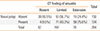
Nasal polyps were found in 134 subjects (41.2%) and were more prevalent in subjects with sinusitis finding (53.7%) compared to subjects without sinusitis finding (9.5%). Nasal symptoms were more prevalent in patients with nasal polyps (22.7%) compared to patients without nasal polyps (8.5%) (P=0.05). The presence of a nasal polyp showed a linear association with the severity of sinusitis (Chi square for linear trend=50.056, P<0.0001, Table 1).
Comparison of clinical characteristics according to co-morbid sinusitis and nasal polyp
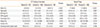
The group suspicious of sinusitis on CT showed male predominance (78.5% vs 54.8%, P<0.001) and was significantly older than the non-sinusitis control group (52.0±0.6 years vs 44.3±2.0 years, P<0.001). Body mass index (BMI) was also slightly higher in the sinusitis group than in the control group (24.6±0.2 kg/m2 vs 23.1±0.5 kg/m2, P=0.015) (Table 2). There were no significant differences in co-morbid conditions and smoking status according to the presence of sinusitis finding on CT.
Subjects with nasal polyps had a male predominance (86.9% vs 68.8%, P<0.001) compared to those without nasal polyps (Table 2). There were no significant differences in age, BMI, and smoking status according to the presence of nasal polyps.
Comparison of lung function parameters and bronchial hyperresponsiveness according to the presence and severity of sinusitis
Lung function was within the normal range; however, there were differences in the FEV1/FVC ratio depending on the presence of sinusitis finding on CT. The mean FEV1/FVC ratio showed a slight but significant reduction in the sinusitis group compared to the control group (78.6±0.5% vs 84.2±1.0%, P<0.001, after adjustment for age, gender, and BMI); however, there were no significant differences in the absolute value of FEV1 and FVC between groups. The methacholine bronchial provocation test was performed on 115 subjects (89 of 242 subjects with sinusitis finding and 26 of 42 subjects without sinusitis finding). None of the 26 sinusitis-free subjects had BHR; however, 13.5% (12/89) of the sinusitis subjects indicated positive BHR.
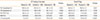
Sinusitis classified according to the severity of CT finding indicated a noticeable difference in lung function (Table 3). FEV1 and FEV1/FVC were significantly lower in subjects with extensive sinusitis than those with limited sinusitis adjusted for age, sex, and BMI. Subgroup analysis according to the severity of sinusitis revealed a linear trend of decreased FEV1 and FEV1/FVC (P=0.034 and P=0.032, respectively). The positive rate of BHR significantly increased in the order of patients without sinusitis, limited sinusitis, and extensive sinusitis (0.0%, 10.9%, and 20.0%).
Subgroup analysis according to the severity of CT finding of sinusitis in subjects without BHR also revealed a linear trend of decreased FEV1 and FEV1/FVC (P=0.022).
Comparison of lung function parameters and bronchial hyperresponsiveness according to the presence of nasal polyps
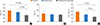
A PFT indicated that subjects with nasal polyps had a lower FEV1 and FEV1/FVC ratio than subjects without nasal polyps (P=0.027, P=0.004 respectively) (Table 3). In addition, patients with sinusitis and nasal polyp indicated a more severe airflow limitation (lower FEV1 and FEV1/FVC) than patients with only sinusitis (Figure A and C). The presence of nasal polyps was not related to BHR positivity (Table 3).
DISCUSSION
The main findings of our study were as follows: 1) decreased lung function was correlated with CT findings that suggested chronic sinusitis and nasal polyp in subjects without lower respiratory disease 2) the severity of CT finding of sinusitis was related to the degree of airway obstruction.
Traditional theories in regards to the association of upper and lower airway disease are well established. In the second century, Galen (130-201 A.D.) noticed the relationship between nasal symptoms and asthma.16 In recent decades, significant medical literature has shown evidence of the association of asthma and rhinosinusitis that has observed significant and frequent sinonasal symptoms in patients with asthma and especially severe asthma.12,17,18 The concept of 'united airway' (i.e. the association between the upper and lower airways) has expanded beyond the scope of asthma and an association with rhinosinusitis has been found in other lung diseases such as bronchiectasis and COPD.7,13
It is not clear whether rhinosinusitis triggers asthma directly or if both conditions are simply manifestations of a common disease process. However, many examples of improvements in asthma after medical treatment for rhinosinusitis support a direct causative relationship between rhinosinusitis and asthma.11 Several hypotheses explain how this relationship might exist. One is that nasopharyngobronchial reflexes may be involved in airway hyperresponsiveness.19 There are studies that indicate that local stimulation by inflammatory mediators might trigger bronchospasm.20,21 Another proposed mechanism for a causal relationship between rhinosinusitis and asthma is that local upper respiratory inflammation may lead to pulmonary inflammation by releasing chemotactic factors and leukocytes that increase cell adhesion receptors.22,23,24 There is evidence that oral breathing caused by rhinosinusitis may also lead to the inhalation of cold dry air and environmental pollutions.25
Our results show that subjects suspicious of chronic sinusitis on CT had lower FEV1/FVC, which reflected a subclinical airway obstruction. The interpretation of the subclinical airflow limitation found in chronic sinusitis subjects without lung disease is complicated. The lung function of the sinusitis group was within the normal range and the difference between the 2 groups was minimal; however, it is possible that this subtle airflow limitation might be prodromal stage of upcoming lower airway disease. More evaluation of the lower airway involvement for chronic sinusitis patients should be done even in cases without overt lower respiratory symptoms.
Generally, the diagnosis of chronic sinusitis based only on symptom criteria is difficult because most symptoms are not specific and do not distinguish between radiographically normal and diseased patients. Therefore, corroborating radiographic evidence is required to diagnose chronic sinusitis.26 Many previous studies diagnosed sinusitis only by symptoms and sinonasal standard X-ray. Plain film radiographies of the sinuses can disclose sinus opacification or reveal an air fluid level associated with maxillary; however, frontal or sphenoid disease (the most commonly infected area is the anterior ethmoid region) is poorly visualized on plain film radiographs.27 OMU CT scan, performed in a coronal plane with cuts of 4 mm or less, can overcome this limitation; consequently, the information afforded by the coronal plane has proven to correlate with the symptoms and endoscopic information. OMU CT scan classify the severity of sinusitis; consequently, a CT scan is considered the gold standard for the radiographic delineation of sinus disease28,29 and CT findings are a key component in severity staging systems for chronic sinusitis. The European Position Paper on Rhinosinusitis and Nasal Polyps, a widely recognized guideline for the diagnosis and management of chronic sinusitis and nasal polyposis, defines rhinosinusitis by the presence of at least 2 symptoms indicating inflammation in the nose and sinuses as well as by the presence of endoscopic findings and/or paranasal sinus computed tomography findings.1 In the current study, chronic sinusitis severity based on OMU CT showed a good correlation with the decrease in lower airway function and the result of the OMU CT seems to reflect upper airway inflammation precisely.
This study has some limitations in the interpretation of the results. First, selection criteria of subjects for OMU CT and bronchial provocation tests are not clear because patients can select any exam from our healthcare screening system. Most patients had no current nasal symptoms at the time to answer the questionnaire; however, it is likely that they had a previous history of nasal symptoms or a family history of respiratory diseases. Second, it is not clear whether subclinical airflow limitation is a result of suffering from chronic sinusitis or a coincidental finding of chronic sinusitis. Although several hypotheses have been proposed,30 the airflow limitation mechanism in chronic sinusitis patients is unclear and it is difficult to conclude the causal relationship between the 2 conditions. Longitudinal studies should be performed to investigate and clarify the relationship of the reversibility of airway limitation after sinusitis treatments in subjects without lower lung disease. Lastly, differences in gender between the groups can represent a bias for evaluation. Males accounted for 87.2%, in the group with CT finding that suggested extensive sinusitis; however, they accounted for 54.8% in the group without a sinusitis finding on CT. Gender differences may affect the differences in lung function.
Despite the limitations, we demonstrated the presence of a subtle airflow limitation in subjects with upper airway inflammatory conditions such as chronic sinusitis and polyps in the absence of concurrent lower airway disease as well as the association of the severity of the upper airway inflammation and the degree of the lower airway limitation.
In conclusion, this study provides evidence that the presence of CT findings suggests chronic sinusitis associated with a subclinical airflow limitation in subjects without lower respiratory disease. A united airway disease hypothesis may be valid for subjects even without definite lung disease.
 XML Download