

 PDF
PDF ePub
ePub Citation
 Print
Print


INTRODUCTION
Exercise-induced bronchoconstriction (EIB) is associated with vigorous physical exertion in 45%-85% of children with asthma.1 EIB is a highly prevalent but discrete clinical phenotype that shares common features with other measures of indirect bronchial hyperresponsiveness (BHR).2 The presence of EIB as a distinct pathophysiology in children may precede the development of other features of asthma, representing an early stage of the disease.3
Exercise causes asthma attacks due to the loss of water from the surface of the airways by evaporation when inspired air is exposed to conditions inside the body.4 This loss of water increases airway osmolarity, which initiates the activation of epithelial and mast cells and releases inflammatory mediators into the airways, leading to bronchoconstriction.5-8 The pathogenesis of EIB involves the release of cysteinyl leukotrienes (CysLTs), demonstrated by the release of CysLTs into the airways,9,10 an increase in leukotriene E4 (LTE4; a marker of CysLTs production and elimination) in the urine,11 and the inhibition of EIB pharmacologically by drugs that antagonize CysLTs.12
Clinicians currently have two main options for the initiation of pharmacotherapy in children with mild-to-moderate persistent asthma. These include inhaled corticosteroids (ICSs) or leukotriene modifiers such as montelukast. Regular antiasthma treatments with ICSs and leukotriene modifiers alleviate EIB.13-15 Clinical trials have shown that ICSs improve asthma control more effectively in both children and adults than leukotriene modifiers.16 However, there is a subset of patients who appear to respond better to leukotriene modifiers with greater improvement in lung function and asthma control.17-19 Two recent studies suggested that high LTE4 levels relative to fractional exhaled nitric oxide (FENO) were associated with a better response to montelukast compared to ICS therapy in the attenuation of albuterol usage20 and a greater forced expiratory volume in one second (FEV1).19
FENO and urinary LTE4 have been used as noninvasive markers of airway inflammation in children with asthma.21-23 The involvement of CysLT and eosinophilic inflammation can be measured by urinary LTE4 and FENO, respectively. In the present study, we investigated the association between the ratio of LTE4 to FENO and the response of therapeutic trials with montelukast or inhaled corticosteroids (fluticasone propionate [FP]) in asthmatic children with EIB.
MATERIALS AND METHODS
Subjects
Subjects were recruited from outpatient clinics of Hanyang University Hospital and screened with an exercise bronchoprovocation test under an approved study protocol by the Hanyang University Hospital Institutional Review Board. All subjects provided written informed consent. The study participants consisted of 24 children with asthma between the ages of 6 and 18 years. Participants with ≥15% postexercise maximum decrease in FEV1 were enrolled. Asthma was defined as the presence of symptoms, either an increased FEV1≥12% after bronchodilator treatment, or ≤16.0 mg/mL, if inhaled methacholine induced a 20% decrease in FEV1.24 The classification of asthma severity was based on the guidelines of the Global Initiative for Asthma using an algorithm including the medication dose and adherence, FEV1, and the symptom levels.25 Asthma was stable in all asthmatic patients at the time of the study. The exclusion criteria included acute exacerbation of asthma within the previous 6 months and parenchymal lung disease apparent on chest radiography within 4 weeks. Participants were excluded if they had used an ICS, leukotriene modifier, long-acting antihistamine, or long-acting β2-agonist 30 days before the study, and those with an FEV1 below 70% of the predicted values were also disqualified. Skin prick tests (SPTs) for common inhalant and food allergens including Dermatophagoides farinae, D. pteronyssinus, cat dander, dog dander, Alternaria alternata, cow milk, egg white, peanut, wheat, and soybean (Allergopharma, Reinbek, Germany), together with negative (saline) and positive (histamine) controls, were performed. A positive response was defined as a mean wheal diameter equal to or greater than one-half of the positive control. Specific IgE against the same common allergens that were used in the SPTs were measured with the Immuno-CAP system (Phadia AB, Uppsala, Sweden). For this analysis, atopy was defined as the presence of at least one positive allergen-specific IgE test result (IgE≥0.35 kU/L) or SPT finding. The spirometry, methacholine, and exercise challenge tests were performed by a trained technician.
Study protocol
This study was a randomized, double-blind, placebo-controlled trial comparing the effects of FP to those of montelukast in children with EIB. Children were randomized to receive either 5 or 10 mg montelukast (Singulair®; Merck/MSD, Whitehouse Station, NJ, USA) with a fluticasone placebo (from training diskus) or fluticasone (Seretide® diskus; GlaxoSmithKline, London, UK) with a montelukast placebo for a 4-week period. The participants had a total of four visits. Two studies were performed prior to treatment at baseline and 30 minutes after exercise challenge on separate days (4-day interval). Two studies were performed after treatment at baseline and 30 minutes after exercise challenge on separate days (4-day intervals). Blood tests were performed for the measurement of eosinophils, serum IgE, eosinophilic cationic protein (ECP), and cytokines. FENO-induced sputum was used to measure LTE4, cytokines, eosinophils, and ECP. Urine was used to measure LTE4. Samples were collected at baseline and 30 minutes after exercise challenge on separate days during a 4-day characterization period prior to treatment. Children were treated for 4 weeks. Following treatment, blood, FENO-induced sputum, and urine were collected at baseline and 30 minutes after exercise challenge on separate days during a 4-day characterization period following treatment.
Spirometry and methacholine challenge
Spirometry and methacholine challenges were conducted in accordance with the American Thoracic Society guidelines.24 Spirometry was performed with a Masterlab® spirometer (Jaeger Co., Freiburg, Germany).26 The bronchodilator response was calculated as the percentage change in FEV1 from baseline 15 minutes after inhaling 0.2 mg salbutamol sulfate (Ventolin®; GlaxoSmithKline) via a metered dose inhaler. Methacholine challenges were performed using a modified Chai procedure.26
Exercise challenge
Exercise challenges were conducted in accordance with the American Thoracic Society guidelines.24 An exercise challenge test was performed by running with the nose clipped on a treadmill (LE 200 CE; Jaeger Co., Hoechberg, Germany) using a standardized protocol. During the test, the heart rate was continuously monitored by a radiographic device (electronic electrocardiogram [ECG] monitor, BCI Autocorr). The ambient temperature in the air-conditioned laboratory was kept constant at 22℃, and the humidity was 40%-50% on each day of the study. The inspired air temperature and humidity were measured. The test began with running at a low speed on the treadmill with a 10% incline. The running speed of the treadmill was increased, raising the heart rate to approximately 85% of the predicted maximum [(220-age)×0.9]. This speed was maintained for a maximum of 6 minutes. Spirometry was conducted 20 and 5 minutes prior to each exercise challenge and repeated 0, 3, 6, 10, 15, and 20 minutes after the end of each exercise event. The better test sample of at least two FEV1 maneuvers within 5% of each other was recorded at each time point. The area under the FEV1-time curve (expressed as the percentage change from baseline values) over a 20-min period after exercise (AUC20)12 was used as an index of the severity of EIB. The AUC20 values were calculated using the trapezoidal rule.
Sputum induction
Induced sputum was conducted with 4.5% hypertonic saline administered through an ultrasonic nebulizer (DeVilbiss, Somerset, PA, USA), as previously described.20 In 2-min intervals, the subjects were asked to clear saliva from their mouth and expectorate sputum. The sputum was collected over 12 minutes and pooled into a single sample container. The induced sputum was placed on ice and processed within 30 minutes of collection. Samples were coded with a subject number, visit number, and date. The link between the clinical characteristics of the participants and the coded label was maintained in a separate file by the principal investigator. The total and differential cell counts were performed by an investigator (MWM) who was blinded to the clinical characteristics of each participant. The levels of LTE4, ECP, interleukin (IL)-4, IL-5, IL-8, interferon (IFN)-γ, and IL-17 were determined in the induced sputum supernatants.
Mediator assays
The concentrations of IL-4, IL-5, IL-8, IFN-γ, and IL-17 in the induced sputum and blood were determined using the cytokine multiplex assay. Enzyme immunoassay analyses of LTE4 were performed in crude urine samples and induced sputum supernatants using competitive enzyme immunoassays for LTE4 (Cayman Chemical Company, Ann Arbor, MI, USA) as described by O'Sullivan et al.27 The concentration of each sample was determined from a standard curve ranging from 7.8 to 1,000 pg/mL. The precision of the EIA for LTE428 was 17.6%. The urine LTE4 levels were reported in picograms and standardized per milligram of creatinine. Urine creatinine analyses were performed using a colorimetric assay (Sigma-Aldrich, St. Louis, MO, USA).
Fractional exhaled nitric oxide
FENO levels were measured with a portable nitric oxide analyzer (NIOX MINO®; Aerocrine, Solna, Sweden) that provided measurements at a 50 mL/sec exhalation flow rate expressed in ppb.29 Determinations made with the device were within the clinically acceptable range with measurements provided by a stationary analyzer according to the guidelines of the American Thoracic Society.30
Statistical analyses
Data analysis was conducted using SPSS software (version 16.0; SPSS Inc., Chicago, IL, USA). The variables measured on a continuous scale are summarized as the mean±standard error (SE). To compare mean values, we used parametric Student's t-tests, or nonparametric Mann-Whitney U and Wilcoxon signed-rank tests where needed. The comparisons of categorical variables were evaluated using a chi-square test or Fisher's exact test.
RESULTS
Characteristics of the study subjects
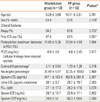
This study included 24 patients, 12 in the montelukast group and 12 in the FP group. Of the 24 subjects with asthma, 8 displayed mild intermittent asthma (4 in the montelukast and 4 in the FP group), 10 had mild persistent asthma (6 in the montelukast and 4 in FP group), and 4 displayed moderate asthma (2 in the montelukast and 2 in the FP group). No significant differences in asthma severity, the mean values of age, sex distribution, the rates of atopy, or prior use of ICSs between the montelukast and FP groups were observed (Table 1). No significant differences were observed in the baseline values of postexercise maximum decrease in FEV1 and PC20 (Table 1). No significant differences in the induced sputum volume (1.46±0.97 mL vs. 1.53±1.05 mL, P=0.185) or the concentration of the lower airway cells in the induced sputum (1.18±0.98×10⁶ cells/mL vs. 1.23±1.17×10⁶ cells/mL, P=0.732) were evident between the two groups. There were no significant differences in the baseline values of the percentage of eosinophils and neutrophils between the montelukast and FP groups (Table 1). No significant differences were observed in the baseline levels of sputum LTE4, urinary LTE4, FENO, or serum and sputum ECP between the groups (Table 1).
Effects of exercise on inflammatory markers
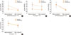
No significant changes were observed in the sputum volume and concentration of the lower airway cells in the induced sputum at baseline and after exercise challenge in either group. No significant differences were detected in eosinophils, macrophages, lymphocytes, and neutrophils in the induced sputum between the baseline and post-exercise samples, or between the two groups (data not shown). The sputum LTE4 significantly increased in the airways after exercise challenge in the montelukast group (347.1±323.4 pg/mL vs. 1,109.5±841.1 pg/mL, P=0.027) and in the FP group (452.6±281.9 pg/mL vs. 1,447.5±921.1 pg/mL, P=0.032) compared to those at baseline, while the urinary LTE4 did not. The sputum LTE4/FENO significantly increased in the airways after exercise challenge in the montelukast group (20.59±15.26 vs. 44.53±39.85, P=0.027) and in the FP group (27.37±17.58 vs. 55.73±44.91, P=0.032) compared to those at baseline, while the urinary LTE4/FENO did not (Fig. 1). The baseline levels of ECP and IFN-γ in the serum and sputum did not differ compared to those after exercise challenge. The levels of IL-5, IL-10, and IL-17A in the airways and blood were below the levels of detection in both groups. No further significant differences were observed in the baseline and postexercise levels of sputum LTE4, urinary LTE4, serum and sputum ECP between the two groups (Table 1).
Treatment effects on the severity of EIB
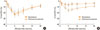
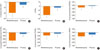
The postexercise maximum decrease in FEV1 and AUC20 decreased significantly following the 4-week treatment in both groups, but were significantly lower in the montelukast group than in the FP group (8.83±7.74% vs. 16.54±7.71%, P=0.024; 232.1±187.9 vs. 374.9±145.0, P=0.035; Table 2 and Fig. 2). There were significant differences in the magnitude of change between the two (Δ; -18.38±14.53% vs. -4.67±8.12% for the montelukast and FP groups, respectively; P=0.021; Fig. 3A). There were also significant differences in the Δ AUC20 between the groups (-21.23±17.53 vs. -3.72±3.81, P=0.028; Fig. 3B).
Effects of treatment on inflammatory markers during EIB
There were no significant differences in the percentage of eosinophils between treatments in the montelukast group (baseline, 2.11±0.54 vs. 1.34±0.41, P=0.077; postexercise, 2.05±2.59 vs. 2.19±0.82, P=0.643) or the FP group (baseline, 2.11±0.54 vs. 1.34±0.41, P=0.062; postexercise, 2.05±2.59 vs. 2.19±0.82, P=0.647). There were no significant differences in the percentage and concentrations of macrophages, lymphocytes, and neutrophils at baseline and postexercise between treatments (data not shown). A significant decrease in the levels of sputum LTE4 at baseline and postexercise after treatment compared to those before treatment was observed in the montelukast group (baseline, 347.1±323.4 pg/mL vs. 253.2±199.4 pg/mL, P=0.021; postexercise, 1,109.5±841.0 pg/mL vs. 554.5±281.9 pg/mL; P=0.018) and the FP group (baseline, 452.6±281.9 pg/mL vs. 203.1±213.7 pg/mL, P=0.038; postexercise, 1,447.5±921.1 pg/mL vs. 778.6±689.7 pg/mL, P=0.017), but there were no further significant differences in the Δbaseline sputum LTE4 or the Δpostexercise sputum LTE4 between the groups. There were no significant differences in the levels of urine LTE4 at baseline and postexercise after treatment compared to those before treatment in either group. The levels of sputum ECP at baseline and postexercise before treatment significantly decreased following treatment in the montelukast group (baseline, 41.28±38.83 ng/mL vs. 27.91±16.61 ng/mL, P=0.032; postexercise, 78.68±78.46 ng/mL vs. 29.38±21.41 ng/mL, P=0.009) and the FP group (baseline, 86.89±75.23 ng/mL vs. 38.56±24.45 ng/mL, P=0.014; postexercise, 87.16±87.73 ng/mL vs. 48.67±59.23 ng/mL, P=0.047). No significant differences were observed in the Δbaseline sputum ECP or the Δpostexercise sputum ECP between the groups. There were no significant differences in the levels of serum ECP at baseline and postexercise after treatment compared to those before treatment. A significant decrease in the levels of FENO at baseline and postexercise after treatment compared to those before treatment was observed in both the montelukast (baseline, 29.36±21.36 ppb vs. 19.00±9.12 ppb, P=0.037; postexercise, 28.09±24.48 ppb vs. 18.17±11.51 ppb, P=0.023) and FP groups (baseline, 31.58±24.46 ppb vs. 20.25±17.63 ppb, P=0.034; postexercise, 28.67±12.78 ppb vs. 18.50±11.37 ppb, P=0.039). There were no significant differences in the Δbaseline levels of FENO or in the Δpostexercise levels of FENO between the two treatment groups.
There were significant differences in the sputum LTE4/FENO ratio at baseline and postexercise after treatment compared to those before treatment in both the montelukast (baseline, 25.66±23.47 vs. 8.42±8.01, P=0.008; postexercise, 45.72±52.94 vs. 16.63±15.26, P=0.001) (Fig. 1A) and FP groups (baseline, 25.24±17.58 vs. 20.41±17.54, P=0.048; postexercise, 51.10±47.91 vs. 45.79±39.86, P=0.044) (Fig. 1B). There were significant differences in the urine LTE4/FENO ratio at baseline and postexercise between treatments in both the montelukast (baseline, 2.36±2.37 vs. 0.88±0.38, P=0.012; postexercise, 2.53±1.64 vs. 1.04±0.68, Fig. 1C) and FP groups (baseline, 2.23±2.01 vs. 1.60±1.48, P=0.038; postexercise, 2.28±1.75 vs. 1.71±1.39, P=0.043) (Fig. 1D).
The Δ logarithmic sputum baseline and postexercise LTE4/FENO ratio were both significantly lower in the montelukast group compared to those in the FP group (baseline, -0.09±0.21 vs. -0.024±0.03, P=0.045, Fig. 3C; postexercise, -0.61±0.33 vs. -0.11±0.28, P=0.023, Fig. 3D), as were the Δ logarithmic urinary baseline and postexercise LTE4/FENO ratio (baseline, -0.60±0.21 vs. -0.084±0.14, P=0.046, Fig. 3E; postexercise, -0.30±0.25 vs. -0.16±0.16, P=0.038, Fig. 3F).
DISCUSSION
Comparing the effects of oral montelukast and inhaled FP in patients with EIB, the postexercise maximum decrease in FEV1, and AUC20 were significantly lower in the montelukast group than in the FP group. These results are consistent with a recent study by Stelmach et al.,31 who observed that montelukast, in combination with budesonide or alone, provided the best protection against EIB compared to other therapeutic options. No differences were seen in the degree of EIB protection between the two groups receiving montelukast, suggesting a lack of additional effects of budesonide on EIB protection in combination therapy. Earlier studies reported that montelukast reduced the immediate and late phases of bronchoconstriction.32-34
This study was conducted using induced sputum, which provided a sample from the airways.35 We showed that the levels of sputum LTE4 significantly increased in the airways 30 minutes after exercise challenge in the montelukast and FP groups. The pathogenesis of EIB involves the release of CysLTs as demonstrated by the release of CysLTs into the airways9,10,36 and the pharmacological inhibition of EIB using drugs that antagonize CysLTs.12 Hallstrand et al.10 demonstrated that treatment with CysLT antagonists reduce the severity of EIB and decrease the release of CysLTs into the airways. The overproduction of CysLTs may increase the susceptibility to EIB through various pro-inflammatory mechanisms, as CysLTs mediate airway smooth muscle constriction, mucus release, and increased vascular permeability.37 Increased levels of CysLTs may be detected in the urine after exercise challenge in subjects with EIB,11,38 but this has not been observed in all studies.39 In the present study, the baseline urinary LTE4 displayed no significant differences compared to the post-exercise urinary LTE4.
Although previous reports have associated eosinophilic airway inflammation with the severity of EIB,40 we observed no significant differences in the eosinophils in the induced sputum between the baseline and post-exercise samples, or between the two treatment groups. Inflammation in asthma is usually associated with eosinophilia, whereas isolated EIB in elite athletes seems to be more associated with neutrophilic or mixed-type airway inflammation.41-45 Airway eosinophilia may not be essential for the development of EIB.9
We showed that the Δ sputum and urinary LTE4/FENO ratio were significantly higher in the montelukast group compared to those in the FP group in children with EIB (Fig. 3).
In the montelukast group, a greater improvement in EIB was observed following treatment compared to the FP group. Understanding the pathogenesis of EIB is key to effective treatment. The pathogenesis of EIB involves the release of LTE4.9,10,36 Thus, treatments that block the activity of LTE4 are logically used as effective therapies for controlling EIB.28,46 In addition, FENO might be considered a marker of allergen-driven, local eosinophilic inflammation (readily targeted by ICS therapy). The mechanisms that trigger EIB seem to involve neutrophilic or mixed-type airway inflammation.41-45 This inflammation is not consistently associated with BHR and does not respond to inhaled steroids as is characteristic of asthma.47,48 The results of the present study are similar to those of earlier studies that reported an association between LTE4/FENO ratios and greater FEV1, and that montelukast led to a better asthma control response than ICS therapy.19 Our results are also similar to another recent study on children with moderate-to-severe asthma who predominantly received ICS therapy, which suggested that increased albuterol usage among schoolchildren was associated with high LTE4 levels, relative to FENO, and was attenuated after randomization for treatment with montelukast.20
Due to the small sample size, the results of the present study have several limitations that must be considered when interpreting the data. As this study excluded patients with unstable asthma and subjects with frequent or unstable asthma exacerbations, we cannot specifically state how our findings relate to poor asthma control. Montelukast can provide significant protection against EIB having an onset within 2 hours following a single oral dose,49 whereas ICSs exert a maximum effect after a few months. As children were treated for 4 weeks in this study, we cannot state that improvement in EIB would extend beyond this study period. A greater improvement in EIB and Δ LTE4/FENO ratio were observed in children with asthma treated with montelukast compared to FP. We speculate that LTE4 is a more general indicator of the inflammatory response to exercise, while the allergic-type triggers eosinophilc inflammation and specifically increases the FENO levels.
These data indicate that the LTE4/FENO ratio is associated with a greater response to montelukast than FP for EIB therapy. These results suggest that LTE4 may play an important role in EIB.

 XML Download