Journal List > Allergy Asthma Immunol Res > v.5(3) > 1052321

Jang, Kim, Kim, Park, Kim, Chang, Lee, Cho, Park, Nahm, Cho, Cho, Yoon, Choi, Moon, Park, and COREA study group: Impact of Atopy on Asthma and Allergic Rhinitis in the Cohort for Reality and Evolution of Adult Asthma in Korea

Abstract

Purpose

Atopy is an important cause of asthma. Few data on the prevalence of atopy or comparisons with clinical characteristics of asthma in Korean patients have been published. We evaluated the effects of atopy on clinical profiles and airway inflammation in Korean asthmatics.

Methods

We retrospectively enrolled 1,492 asthmatics from the Cohort for Reality and Evolution of Adult Asthma in Korea (COREA) cohort who had undergone skin prick tests for aeroallergens. The patients' clinical characteristics, lung function, PC20, and sputum and blood inflammatory cell counts were compared based on the presence or absence of atopy. Atopy was defined as one or more positive reactions (A/H ratio >1) on a skin prick test.

Results

Among 11 aeroallergens, house dust mites (Dermatophagoides farinae and Dermatophagoides pteronyssinus) were the most prevalent cause of a positive skin prick test. As compared with non-atopic asthmatics, atopic asthmatics showed early onset of the disease. Atopic patients with asthma had a higher FEV1, FVC, and FEV1/FVC as compared with non-atopic patients with asthma. In addition, asthmatics without atopy had more uncontrolled asthma (P=0.001) and severe rhinitis (P<0.05) as compared with atopic asthmatics. Smoking, as measured in pack years, was higher in the non-atopic asthmatics than in the atopic asthmatics. The erythrocyte sedimentation rate was higher in non-atopic asthmatics than in the atopic asthmatics and patients with non-atopic asthma had a higher sputum neutrophil count than did those with atopic asthma.

Conclusions

Our data indicate that atopic asthmatics had an early onset of disease and high IgE levels, while the non-atopic asthmatics had decreased lung function and a high sputum neutrophil count, suggesting that a different approach is needed to treat atopic asthma.

INTRODUCTION

Asthma is an increasingly common health problem in adults and children.1,2 The prevalence of asthma has increased every year for the last five decades.3-5 A Korean survey showed that 8.9% of the population has been diagnosed with asthma.6 Further, the prevalence of asthma in Korea has increased from 5.7% in 1980 to 10.1% in 1990.6
Genetic (cytokine and immune response genes),7,8 developmental, and environmental factors such as viral infections,9 allergens,10 and occupational exposure11 have been associated with asthma susceptibility, age at onset, and severity. Although the pathogenesis of the disease has not been fully elucidated, a major risk factor is the development of an immune response to a foreign antigen that is characterized by the production of antigen-specific immunoglobulin E (IgE).12 IgE-mediated allergic reactions to environmental allergens play an important role in the pathophysiology of asthma. Atopic persons have high levels of IgE that is reactive against one or more specific allergens and they often have increased plasma total IgE concentrations.13 Moreover, the prevalence of asthma is closely related to serum IgE levels standardized for age and sex.14 Overwhelming evidence has confirmed the role of IgE in atopic asthma, while a link between IgE and non-atopic asthma has also been reported.15
Several studies have reported a strong relationship between exposure to house dust mites, the presence of serum IgE directed against mite allergens, and asthma.16-18 Epidemiological evidence indicates that atopic asthma is correlated with a high serum IgE level, although the contribution of allergen-specific IgE antibodies to the pathogenesis and severity of this disease is unclear. In the present study, we examined the prevalence of atopy and compared clinical characteristics and the presence of airway inflammation by atopy in the Cohort for Reality and Evolution of Adult Asthma in Korea (COREA) cohort asthmatics.

MATERIALS AND METHODS

Subjects

The Corea is an eight-year (2005-2012), multicenter, prospective cohort study of asthmatics 19 years of age or older.19
Patients were enrolled at 11 clinical centers in various regions of South Korea. The participating institutions were the Asan Medical Center at the University of Ulsan and the hospitals of Seoul National University, Bundang Seoul National University, Yonsei University, Chung-Ang University, Ewha Womans University, Hanyang University, Soonchunhyang University, Ajou University, The Catholic University of Korea, and Chonbuk National University. We retrospectively studied the clinical data of 2,588 asthma patients who were registered in the COREA cohort. Asthma was diagnosed based on GINA guidelines.20 All subjects had a clinical diagnosis of asthma that was supported by one or more of the following criteria: 1) variability in the maximum diurnal peak expiratory flow of >20% over the course of 14 days, 2) an increase in FEV1 of >15% after the inhalation of 200-400 µg of albuterol, or 3) a 20% reduction in FEV1 in response to a provocative concentration of inhaled methacholine (PC20) of <25 mg/mL. Asthma control status and allergic rhinitis severity were classified to GINA guideline20 and ARIA guideline.21 The subjects underwent a standardized assessment, including an analysis of induced-sputum specimens, complete blood cell counts with differential counts, IgE concentrations, chest posteroanterior radiography, allergy skin prick tests, and spirometry. The body mass index (BMI) for each patient was calculated as weight (kg)/height (m2).22,23
The exclusion criteria were respiratory infection, COPD, vocal cord dysfunction, obstructive sleep apnea, Churg-Strauss synsyndrome, cardiac dysfunction, allergic bronchopulmonary aspergillosis, and poor adherence to treatment. All subjects were ethnic Koreans and required to provide written informed consent. The research protocol for this cohort study was approved by the Institutional Review Board (IRB) of each center.

Lung function

During a baseline visit, information regarding demographic characteristics was collected from all subjects, and spirometry was performed before and after the use of a bronchodilator. Baseline FVC and FEV1 measurements were obtained in the absence of recent bronchodilator use (within 8 hours). The basal and post-bronchodilator FEV1, FVC, forced expiratory flow between 25 and 75% FVC (FEF25-75%), and DLCO were measured. FVC and FEV1 were measured at regular intervals of one or two months. We used Vmax Series 2130 Autobox Spirometry (Sensor Medics, Yorba Linda, CA, USA) and applied a calibration check every morning at 8:00 AM.

Allergy skin tests

Common inhalant allergens, including dust mites (Dermatophagoides farinae and Dermatophagoides pteronyssinus), Cat fur, Dog fur, Cockroache, Grass, Tree pollen (mixed), Ragweed, Mugwort, Alternaria, and Aspergillus spp. (Bencard Co., Brentford, UK) were used for the skin prick test.24 Atopy was defined as having an allergen-induced wheal reaction equal to or greater than that caused by histamine (1 mg/mL) or equal to or greater than 3 mm in diameter. Total IgE was measured using the UniCAP system (Pharmacia Diagnostics, Uppsala, Sweden).

Sputum examination

Sputum samples were obtained for differential cell counts for patients in a stable state. Sputum was induced using isotonic saline containing a short-acting bronchodilator, and the samples were treated within 2 hours of collection as described previously.25 Briefly, all visible portions with greater solidity were carefully selected and placed in a pre-weighed Eppendorf tube. The samples were treated by adding eight volumes of 0.05% dithiothreitol (Sputolysin; Calbiochem Corp., San Diego, CA, USA) in Dulbecco's phosphate-buffered saline (PBS). One volume of protease inhibitor (0.1 M EDTA and 2 mg/mL phenylmethylsulfonylfluoride) was added to 100 volumes of the homogenized sputum, and the total cell count was determined with a hemocytometer. The cells of the homogenized sputum were collected by cytocentrifugation, and 500 cells were examined on each sputum slide after staining with Diff-Quick (American Scientific Products, Chicago, IL, USA).

Statistical analysis

The data were double-entered into a statistical software package (SPSS, version 14.0; SPSS Inc., Chicago, IL, USA). The data are expressed as the mean±standard deviation (SD) or standard error of the mean (SEM). Group differences in atopy were compared using a two-sample t-test, Mann-Whitney test, or Pearson's χ2 test for normally distributed, skewed, or categorical data, respectively. Differences in the proportions of the patient populations were analyzed by the χ2 test with Fisher's exact test when low expected cell counts were encountered. A P value<0.05 was considered to be statistically significant.

RESULTS

Common inhalant allergen sensitization

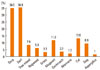
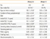
House dust mites (D. farinae and D. pteronyssinus) were the most prevalent allergen on the skin prick test (Fig. 1). The percentage of atopic patients with sensitization to two or more allergens was 39.3%. Total IgE levels were higher in atopic asthmatics than in non-atopic asthmatics (514.2±32.2 vs. 293.7±26.9 ku/L, P=0.001, Table 1).

Relationship between atopy and clinical variables

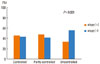
BMI was lower in atopic asthmatics than in non-atopic asthmatics (23.5±0.11 vs. 24.4±0.25 kg/m2, P=0.001). Although the allergen sensitization number was correlated with the total IgE level (r=0.351, P=0.001) and total IgE was correlated with asthma severity (r=0.101, P=0.005), allergen sensitization was negatively correlated with asthma severity (r=-0.102, P=0.001). As compared with non-atopic asthmatics, atopic asthmatics showed early onset of the disease (30.2±0.45 vs. 43.1±0.65 years, P<0.05). Cigarette smoking (in pack years) was higher in the non-atopic asthmatics than in the atopic asthmatics (23.3±0.76 vs. 13.8±0.59, P=0.001). The erythrocyte sedimentation rate (ESR) was higher in the non-atopic asthmatics than in the atopic asthmatics (20.1±2.68 vs. 12.5±1.03 mm/hr, P=0.004, Fig. 2).

Relationship between atopy and severity of asthma and allergic rhinitis

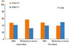
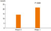
Atopic patients with asthma had a higher FEV1 (83.5±0.68 vs. 79.7±0.81% predicted, P=0.001), FVC (91.1±0.61 vs. 87.2±0.65% predicted, P=0.001), and FEV1/FVC (90.8±0.81 vs. 84.3±1.17%, P=0.001) as compared with non-atopic patients with asthma. Asthmatics without atopy had more uncontrolled asthma (control status [n=atopy/non-atopy], controlled=185/177 vs. partly controlled=326/289 vs. uncontrolled=127/200, P=0.001, Fig. 3) and severe rhinitis as compared with atopic asthmatics (severity [n=atopy/non-atopy], mild intermittent=99/87 vs. moderate to severe intermittent=59/35 vs. mild persistent=232/197 vs. moderate to severe persistent=68/83, P<0.05, Fig. 4).

Relationship between atopy and sputum inflammatory cells in patients with asthma

The atopic patients with asthma had a higher percentage of sputum lymphocytes (4.46±1.49 vs. 2.44±0.47%, P=0.001) than the non-atopic patients with asthma, and those patients with non-atopic asthma had a higher percentage of sputum neutrophils (36.0±2.28 vs. 25.3±2.02%, P=0.001) than those with atopic asthma (Table 2). There were no differences in PC20 and blood eosinophils counts between the two groups.

DISCUSSION

The results of this study suggest that atopy is associated with the severity of adult asthma and rhinitis, independent of airway hyperresponsiveness.
Asthmatics are typically classified as atopic or non-atopic based on sensitization to environmental aeroallergens. However, atopic and non-atopic asthma are not well-separated disease entities.26 Although atopy is clearly a risk factor for the development of asthma, only a small subset of subjects sensitized to aeroallergens express persistent symptoms, suggesting that additional pathogenic mechanisms such as respiratory viral infections are involved as cofactors.27 In this study, house dust mites were the most prevalent allergen and atopy was related with the severity of asthma and allergic rhinitis in the COREA cohort. These results are consistent with the notion that the quantification of IgE antibodies may serve as a marker of asthma severity.22 In this study, although allergen sensitization was correlated with total IgE levels and total IgE was correlated with asthma severity, the allergen sensitization number was negatively correlated with asthma severity, indicating that not only IgE antibody levels, but also the number of allergens reacting positively when tested, are involved in asthma severity.23 In this study, the atopic asthmatics showed early disease onset and high IgE levels, indicating a role for allergen-specific IgE antibodies in the pathogenesis of asthma and suggesting preventive and therapeutic measures for asthma control.
Asthma and allergies in developing countries may be associated with the adoption of an urbanized "Western" lifestyle. Total caloric intake is increasing with the industrialization of Korea, and BMI has been shown to be associated with wheezing in older adults living in rural areas of Korea.28 General obesity and central obesity are potential risk factors for asthma in relatively non-obese Korean adults.29 Further, BMI has been shown to be associated with airway hyperresponsiveness in school-age boys. In this study, the BMI was lower in atopic asthmatics than in non-atopic asthmatics. In addition, the non-atopic asthmatics were older and most were not obese, suggesting that Korean adult asthmatics are relatively non-obese.
Limited data regarding systemic inflammatory markers exist with respect to asthma. Some studies reported that systemic inflammatory markers were elevated in patients with non-allergic asthma and obese patients with asthma.30
Measured systemic inflammation markers, including C-reactive protein (CRP), total leukocyte count, and ESR were higher in patients with asthma and reduced by inhaled steroids plus an inhaled β2 agonist.31 One study showed that CRP was higher in patients with non-allergic asthma than in patients with allergic asthma or healthy controls.32 Another study investigated the role of obesity in local and systemic inflammation in patients with asthma.33 In this study, although the number of white blood cells did not differ between the atopic and non-atopic asthmatics, the ESR was higher in non-atopic asthmatics than in atopic asthmatics, indicating that systemic inflammation is involved in non-atopic asthma.
Cigarette smoking and asthma are associated with poor symptom control and impaired therapeutic responses to anti-asthma drugs.34 In the developed world, approximately 25% of the population smokes cigarettes35,36 and an additional 25% are former smokers. Smoking rates are similar amongst adults with asthma.37-39 Higher levels are seen in patients with asthma that present to emergency departments with exacerbations.39 As compared with nonsmokers with asthma, those who smoke have worse symptom control,37,38 an accelerated decline in lung function,40,41 and an increased mortality rate.42 Asthmatic smokers have more severe symptoms,36,37 a greater need for rescue medication,43 and worse health status indices as compared with never-smokers.43,44 In this study, non-atopic asthmatics had higher pack year values and more severe asthma, suggesting that smoking contributes to difficulties in controlling asthma.
Previous immunopathologic studies of patients with mild asthma that underwent bronchoscopic biopsy suggested that eosinophilic airway inflammation is a characteristic abnormality in both atopic and non-atopic asthma.45-47 However, other pathologic studies have suggested that neutrophils are present in higher quantities in the airways of patients with severe asthma than in the airways of patients with mild asthma or in normal control subjects.48-50 In this study, although small group of asthmatic patients of total patients were included, non-atopic asthmatics had high sputum neutrophil counts, suggesting that sputum inflammatory cell numbers can be used as a guide in the treatment of asthmatics, and that the pathology in non-atopic asthmatics is different from that in atopic asthmatics.
In conclusion, our study of a Korean cohort indicates that atopic asthmatics had an early onset of disease and high IgE levels, while non-atopic asthmatics had decreased lung function and a high sputum neutrophil count, suggesting that the treatment of asthma may be different by the atopic status of the patient.

Figures and Tables

Fig. 1
Allergen prevalence according to an allergy skin test.
aair-5-143-g001
Fig. 2
Differences in erythrocyte sedimentation rate (ESR) between atopic and non-atopic asthmatics.
aair-5-143-g002
Fig. 3
Relationship between asthma severity and atopy status.
1=mild intermittent, 2=mild persistent, 3=moderate persistent, 4=severe persistent.
aair-5-143-g003
Fig. 4
Relationship between rhinosinusitis severity and atopy status.
Symptoms of rhinosinusitis: 1=absence, 2=1x/week, 3=2x/week, 4=3-4x/week, 5=>5x/week.
aair-5-143-g004
Table 1
Clinical and physiological variables in patients with bronchial asthma by atopy
aair-5-143-i001
Atopy (+) Atopy (-)
No. 751 741
Sex, male (%) 47.1%* 41.4%
Age (at initial visit) (yr) 39.7±0.50 53.5±0.99
Duration of lung symptoms (yr) 9.53±0.41 10.4±0.52
BMI 23.5±0.11 24.4±0.25
Initial FEV1, % pred. 83.5±0.68 79.7±0.81
Initial FVC, % pred. 91.1±0.61 87.2±0.65
Initial FEV1/FVC, % pred. 90.8±0.81 84.3±1.17
Smoking status (none/ex/current) 359/238/154 357/278/106
Smoking (pack-yr) 13.8±0.59 23.3±0.76
PC20, mg/mL 5.91±0.51 6.61±0.40
Total IgE, ku/L 514.2±32.2 293.7±26.9
Blood eosinophils, % 9.0±2.53 4.7±0.20

Plus-minus values are the mean±standard error (SE). PC20 methacholine, the concentration of methacholine required to decrease the FEV1 by 20%.

*Compared with asthmatic patients without atopy, P<0.05. Compared with asthmatic patients without atopy, P<0.001.

FEV1, forced expiratory volume in one second; FVC, forced vital capacity; BMI, body mass index.

Table 2
Sputum differentials in patients with bronchial asthma by atopy
aair-5-143-i002
Atopy (+) Atopy (-)
No. 163 189
Total cells, no. 362.4±6.84* 393.6±13.1
Macrophages, % 50.8±2.54 35.6±2.2
Neutrophils, % 25.3±2.0 36.0±2.28
Eosinophils, % 11.0±1.29 10.8±1.31
Lymphocytes, % 4.46±1.49 2.44±0.47

ACKNOWLEDGMENTS

This study was supported by a grant from the Korean Healthcare Technology R&D Project, Ministry for Health and Welfare, Republic of Korea (A102065).

Notes

There are no financial or other issues that might lead to conflict of interest.

References

1. Mokdad AH, Serdula MK, Dietz WH, Bowman BA, Marks JS, Koplan JP. The spread of the obesity epidemic in the United States, 1991-1998. JAMA. 1999; 282:1519–1522.
2. Mannino DM, Homa DM, Akinbami LJ, Moorman JE, Gwynn C, Redd SC. Surveillance for asthma--United States, 1980-1999. MMWR Surveill Summ. 2002; 51:1–13.
3. Fanta CH. Asthma. N Engl J Med. 2009; 360:1002–1014.
4. Jansson SA, Rönmark E, Forsberg B, Löfgren C, Lindberg A, Lundbäck B. The economic consequences of asthma among adults in Sweden. Respir Med. 2007; 101:2263–2270.
5. Seaton A, Godden DJ, Brown K. Increase in asthma: a more toxic environment or a more susceptible population? Thorax. 1994; 49:171–174.
6. Shin TS, Lee GJ, Yoon HS. A survey of the distribution of allergic diseases in primary school children. Allergy. 1990; 10:201–212.
7. Cookson W. The alliance of genes and environment in asthma and allergy. Nature. 1999; 402:B5–B11.
8. Prescott SL, Macaubas C, Holt BJ, Smallacombe TB, Loh R, Sly PD, Holt PG. Transplacental priming of the human immune system to environmental allergens: universal skewing of initial T cell responses toward the Th2 cytokine profile. J Immunol. 1998; 160:4730–4737.
9. Stein RT, Sherrill D, Morgan WJ, Holberg CJ, Halonen M, Taussig LM, Wright AL, Martinez FD. Respiratory syncytial virus in early life and risk of wheeze and allergy by age 13 years. Lancet. 1999; 354:541–545.
10. Halonen M, Stern DA, Lohman C, Wright AL, Brown MA, Martinez FD. Two subphenotypes of childhood asthma that differ in maternal and paternal influences on asthma risk. Am J Respir Crit Care Med. 1999; 160:564–570.
11. Venables KM, Chan-Yeung M. Occupational asthma. Lancet. 1997; 349:1465–1469.
12. Platts-Mills TA. The role of immunoglobulin E in allergy and asthma. Am J Respir Crit Care Med. 2001; 164:S1–S5.
13. Gould HJ, Sutton BJ, Beavil AJ, Beavil RL, McCloskey N, Coker HA, Fear D, Smurthwaite L. The biology of IGE and the basis of allergic disease. Annu Rev Immunol. 2003; 21:579–628.
14. Burrows B, Martinez FD, Halonen M, Barbee RA, Cline MG. Association of asthma with serum IgE levels and skin-test reactivity to allergens. N Engl J Med. 1989; 320:271–277.
15. Menz G, Ying S, Durham SR, Corrigan CJ, Robinson DS, Hamid Q, Pfister R, Humbert M, Kay AB. Molecular concepts of IgE-initiated inflammation in atopic and nonatopic asthma. Allergy. 1998; 53:15–21.
16. Platts-Mills TA, Erwin EA, Heymann PW, Woodfolk JA. Pro: The evidence for a causal role of dust mites in asthma. Am J Respir Crit Care Med. 2009; 180:109–113.
17. Tovey ER, Almqvist C, Li Q, Crisafulli D, Marks GB. Nonlinear relationship of mite allergen exposure to mite sensitization and asthma in a birth cohort. J Allergy Clin Immunol. 2008; 122:114–118.
18. Granada M, Wilk JB, Tuzova M, Strachan DP, Weidinger S, Albrecht E, Gieger C, Heinrich J, Himes BE, Hunninghake GM, Celedón JC, Weiss ST, Cruikshank WW, Farrer LA, Center DM, O'Connor GT. A genome-wide association study of plasma total IgE concentrations in the Framingham Heart Study. J Allergy Clin Immunol. 2012; 129:840–845.e21.
19. Kim TB, Park CS, Bae YJ, Cho YS, Moon HB. COREA Study Group. Factors associated with severity and exacerbation of asthma: a baseline analysis of the cohort for reality and evolution of adult asthma in Korea (COREA). Ann Allergy Asthma Immunol. 2009; 103:311–317.
20. National Asthma Education and Prevention Program. Expert Panel Report: Guidelines for the Diagnosis and Management of Asthma Update on Selected Topics--2002. J Allergy Clin Immunol. 2002; 110:S141–S219.
21. Bousquet J, Khaltaev N, Cruz AA, Denburg J, Fokkens WJ, Togias A, Zuberbier T, Baena-Cagnani CE, Canonica GW, van Weel C, Agache I, Aït-Khaled N, Bachert C, Blaiss MS, Bonini S, Boulet LP, Bousquet PJ, Camargos P, Carlsen KH, Chen Y, Custovic A, Dahl R, Demoly P, Douagui H, Durham SR, van Wijk RG, Kalayci O, Kaliner MA, Kim YY, Kowalski ML, Kuna P, Le LT, Lemiere C, Li J, Lockey RF, Mavale-Manuel S, Meltzer EO, Mohammad Y, Mullol J, Naclerio R, O'Hehir RE, Ohta K, Ouedraogo S, Palkonen S, Papadopoulos N, Passalacqua G, Pawankar R, Popov TA, Rabe KF, Rosado-Pinto J, Scadding GK, Simons FE, Toskala E, Valovirta E, van Cauwenberge P, Wang DY, Wickman M, Yawn BP, Yorgancioglu A, Yusuf OM, Zar H, Annesi-Maesano I, Bateman ED, Ben Kheder A, Boakye DA, Bouchard J, Burney P, Busse WW, Chan-Yeung M, Chavannes NH, Chuchalin A, Dolen WK, Emuzyte R, Grouse L, Humbert M, Jackson C, Johnston SL, Keith PK, Kemp JP, Klossek JM, Larenas-Linnemann D, Lipworth B, Malo JL, Marshall GD, Naspitz C, Nekam K, Niggemann B, Nizankowska-Mogilnicka E, Okamoto Y, Orru MP, Potter P, Price D, Stoloff SW, Vandenplas O, Viegi G, Williams D. World Health Organization. GA(2)LEN. AllerGen. Allergic Rhinitis and its Impact on Asthma (ARIA) 2008 update (in collaboration with the World Health Organization, GA(2)LEN and AllerGen). Allergy. 2008; 63:Suppl 86. 8–160.
22. Wickman M. Experience with quantitative IgE antibody analysis in relation to allergic disease within the BAMSE birth cohort--towards an improved diagnostic process. Allergy. 2004; 59:Suppl 78. 30–31.
23. Wickman M, Lilja G, Söderström L, van Hage-Hamsten M, Ahlstedt S. Quantitative analysis of IgE antibodies to food and inhalant allergens in 4-year-old children reflects their likelihood of allergic disease. Allergy. 2005; 60:650–657.
24. Park CS, Kim YY, Kang SY. Collection between RAST and skin test for inhalant offending allergens. Allergy. 1983; 3:1–9.
25. Park SW, Lee YM, Jang AS, Lee JH, Hwangbo Y, Kim DJ, Park CS. Development of chronic airway obstruction in patients with eosinophilic bronchitis: a prospective follow-up study. Chest. 2004; 125:1998–2004.
26. Lötvall J, Akdis CA, Bacharier LB, Bjermer L, Casale TB, Custovic A, Lemanske RF Jr, Wardlaw AJ, Wenzel SE, Greenberger PA. Asthma endotypes: a new approach to classification of disease entities within the asthma syndrome. J Allergy Clin Immunol. 2011; 127:355–360.
27. Holt PG, Sly PD. Interaction between adaptive and innate immune pathways in the pathogenesis of atopic asthma: operation of a lung/bone marrow axis. Chest. 2011; 139:1165–1171.
28. Jang AS, Lee JH, Park SW, Shin MY, Kim DJ, Park CS. Severe airway hyperresponsiveness in school-aged boys with a high body mass index. Korean J Intern Med. 2006; 21:10–14.
29. Jang AS, Son MH, Choi IS, Koh YI. High body mass index is associated with wheezing among older adults living in high-altitude area in Korea. J Korean Med Sci. 2002; 17:479–482.
30. Magnussen H, Watz H. Systemic inflammation in chronic obstructive pulmonary disease and asthma: relation with comorbidities. Proc Am Thorac Soc. 2009; 6:648–651.
31. Girdhar A, Kumar V, Singh A, Menon B, Vijayan VK. Systemic inflammation and its response to treatment in patients with asthma. Respir Care. 2011; 56:800–805.
32. Olafsdottir IS, Gislason T, Thjodleifsson B, Olafsson I, Gislason D, Jögi R, Janson C. C reactive protein levels are increased in non-allergic but not allergic asthma: a multicentre epidemiological study. Thorax. 2005; 60:451–454.
33. Sutherland TJ, Cowan JO, Young S, Goulding A, Grant AM, Williamson A, Brassett K, Herbison GP, Taylor DR. The association between obesity and asthma: interactions between systemic and airway inflammation. Am J Respir Crit Care Med. 2008; 178:469–475.
34. Thomson NC, Spears M. The influence of smoking on the treatment response in patients with asthma. Curr Opin Allergy Clin Immunol. 2005; 5:57–63.
35. Centers for Disease Control and Prevention (CDC). State-specific prevalence of current cigarette smoking among adults--United States, 2002. MMWR Morb Mortal Wkly Rep. 2004; 52:1277–1280.
36. Rickards L, Fox K, Roberts C, Fletcher L, Goddard E. Living in Britain: results from the 2002 General Household Survey [Internet]. London: Office for National Statistics;2004. accessed 2004 Aug 20. Available from: http://onstest.landmarkgovernment.co.uk/ons/rel/ghs/general-household-survey/2002-edition/living-in-britain--full-report.pdf.
37. Althuis MD, Sexton M, Prybylski D. Cigarette smoking and asthma symptom severity among adult asthmatics. J Asthma. 1999; 36:257–264.
38. Siroux V, Pin I, Oryszczyn MP, Le Moual N, Kauffmann F. Relationships of active smoking to asthma and asthma severity in the EGEA study. Epidemiological study on the Genetics and Environment of Asthma. Eur Respir J. 2000; 15:470–477.
39. Silverman RA, Boudreaux ED, Woodruff PG, Clark S, Camargo CA Jr. Cigarette smoking among asthmatic adults presenting to 64 emergency departments. Chest. 2003; 123:1472–1479.
40. Apostol GG, Jacobs DR Jr, Tsai AW, Crow RS, Williams OD, Townsend MC, Beckett WS. Early life factors contribute to the decrease in lung function between ages 18 and 40: the Coronary Artery Risk Development in Young Adults study. Am J Respir Crit Care Med. 2002; 166:166–172.
41. Lange P, Parner J, Vestbo J, Schnohr P, Jensen G. A 15-year follow-up study of ventilatory function in adults with asthma. N Engl J Med. 1998; 339:1194–1200.
42. Marquette CH, Saulnier F, Leroy O, Wallaert B, Chopin C, Demarcq JM, Durocher A, Tonnel AB. Long-term prognosis of near-fatal asthma. A 6-year follow-up study of 145 asthmatic patients who underwent mechanical ventilation for a near-fatal attack of asthma. Am Rev Respir Dis. 1992; 146:76–81.
43. Gallefoss F, Bakke PS. Does smoking affect the outcome of patient education and self-management in asthmatics? Patient Educ Couns. 2003; 49:91–97.
44. Sippel JM, Pedula KL, Vollmer WM, Buist AS, Osborne ML. Associations of smoking with hospital-based care and quality of life in patients with obstructive airway disease. Chest. 1999; 115:691–696.
45. Wardlaw AJ, Dunnette S, Gleich GJ, Collins JV, Kay AB. Eosinophils and mast cells in bronchoalveolar lavage in subjects with mild asthma. Relationship to bronchial hyperreactivity. Am Rev Respir Dis. 1988; 137:62–69.
46. Djukanović R, Wilson JW, Britten KM, Wilson SJ, Walls AF, Roche WR, Howarth PH, Holgate ST. Quantitation of mast cells and eosinophils in the bronchial mucosa of symptomatic atopic asthmatics and healthy control subjects using immunohistochemistry. Am Rev Respir Dis. 1990; 142:863–871.
47. Humbert M, Durham SR, Ying S, Kimmitt P, Barkans J, Assoufi B, Pfister R, Menz G, Robinson DS, Kay AB, Corrigan CJ. IL-4 and IL-5 mRNA and protein in bronchial biopsies from patients with atopic and nonatopic asthma: evidence against "intrinsic" asthma being a distinct immunopathologic entity. Am J Respir Crit Care Med. 1996; 154:1497–1504.
48. Wenzel SE, Szefler SJ, Leung DY, Sloan SI, Rex MD, Martin RJ. Bronchoscopic evaluation of severe asthma. Persistent inflammation associated with high dose glucocorticoids. Am J Respir Crit Care Med. 1997; 156:737–743.
49. Jatakanon A, Uasuf C, Maziak W, Lim S, Chung KF, Barnes PJ. Neutrophilic inflammation in severe persistent asthma. Am J Respir Crit Care Med. 1999; 160:1532–1539.
50. Wenzel SE, Schwartz LB, Langmack EL, Halliday JL, Trudeau JB, Gibbs RL, Chu HW. Evidence that severe asthma can be divided pathologically into two inflammatory subtypes with distinct physiologic and clinical characteristics. Am J Respir Crit Care Med. 1999; 160:1001–1008.
TOOLS
ORCID iDs

Yoon-Seok Chang
https://orcid.org/http://orcid.org/0000-0003-3157-0447

You Sook Cho
https://orcid.org/http://orcid.org/0000-0001-8767-2667

Dong-Ho Nahm
https://orcid.org/http://orcid.org/0000-0001-5253-6577

Choon-Sik Park
https://orcid.org/http://orcid.org/0000-0003-2977-0255

Similar articles