

 PDF
PDF ePub
ePub Citation
 Print
Print


INTRODUCTION
Drugs, latex, and disinfectants are major causes of occupational allergic diseases in health care workers (HCW) in hospitals.1,2 Drugs are one of the most common causes of allergic diseases in HCW, including nurses and pharmacists, who both work with drugs.3-9 HCW in hospitals are commonly exposed to beta-lactam antibiotics, such as penicillins and cephalosporins. Occupational allergic diseases caused by these agents, including bronchial asthma, allergic contact dermatitis, and contact urticaria syndrome, have been frequently reported.3-7,10,11 An IgE-mediated mechanism has been suggested for contact urticaria syndrome, which includes urticaria and anaphylaxis induced by beta-lactam antibiotics.3,4
A few published studies have used skin prick tests (SPTs) to determine sensitization rates to beta-lactam antibiotics in HCW. The sensitization rate to beta-lactam antibiotics by SPT was 2.6% Koreain hospital nurses, and the prevalence of contact urticaria syndrome associated with beta-lactam antibiotics was 8.9%.12 In hospital nurses without a history of penicillin allergy, the sensitization rate by SPT to penicillins was 12%.13 In recent years, cephalosporins have been more commonly prescribed than penicillins in major hospitals in this country. Therefore, we investigated the sensitization rates to commonly used cephalosporins in HCW in a single university hospital. We used two methods: SPT and measurement of specific IgE antibodies to cephalosporins conjugated with human serum albumin (HSA).
MATERIALS AND METHODS
Subjects
The study subjects included 161 HCW and 86 non-atopic healthy controls that were recruited from a single tertiary hospital and the general population. The HCW consisted of 138 nurses and 23 pharmacists. The nurses were divided into the following two groups: group I included 125 nurses working in wards where they were exposed to cephalosporins (current-exposure group) and group II included 13 nurses working in outpatient departments or in offices, who had a prior history of working in general wards at least six months before the study (previously-exposed group). The pharmacists-who were employed in the hospital pharmacy-might have been exposed to cephalosporins. The control subjects were healthy volunteers who had no history of allergic diseases or antibiotic allergy. Informed consent was obtained from each subject, and the study protocol was approved by the Institutional Review Board of the hospital (AJIRB-GEN-SUR-09-195).
Questionnaire
The questionnaire was a modification of that used in the International Study on Asthma and Allergies in Childhood (ISAAC),14 which included workplace, use of protective equipment when handling cephalosporins, smoking status, duration of employment, personal history of allergic diseases, presence of work-related symptoms, and duration of the latent period. Personal history of allergic diseases included asthma, rhinitis, atopic dermatitis, or any kind of antibiotic allergy. Work-related symptoms were defined as those that developed after handling any cephalosporin and consisted of upper and/or lower respiratory symptoms, cutaneous symptoms, and anaphylaxis. Upper and/or lower respiratory symptoms included nasal itching, runny nose, sneezing, nasal congestion, cough, sputum, shortness of breath, or wheezing, and cutaneous symptoms included urticaria or dermatitis.
Cephalosporins tested
The most commonly prescribed intravenous cephalosporins, i.e., cefotiam, ceftriaxone, and ceftizoxime, were selected for use in this study. Cefotiam was purchased from CJ Pharmaceutical Co., Ltd (Seoul, Korea), ceftriaxone from Hanmi Pharmaceutical Co., Ltd. (Seoul, Korea), and ceftizoxime from Dong-A Pharmaceutical Co., Ltd. (Seoul, Korea).
Skin prick tests with aeroallergens and cephalosporins
HCW and healthy controls were tested by SPTs using common aeroallergens and the three cephalosporins. The aeroallergens included Dermatophagoides farinae, tree pollens, weed pollens, Japanese hops, grass pollens, and cat dander (Bencard, Brentford, UK). The concentration of each cephalosporin solution used was 10 mg/mL. Saline and 1 mg/mL histamine were used as negative and positive controls, respectively. Atopy was defined as those subjects who showed positive responses to at least one common inhalant allergen on SPT.
Measurements of serum total and specific IgE antibodies to three cephalosporin-HSA conjugates (cefotiam, ceftriaxone, and ceftizoxime)
Serum total IgE antibody levels were measured using an ImmunoCAP System (Phadia, Uppsala, Sweden). Serum specific IgE antibodies to cephalosporins were detected by capture enzyme-linked immunosorbent assay (ELISA) using cephalosporin-HSA conjugates. Three cephalosporin-HSA conjugates (cefotiam, ceftriaxone, and ceftizoxime) were prepared according to methods described previously.3,10,15
Cephalosporin-HSA conjugates or mock-HSA conjugate were dissolved in phosphate buffered saline (PBS, pH 7.4) at 10 µg/mL. This solution (100 µL) was coated onto 96-well ELISA microplates (Corning, New York, NY, USA) for two hours at 37℃ and the plates were placed at 4℃ overnight. After washing four times with 350 µL PBS containing 0.05% Tween 20 (PBS-T; Sigma-Aldrich, St. Louis, MO, USA), unoccupied antigen binding sites on the plate were blocked using 200 µL of blocking buffer that contained 10% fetal bovine serum (FBS; GIBCO; Invitrogen, Carlsbad, CA, USA) in PBS at 37℃ for two hours. The plates were again washed four times with PBS-T, and 50 µL of three-fold diluted serum samples in blocking buffer were added to the wells, and the plates were incubated for two hours at room temperature with shaking. The plates were washed four times with PBS-T, and 100 µL of goat anti-human IgE antibody (Kirkegaard and Perry Laboratories, Inc., Gaithersburg, MD, USA), diluted 1:1,000 v/v in 10% FBS-PBS, was added to each well. The plates were then incubated for one hour at room temperature, followed by washing four times with PBS-T. A total of 100 µL of alkaline phosphatase-conjugated rabbit anti-goat IgG antibody (ReserveAP; Kirkegaard and Perry Laboratories, Inc.) diluted 1:500 v/v in 10% FBS-PBS was added to each well, and plates were again incubated for one hour at room temperature. PNPP (100 µL; p-nitro-phenyl phosphate; Sigma-Aldrich) was added as the substrate. The reaction was stopped by the addition of 100 µL of 1 N NaOH. Optical densities were measured at 405 nm on a microplate reader (Synergy HT; Bio Tek Instruments, Inc., Winooski, VT, USA). The final absorbance values of samples tested using cephalosporin-HSA conjugates were determined by subtracting the absorbance values of the same samples tested on mock-HSA conjugate. Values were then multiplied by 1,000. Positive cutoff values for the ELISAs were determined from the mean plus three times the standard deviation of the absorbance values of the non-atopic healthy controls (n=86).
IgE ELISA inhibition test
Binding specificities were confirmed by an ELISA inhibition test. Increasing amounts (1-100 µg/mL) of free and conjugated cephalosporins and HSA alone were incubated overnight with 50 µL of three-fold diluted serum samples from subjects who had high levels of specific IgE antibodies to any cephalosporin-HSA conjugate. These mixtures were subjected to ELISAs, as described previously. The percentage of inhibition was calculated as follows: % inhibition=100×(1-[absorbance with inhibitor/absorbance without inhibitor]).
Statistical analysis
All statistical analyses were performed using SPSS ver. 12.0 (SPSS Inc., Chicago, IL, USA). The χ2 test, Fisher's exact test, and one-way analysis of variance (ANOVA) followed by a post-hoc Bonferroni correction, were used to compare data between the three study groups. Cross-tabulation analysis and independent t-tests were used to evaluate associations between the characteristics
of study subjects, work-related symptoms, SPT results, and specific IgE antibodies to cephalosporins. Multivariate logistic
regression analysis was used to evaluate the risk factors for work-related symptoms, positive SPTs, and presence of serum
specific IgE antibody to cephalosporin-HSA conjugates. A value of P<0.05 was considered statistically significant.
RESULTS
Clinical characteristics of the study subjects
The number of male subjects was significantly higher in the healthy controls (25/86; 29.1%) than in HCW (1/161; 0.6%). The mean age was significantly younger in the healthy controls (22.2±3.2 years) than in HCW (27.4±4.3 years). Among HCW, the mean age was significantly greater in group II nurses (32.1±3.5 years) than in group I nurses (26.8±4.2 years) or pharmacists (27.6±3.6 years). No subject used any protective equipment when handling cephalosporins. All subjects were non-smokers. The duration of employment was also significantly longer in group II nurses (298±93 months) than in group I nurses (151±127 months) or pharmacists (133±135 months). The personal histories of allergic diseases or antibiotic allergies in the three groups did not differ significantly (Table 1).
Work-related symptoms
The overall prevalence of work-related symptoms associated with cephalosporin exposure in HCW was 17.4%. The most common symptoms were upper and/or lower respiratory (14.4%), followed by cutaneous (4.3%) and anaphylaxis (0.6%). The prevalence of work-related symptoms and the duration of latent periods of HCW did not differ significantly between the three groups (Table 1). There were no significant associations between the presence of cephalosporin-associated work-related symptoms and the results of SPT or serum specific IgE antibodies to any cephalosporin (data not shown).
Skin prick tests and atopic status
There was no significant difference in the atopy rate between the three groups of HCW. SPT using cephalosporins demonstrated that five subjects (3.1%) had positive responses to ceftriaxone, which was the only cephalosporin that elicited positive SPT responses. There was no significant difference in the prevalence of positive SPT responses between the three groups. There was no significant difference in atopy rate between positive and negative groups on SPT to the cephalosporins (60% and 38.5% respectively, Fisher's exact test).
Serum total and specific IgE antibodies to cephalosporin-HSA conjugates and competitive ELISA inhibition tests
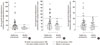
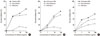
The mean serum total IgE level was 158±433 kU/L, and there was no significant difference in serum total IgE levels among the three groups (Table 1). The serum specific IgE levels to the three cephalosporin-HSA conjugates are shown in Fig. 1. The prevalence of detection of specific IgE antibodies to cephalosporin-HSA conjugates were 17.4% for any cephalosporin, 10.4% for cefotiam, 6.8% for ceftriaxone, and 3.7% for ceftizoxime (Table 1). There was no significant difference in the atopy rate between the positive and negative groups for specific IgE to the cephalosporin-HSA conjugates. The prevalence of specific IgE to cefotiam tended to be higher in group I than in group II, but no significant difference was found in the prevalence of specific IgE to cefotiam and ceftriaxone-HSA conjugates among the three groups. The prevalence of serum specific IgE to ceftizoxime-HSA conjugate was significantly higher in pharmacists (12.0%) than in groups I or II (1.6 and 7.7%, respectively; Fisher's exact test). ELISA inhibition tests of the three cephalosporin-HSA conjugates demonstrated significant dose-dependent inhibition after addition of both free and conjugated forms of the corresponding cephalosporins (Fig. 2).
Risk factors for cephalosporin-associated work-related symptoms and for presence of serum specific IgE antibodies to cephalosporins
Based on a multivariate logistic regression analysis, a personal history of antibiotic allergy was a significant risk factor for the presence of cephalosporin-associated work-related symptoms (OR, 24.93; 95% CI, 2.61-238, Table 2), but not for the presence of serum specific IgE antibodies to cephalosporins (OR, 0.9; 95% CI, 0.18-4.53, data not shown). Additionally, a personal history of atopic dermatitis was a significant risk factor for the presence of serum specific IgE antibody to cefotiam-HSA conjugate (OR, 6.30; 95% CI, 1.23-32.3, Table 3), while no risk factor was significantly associated with the other cephalosporin-HSA conjugates or positive cephalosporin SPT results (data not shown).
DISCUSSION
A beta-lactam antibiotic sensitization rate of 2.6% as measured by SPT was reported in nurses working in a tertiary hospital.12 One study reported that the sensitization rate by SPT and intradermal (ID) tests to benzylpenicilloyl polylysine (PPL) and a mixture of minor antigenic determinants (MDM) was 12% in 83 asymptomatic Turkish nurses.13 Prick and intradermal penicillin sensitivity tests reported rates of 22% for PPL, 21% for MDM, 43% for amoxicillin, and 33% for ampicillin in patients with a clinical history of urticaria and/or anaphylaxis.16 Regarding skin test sensitivity to cephalosporin, to our knowledge there is no published report based on a large cohort.17 In this study, we did not perform the ID test as it is too invasive and time-consuming to be used in a large cohort; moreover, its sensitivity is inadequate for the detection of sensitized subjects.18 Instead, we developed an ELISA system for detecting serum IgE specific to commonly prescribed cephalosporins, such as cefotiam, ceftriaxone, and ceftizoxime, and found an overall sensitization rate of 17.4%, while the sensitization rate by SPT was 3.1%. This indicates that the ELISA is sufficiently sensitive to screen for symptomatic or asymptomatic sensitized HCW. We focused on the rate of sensitization to cephalosporins, not penicillin, because the rate of penicillin prescription has been low in recent years in our hospital.
The sensitization rate of cephalosporin by SPT may be lower than that of penicillin. This may be due to the structural differences between these compounds. Penicillin can be easily haptenized with proteins through its unstable pentagonal thiazolidine ring, while cephalosporin shows relatively slow haptenization because of its stable hexagonal dihydrothiazine ring.19 Therefore, the rate of positive cephalosporin SPT was lower in this study because the three cephalosporins tested may not have formed conjugates in the skin during the SPT. In contrast, use of cephalosporins already conjugated to HSA to detect serum specific IgE antibodies resulted in a higher prevalence. The discrepancy between specific IgE measurements and SPT may be derived from differences in the protein binding properties of the cephalosporins tested. In the present study, ceftriaxone was the only cephalosporin that generated a positive SPT response, likely because the protein binding ratio of ceftriaxone in plasma is higher (90-95%) than that of the other cephalosporins, which may more readily induce a positive response during SPT. Moreover, the specificities of the IgE antibodies to the three cephalosporins could be confirmed by ELISA inhibition tests, in which significant inhibition by both the free and conjugated forms of each cephalosporin were noted. These findings indicate that the measurement of specific IgE antibodies to cephalosporins using various cephalosporin-HSA conjugates is a useful method screening for sensitized subjects in a large HCW cohort.
When we determined the prevalence of serum specific IgE to three cephalosporins, that of cefotiam was the highest (9.3%), followed by ceftriaxone (6.8%) and ceftizoxime (3.7%). When we analyzed the cephalosporin prescription pattern in the months prior to this investigation, ceftriaxone was the most frequently prescribed (24%), followed by cefotiam (9%), and ceftizoxime (7%). Moreover, exposed workers could be sensitized to cefotiam via two routes, inhalation and skin contact, as suggested by previous investigations,3-5 because cefotiam develops more foam and evaporates more easily than other intravenous antibiotics during preparation of injections. Among nurses, the prevalence of specific IgE to cefotiam-HSA conjugate tended to be higher in group I than in group II, but this difference was not statistically significant. Although there have been a few case reports describing the detection of serum specific IgE to cefotiam-HSA conjugate in contact urticaria or anaphylaxis patients,13 this study is the first to investigate the cefotiam sensitization status of HCW in a single hospital setting. Our results showed that cefotiam is the most common sensitizing cephalosporin in exposed HCW.
Although we were unable to measure dust concentrations in the workplace using personal air samplers, many workers might be sensitized via either inhalation or skin contact. Additional details regarding the relative contributions of these routes may have been obtained if the degree of exposure intensity in the different work environments could have been determined. Moreover, this is the first investigation in exposed HCW to measure serum specific IgEs levels to ceftriaxone and ceftizoxime by ELISA. The prevalence of specific IgE antibodies to ceftizoxime-HSA conjugate was significantly higher in pharmacists than in nurses. Pharmacists are likely exposed to cephalosporins while performing procedures such as grinding tablets or pouring syrup. They may also be sensitized while handling cephalosporins that share similar structures and cross-react with ceftizoxime. However, this conclusions may be a type I error, because only three pharmacists showed high serum specific IgE antibody levels to ceftizoxime-HSA conjugate. Further studies are needed to monitor changes in serum specific IgE to each cephalosporin in addition to symptom development.
Exposure to cephalosporin powders may have induced various occupational IgE-mediated allergies, as reported for workers with positive SPT or high levels of serum specific IgE antibodies to corresponding cephalosporin-HSA conjugates.3-7,10,11 Contact urticaria syndrome was associated with positive SPT to beta-lactam antibiotics in nurses.12 Although we previously reported a patient with occupational anaphylaxis who had a high serum level of cefotiam-HSA conjugate,3 no significant association between the presence of cephalosporin-associated work-related symptoms and the results of SPT or serum specific IgE antibodies to any cephalosporin was found. Furthermore, no significant associations with duration of employment, intensity of exposure, and personal history of allergic diseases were found.
In this study, a significant risk factor for the presence of cephalosporin-associated work-related symptoms was a personal history of previous antibiotic allergies. This is similar to previous studies, wherein a past history of drug allergy was a risk factor for contact urticaria syndrome due to beta-lactam antibiotics. 12 Our analysis of the factors associated with the presence of serum specific IgE antibodies to cefotiam-HSA conjugate determined that a personal history of atopic dermatitis was a risk factor. Contact urticaria syndrome is a major manifestation of cefotiam-induced occupational allergic diseases.4,12,20,21 Atopic dermatitis was seen in most of these reports, since it is a major clinical manifestation of cefotiam contact and may have facilitated sensitization. In this study, we confirmed by personal interview that nurses with a history of atopic dermatitis had the condition a number of years before beginning their nursing career. In some nurses, atopic dermatitis recurred or a pre-existing atopic dermatitis became worse after beginning their employment. Additional investigations are needed to evaluate the role of protective equipment, especially gloves, in preventing sensitization and development of contact urticaria syndrome after cefotiam exposure.
In conclusion, we developed an ELISA for the detection of serum specific IgE antibodies to three cephalosporins and found an IgE sensitization rate of 17.4% in HCW exposed to cephalosporins. Atopic dermatitis may be a risk factor for cefotiam sensitization.


 XML Download