

 PDF
PDF ePub
ePub Citation
 Print
Print


INTRODUCTION
Allergic bronchopulmonary aspergillosis (ABPA) is a complex disease that occurs when the pulmonary immune system reacts to antigens of Aspergillus fumigatus. Aspergillus species are thermophilic fungi that are ubiquitous in the environment.1 Airway colonization of A. fumigatus usually occurs in patients with underlying chronic airway disease, such as asthma, chronic obstructive pulmonary disease, bronchiectasis, and cystic fibrosis.2 Eight diagnostic criteria for ABPA, consisting of presence of asthma, transient chest roentgenographic infiltrates, immediate cutaneous reactivity to A. fumigatus, presence of precipitating antibodies, elevated IgG or IgE to A. fumigates, and central bronchiectasis, have been defined.3 However, all eight criteria are rarely found in a single patient, and ABPA is sometimes diagnosed in the absence of typical proximal bronchiectasis. Therefore, minimum criteria were suggested for ABPA-central bronchiectasis (ABPA-CB) and APBA-seropositive (APBA-S), which has been more widely accepted in recent years.4
Tuberculosis (TB) is still a fatal infectious disease in Asia. South Korea has a relatively high burden of TB for an industrialized country (73.5 cases per 100,000 persons in 2009), which is a major cause of mortality and morbidity.5,6 A variety of sequelae and complications, such as parenchymal scarring and bronchiectasis, occur in the lungs of patients with pulmonary TB,7 which may also be sites for colonization of A. fumigatus.8 The prevalence of ABPA ranges from 2% to 25% in patients with cystic fibrosis and 1% to 8% in patients with severe asthma.2 However, there are limited studies on the prevalence of ABPA in Far East countries, although there have been several case series of ABPA in China9 and Japan.10 In the present study, we describe the clinical and immunologic features of 10 patients with ABPA in Korea.
MATERIALS AND METHODS
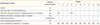
We retrospectively analyzed 10 adult patients diagnosed with ABPA between January 2001 and December 2009. Diagnosis was based on Greenberger's criteria2: (1) asthma, (2) immediate cutaneous reactivity to Aspergillus species or A. fumigatus, (3) elevated total serum IgE (>417 kU/L or 1,000 ng/mL), (4) elevated serum A. fumigates-specific IgE and/or serum IgG-A. fumigatus, and (5) proximal bronchiectasis. Patients with asthma who lacked demonstrable lung disease by computed tomography (CT), but otherwise fulfilled the diagnostic criteria for ABPA, were diagnosed as ABPA-S, as compared to ABPA-central bronchiectasis (ABPA-CB) (Table 1). Eight patients had ABPA-CB, while two had ABPA-S.
Skin-prick tests were performed using commercial fungal allergen extracts, including A. fumigatus, Penicillium notatum, Cladosporium, Alternaria, and Fusarium spp. (Bencard, Bradford, UK), which are the major fungal allergens in asthma. Responses were considered positive if they produced a wheal with a diameter exceeding the negative control wheal by 3 mm. Atopy was determined by a positive skin test response to at least one common inhalant allergen, including house dust mite, tree and pollen mixtures, mugwort, ragweed pollens, and Alternaria (Bencard, Bradford, UK). Total IgE and A. fumigatus-specific IgE and IgG antibodies were measured using the ImmunoCAP system (Phadia, Uppsala, Sweden). A specific IgE level greater than or equal to class II (≥0.7 kU/L) was considered a positive response. A. fumigatus-specific IgG antibodies were measured instead of serum precipitins to Aspergillus antigen. High-resolution computed tomography (HRCT) was used to detect central bronchiectasis. The stage of disease was categorized as follows: stage I, acute; stage II, remission; stage III, exacerbation; stage IV, corticosteroid-dependent asthma; stage V, end-stage lung disease with fibrosis.2
Statistical analysis was performed using a statistical software package (SPSS for Microsoft Windows, package version 16.0; SPSS Inc., Chicago, IL, USA). The data are presented as median values and ranges. The Mann-Whitney U test was used to compare the mean levels of total IgE, IgE specific to A. fumigates, and peripheral eosinophil count between the ABPA patients with post-TB destructive lung lesions and those without. P values of less than 0.05 were considered statistically significant.
RESULTS
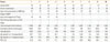
The clinical characteristics of 10 patients diagnosed with ABPA are summarized in Table 1. Most patients were middle-aged men (median, 62.5; range, 19.0-79.0 years) at diagnosis of ABPA with a long duration of asthma (median, 15.0; range, 1.0-48.0 years). Approximately 40% of the study subjects were diagnosed with pulmonary TB, finished anti-TB medications more than 10 years ago (median 23.5; range, 10.0-31.0 years), and have remained in the inactive stage to date. These patients had post-tuberculous destructive lung lesion sequels, such as a cavity or fibrosis on chest X-ray or HRCT. All had moderate to severe airflow obstruction; their forced exhale vital capacity within the first minute (FEV1) ranged from 28.0% to 77.0% (median FEV1, 62.3%). The median total IgE level was 926.5 kU/L, ranging from 127.0 to 3,336.0 kU/L. IgE and IgG levels specific to A. fumigatus ranged from 1.2 to 72.9 kU/L (median, 6.05 kU/L) and 17.5 to 156.0 kU/L (median, 56.45 kU/L), respectively (Table 2).
Comparison of the clinical features of the subjects according to the presence of radiological pulmonary TB sequels highlighted several findings. First, patients were diagnosed with bronchial asthma after the diagnosis of pulmonary TB. Second, all patients had atopy and other allergic diseases, such as allergic rhinitis, conjunctivitis, or drug allergies. Third, immunologic markers, such as peripheral eosinophil count and A. fumigates-specific IgE, tended to be higher in patients with tuberculous sequels compared to those without cavitary or fibrotic changes (peripheral eosinophil count, 3,250.0±5,216.8 vs. 1,229.2±677.2 cells/µL, P=0.83; A. fumigates-specific IgE, 35.5±27.35 vs. 11.24±21.4 kU/L, P=0.05).
Anti-asthmatic medications were continuously required by all patients, who used high-dose inhaled corticosteroids and other controller medications. Two patients were diagnosed with stage IV-ABPA (patients 5 and 6). These patients suffered from steroid-dependent ABPA and experienced several side effects, including osteoporosis and uncontrolled glucose levels. We treated them with anti-IgE antibody (omalizumab, 300 mg every 2 weeks). After the second injection, the patients showed clinical improvement, and the dose of daily oral corticosteroids was reduced; however, there were no significant changes in FEV1 before and after anti-IgE antibody therapy in patient 5.
DISCUSSION
This is the first report describing the clinical features of ABPA in Korea. Clinical features in general were not significantly different from those described in previous studies.11,12 The present study's subjects had a long asthmatic history of more than 20 years, and the majority of them were middle-aged men, consistent with previous reports from Western countries. Moreover, other immunologic parameters, such as total IgE levels, A. fumigates-specific IgE, and total eosinophil count, were also comparable to those of a previous study.12 However, in this report, we found four cases of ABPA who had previous histories of post-TB destructive lung lesions several years prior, who then developed ABPA with presenting severe obstructive patterns, as compared to the other ABPA patients without any previous lung lesions. The development of ABPA after the formation of post-TB sequelae has not previously been reported, and the underlying mechanisms of how post-TB sequelae contribute to the development of ABPA are not understood.
Our findings suggest several such mechanisms. First, the patients were diagnosed with pulmonary TB approximately 17 years before development of asthma and were considered to be cured after chemotherapy with pulmonary sequels, such as cavitary or fibrotic changes. History of TB is a well known independent risk factor for airflow obstruction, even in patients treated with anti TB medications.13,14 As a consequence of Mycobacterium tuberculosis infection, excess matrix metalloproteinase activity by lung epithelial cells has been implicated in the destructive pulmonary pathology of TB, leading to impaired ciliary function and adhesion of the mucus gel layer to the epithelial surface.15 Consequently, cellular debris, mucus impaction, cavities, and ectatic bronchi can trap Aspergillus spores, enabling Aspergillus colonization. We postulated that the pathogenesis of ABPA after pulmonary TB may be similar to patients with cystic fibrosis,16 which is a common risk factor for ABPA. Moreover, these patients showed central types of bronchiectasis in the middle and lower lobes of their lungs, which were different from those of post-TB bronchiectatic changes presenting as the peripheral type commonly found in the upper lobes. Therefore, we suggest a possible association between post-TB destructive lung lesions and the development of ABPA.
Second, all patients with pulmonary sequels after TB had atopy in this study. Moreover, peripheral eosinophil counts and IgE antibodies specific to A. fumigatus tended to be higher in these patients. Considering that pulmonary TB is a common infectious disease in the Far East region, patients who have been cured of TB and who have long-standing atopic asthma may be predisposed to developing ABPA.
Third, anti-IgE therapy can be an effective treatment for ABPA. In this study, two patients who suffered from steroid-dependent ABPA and the severe side effects of long-term steroid use were treated with anti-IgE therapy. After the second injection, these patients experienced significant, sustained clinical improvement and the dosage of oral corticosteroids could be reduced, similar to previous reports.17,18 An enhanced IgE allergic response to A. fumigatus is important for the pathogenesis of ABPA. Patients with ABPA have higher A. fumigates-induced Th2 responses, which promote B-cell stimulation and production of IgE. Omalizumab reduces unbound IgE antibodies and downregulates IgE receptors, decreasing exacerbations of severe allergic asthma as well as ABPA, suggesting that omalizumab may be an alternative therapy in patients with steroid-dependent ABPA.19
Our study has a limitation. We included a relatively small number of patients from a single tertiary medical center. To better evaluate clinical features, immunological profiles, and prevalence of ABPA in Korea, multicenter studies should be performed.
In conclusion, the present study showed that ABPA occurs in patients with post TB destructive lung lesions, suggesting that a history of pulmonary TB with pathological consequences is a risk factor for developing ABPA in TB-endemic areas. In particular, ABPA should be considered in severe asthma patients with high serum IgE levels and lung damage caused by previous TB infection. Furthermore, anti-IgE antibody therapy may be an alternative strategy for patients with steroid-dependent ABPA.
 XML Download