

 PDF
PDF ePub
ePub Citation
 Print
Print


INTRODUCTION
Stinging insects belong to the order Hymenoptera. The Hymenoptera families most relevant to allergies are the Apidae, Vespidae, and Formicidae. The Apidae and Vespidae are responsible for hymenoptera venom allergy, and stings by these bees and wasps can lead to local or systemic reactions, including anaphylaxis. Systemic allergic reactions to hymenoptera stings have been reported in ≤3% of adults, and nearly 1% of children have a medical history of severe hymenoptera sting reactions.1
A patient's medical history is the primary and most important tool used to diagnose hymenoptera venom allergy. However, this information has limited value for identifying the culprit species. An exact diagnosis is needed for the development of an effective strategy for immunotherapy, and thus, other methods, such as skin prick tests and serum-specific IgE detection, are also used.2 Serological measurement of venom-specific IgE antibodies can confirm sensitization in hymenoptera venom allergy patients. Although conventional venom extracts are used to measure serum-specific IgE in commercially available immunoassay systems, venom is a complex of many substances. For example, the major component allergens of honeybee venom include phospholipase A2 and hyaluronidase,3,4 and those of Vespidae include phospholipase A1, antigen 5, and hyaluronidase.5 Despite the component allergen differences between honeybee and Vespidae venoms, >50% of hymenoptera venom allergy patients react to both venoms in serologic tests.6
Component-resolved diagnosis (CRD) was introduced after the development of microarrays that test for 94 purified allergens, including hymenoptera venoms.7 CRD uses defined allergens as antigens, instead of whole allergen extracts, which are mixtures of both allergenic and non-allergenic components. Some studies have demonstrated the highly sensitive and specific diagnostic value of component allergen-specific IgE detection for food, cat, birch, and grass pollen allergies.8,9 A recent study using recombinant component allergens to investigate the cross-reactivity of IgE antibodies for different hymenoptera venoms found that cross-reacting carbohydrate determinants (CCDs) were the main cause of double sensitization.10
In this study, we compared the diagnostic value of component allergen-specific IgE to that of conventional venom extract-specific IgE and evaluated antibody cross-reactivity for Apidae and Vespidae species allergen components.
MATERIALS AND METHODS
Patients
Fifty-six patients who had been diagnosed with hymenoptera venom allergy and who had received venom immunotherapy at Ajou University Hospital in Suwon, Korea, between April 1998 and April 2011 were enrolled in the study. The diagnosis of hymenoptera venom allergy and the choice of venom allergen (Hollister-Stier, Spokane, WA, USA) used for allergen immunotherapy were made based on patient history and the levels of serum-specific IgE to major venoms measured at the time of diagnosis.
Clinical manifestations
The demographic characteristics of the enrolled patients were reviewed, and information regarding age, gender, atopy status (based on skin prick tests or in vitro allergen-specific IgE results), acupuncture history using honeybee extract, cross-reactivity with fire ant (Solenopsis invicta) allergen, and the severity of clinical manifestations were recorded retrospectively. We identified anaphylaxis patients following National Institute of Allergy and Infectious Disease (NIAID)/Food Allergy and Anaphylaxis Network (FAAN) criteria11 and classified these patients as severe or moderate based on the grading system for generalized hypersensitivity reactions.12
Measurement of serum-specific IgE
Serum levels of IgE specific for major hymenoptera venom extracts (i.e., honeybee, yellow jacket, yellow hornet, white-faced hornet, and paper wasp) and three hymenoptera venom component allergens (recombinant phospholipase A2 from honeybee [rApi m 1], recombinant antigen 5 from yellow jacket [rVes v 5], and recombinant antigen 5 from paper wasp [rPol d 5]) were measured using an ImmunoCAP system (Pharmacia, Uppsala, Sweden). Patient blood samples were obtained at the time of diagnosis and 3 years after immunotherapy. Allergen-specific IgE levels >0.35 kUA/L were considered positive, and individuals with positive IgE levels to any two of the major bee venoms were considered to have double sensitization.
Statistical analysis
Clinical features were reported by descriptive analysis. Correlations between results for conventional venom extracts and component allergens were calculated by Pearson's correlation test. Wilcoxon's rank test was used for nonparametric analysis of changes in serum allergen-specific IgE after hymenoptera venom allergen immunotherapy. Differences were considered statistically significant at P<0.05.
RESULTS
Clinical characteristics of study subjects
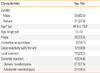
The clinical characteristics of the study subjects are summarized in Table 1. Male patients (62.5%) were predominant, consistent with other studies, and the mean patient age was 44.9±13.8 years (range, 11-73 years). Based on skin prick tests or serum levels of IgE specific to common aeroallergens, 73.3% of the study population had atopic tendencies. Of note, 10.7% of the patients developed allergies due to honeybee extract acupuncture therapy, which is widely used in oriental medicine to relieve pain.
As our study design selected for patients who had received venom allergen immunotherapy, few patients (17.3%) had local reactions, such as redness or swelling around the stinging site; 82.7% of the patients had systemic reactions. The most frequent clinical manifestation was skin involvement (78.6%), including urticaria and/or angioedema, followed by respiratory (44.6%), cardiovascular (37.5%), central nervous system (26.8%), and gastrointestinal (12.5%) symptoms. According to NIAID/FAAN criteria, anaphylaxis occurred in 76.8% of the study population; 39.3% of these cases were classified as moderate and 37.5% were severe (Table 1). We checked all patients' histories after venom immunotherapy. We could not evaluate the outcome, as it is impractical to perform sting challenge tests after venom immunotherapy because of the risk.
Bee venom-specific IgE levels at diagnosis
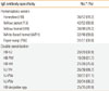
Yellow jacket venom (80.8%) was the most common sting allergen at the time of diagnosis, as determined by serum-specific IgE detection with conventional Hymenoptera venom extracts, followed by yellow hornet (79.2%), white-faced hornet (69.6%), honeybee (69.2%), and paper wasp (60.8%) venoms. Given that cross-reactivity exists between and within the Apidae and Vespidae Hymenoptera families, we determined the number of patients with double sensitization at the initial visit. Double sensitization most frequently involved yellow jacket and yellow hornet venoms (89.7%), whereas the least frequent combination was honeybee and paper wasp venoms (51.7%). In total, 70.9% patients had double sensitization at the time of diagnosis (Table 2).
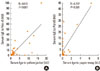
We measured serum IgE specific to venom component allergens, including recombinant phospholipase A2 from honeybee (rApi m 1), recombinant antigen 5 from yellow jacket (rVes v 5), and recombinant antigen 5 from paper wasp (rPol d 5). The positive predictive values (PPVs) of rVes v 5-specific and rPol d 5-specific IgE detection were 85.7% and 87.5%, respectively. These were positively correlated with conventional venom extract-specific IgE detection (r=0.762 and r=0.757, respectively). In contrast, the PPV of rApi m 1 was 34.8% at the time of diagnosis (Fig. 1). Next, we analyzed the status of combined sensitization using component allergens at diagnosis. The combined sensitization within the Vespidae family (Ves v 5 and Pol d 5) was 80%, and there was only 27.3% cross-reactivity between the Apidae and Vespidae families (Api m 1 and Ves v 5, or Api m 1 and Pol d 5).
Serum-specific IgE levels with venom immunotherapy
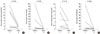
After immunotherapy for 3 years, we observed the change in serum-specific IgE to venom allergens and three components of venom allergen. Specific IgE levels to Vespidae tended to decline compared to those collected before allergen immunotherapy; however, those to Apidae did not change (P=0.18). Among the specific IgE to Vespidae, rVes v 5 and rPol d 5 significantly decreased after 3 years of immunotherapy (P=0.046 and P=0.028, respectively) compared to whole yellow jacket and paper wasp venom extracts (P=0.075 and P=0.116, respectively). Together, these results indicate that for detecting changes in antibody levels in response to allergen immunotherapy, the measurement of IgE specific for rVes v 5 or rPol d 5 is as sensitive as the measurement of IgE specific for conventional Vespidae venom extract (Fig. 2).
DISCUSSION
The worldwide incidence of systemic reactions to Hymenoptera is estimated to be about 0.8%-5%,2 and the prevalence of serum IgE specific for Hymenoptera is even higher.13,14 Hymenoptera stings are one of the most common causes of anaphylaxis and result in more than 40 deaths per year in the United States.15 Venom immunotherapy is the primary treatment for preventing systemic allergic reactions, and its reported success rate is >95%, although this number is biased because of the careful selection of patients and immunotherapy regimens.16,17 Hymenoptera venom provocation tests are definitive diagnostic tools, but are often impractical owing to the risk for inducing dangerous symptoms. Thus, clinicians rely on patient history, skin prick tests, and venom-specific IgE detection to determine which allergens to use for immunotherapy.
Many Hymenoptera allergy patients also have IgE specific for other venoms, due to antibody cross-reactivity with homologous peptide sequences in different protein allergens, or to CCDs, which are present in the majority of Hymenoptera venom allergens.18-21 Cross-reactive double positivity presents significant challenges in choosing venoms to be used for immunotherapy. After the introduction of the ImmunoCAP system, a highly sensitive test for allergy diagnosis, the reported prevalence of double positivity increased, presumably because of its better detection.22 Similarly, an increased incidence of food allergies in pollen allergy patients was also reported.23
To more definitively identify clinically relevant allergenic components, purified allergens and individual peptides have been developed for use in CRD.7,24 CRD has excellent diagnostic and monitoring value for allergies to cats, birch, and grass pollens.8,9 So far, however, the reported values of CRD are comparable to tests using conventional venom extracts. Our data indicated that the PPVs of Vespidae allergen components were also comparable to those of whole venom extracts. In contrast, the PPVs of honeybee allergens were very low at diagnosis, consistent with previous results showing that CRD using honeybee allergens had a markedly lower diagnostic value compared to basophil activation tests and Western blot analysis, indicating minimal sensitivity to rApi m 1.25 These results suggest that determining the sensitivity to rApi m 1 alone is not sufficient for accurate diagnosis of honeybee allergies, and CRD for this allergy may require testing for additional component allergens. Other honeybee component allergens have been identified, specifically hyaluronidase (Api m 2) and acid phosphatase (Api m 3), but their clinical relevance is unknown. We determined that Api m 1 was not sensitive enough for proper diagnosis of honeybee allergy, and additional component allergens may be needed for better diagnosis. In the case of birch pollen allergy, the addition of rBet v 1 and rBet v 2 component allergens to CRD in patients increased its sensitivity, compared to ImmunoCAP analysis using whole allergen extract.8 The lack of commercially available component allergens, especially from the Apidae family, is a major limiting factor for the use of CRD.
Immunotherapy with venom extracts is well known to induce a gradual decline in allergen-specific IgE, with a concomitant increase in allergen-specific IgG. ImmunoCAP analysis is not always the ideal method to measure changes in conventional venom extract-specific IgE, particularly in patients who are sensitized to more than one Hymenoptera allergen. For example, in our study, only IgE antibodies against the component Vespidae allergens (i.e., rVes v 5 and rPol d 5) significantly declined after immunotherapy. Although our data do not demonstrate the sensitivity of allergen component-specific IgE with regard to clinical outcomes, the data do suggest that measuring allergen component-specific IgE is a highly sensitive tool for monitoring IgE levels after immunotherapy.
The clinical features of allergy in this study were similar to those reported in other studies.26-28 Interestingly, nearly 10% of the Hymenoptera venom patients in the present study had allergies caused by acupuncture therapy with honeybee extract, and this number might have been much higher if minor reactions had been included. Honeybee acupuncture is a traditional method for pain relief in Oriental medicine, but the prevalence of allergic reactions among patients of this therapy has not been reported previously.
In conclusion, Vespidae component allergen-specific IgE was as good as, if not better than, conventional venom extract-specific IgE for identifying and tracking allergen-specific IgE in Hymenoptera allergy patients.
 XML Download