

 PDF
PDF ePub
ePub Citation
 Print
Print


INTRODUCTION
Peanut allergy is one of the most common and severe IgE-mediated food reactions.1 However, reports of severe anaphylaxis and fatalities from peanuts in Asia are relatively rare.2 In addition, there are differences in the prevalence and severity of peanut allergy in different parts of the world.1,2 Many genetic and environmental factors could be responsible for the epidemiologic characteristics of peanut allergy.3 Regional dietary habits and cooking methods also play a role in the prevalence of specific food allergies in various countries, as differences in the methods of peanut preparation affect their final allergenicity.1,4 For example, the amount of personal peanut consumption in China and the United States are similar, but there is virtually no peanut allergy in China.5 This is likely because the Chinese predominantly eat boiled or fried peanuts, while Americans eat almost exclusively dry-roasted peanuts.5 The higher heat of dry roasting and the process of maturation and curing have been shown to increase the allergenicity of peanut proteins.4,6,7 Koreans occasionally eat pickled peanuts. In our previous study, bands corresponding to peanut allergens such as Ara h 1, Ara h 2, and Ara h 3, as well as the IgE-binding intensities to these allergens, were shown to change after treatment of the peanuts with vinegar.8 The concentration of acetic acid, which is a key ingredient of vinegar, typically ranges from 4% to 8% by volume for table vinegar and up to 18% for pickling vinegar.
To our knowledge, no previous studies have evaluated changes in food allergens under acidic conditions; therefore, we attempted to determine the effects of various pH conditions on major peanut allergens.
MATERIALS AND METHODS
Patients' sera
Sera from seven patients sensitized to peanuts were pooled. The patients had a peanut-specific IgE levels>15 kUA/L, as measured using the Pharmacia CAP system (Uppsala, Sweden). This cutoff value was considered to be predictive of clinical reactivity.9 Normal human sera were used as negative controls. After receiving informed consent, blood samples were obtained and the sera were frozen at -80℃ until use. This study was approved by the Samsung Medical Center Institutional Review Board.
Preparation of peanut extracts
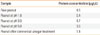
Raw in-shell peanuts grown in Korea were obtained from the Korean Rural Development Administration.10 Peanut kernels were soaked overnight in commercial vinegar (pH 2.3), and in acetic acid solutions with pH values of 1.0, 3.0, or 5.0. The peanuts were then ground using a mortar and pestle until a smooth paste was achieved. The paste was defatted by washing with n-hexane and dried overnight at room temperature. Proteins were then extracted from the dried powder by agitating the samples in phosphate-buffered saline (PBS) for 2 hours at 4℃. Following centrifugation at 3,000×g for 20 minutes at 4℃, supernatants were collected, filtered (0.45 µm pore size), and lyophilized. Protein concentrations were then determined using a 2-D Quant kit (Amersham Biosciences, Piscataway, NJ, USA) and a microplate reader (Bio-Rad, Hercules, CA, USA). Table shows the protein concentration of each peanut sample. All extracts were stored at -80℃.
SDS-PAGE and immunoblot analysis
Extracts were separated by sodium dodecyl sulfate polyacrylamide gel electrophoresis (SDS-PAGE; Tricine System; Invitrogen, Carlsbad, CA, USA) according to the manufacturer's instructions. Extracts were then loaded into each well of a 96-well plate at 6 µg/well. Precision Plus Protein standards (Bio-Rad) were used as molecular weight markers to estimate protein size. The separated proteins were transferred from the gel to a polyvinylidene difluoride membrane (PVDF) using the iBlot™ Dry Blotting System (Invitrogen). The PVDF membranes were then blocked with 2% non-fat dried milk (NFDM) for 1 hour at room temperature, and then incubated overnight with the patients' pooled sera, which was diluted 1:10 in 2% NFDM. After washing with PBS-Tween (PBS-T), the membrane was incubated with biotin-labeled goat IgG-anti-human IgE (KPL, Gaithersburg, MD, USA), which was diluted 1:4,000 in 2% NFDM, for 1 hour at room temperature. After rinsing with PBS-T, the membrane was incubated with NeutrAvidin-HRP (Pierce Chemical Co., Rockford, IL, USA) for 30 minutes at room temperature. After washing with PBS-T, the membrane was reacted with Amersham ECL reagent (GE Healthcare Bio-Sciences AB, Uppsala, Sweden) for 2 minutes. The blotted membrane was then exposed to a high performance chemiluminescence film (GE Healthcare Limited, Buckinghamshire, UK) and the film was developed. The density of SDS bands and immunoblots were compared using a reflective densitometer (Bio-Rad) and Quantity One software (Bio-Rad).
RESULTS
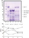
Almost all of the peanut allergens were altered after incubation in the acidic solutions (Fig. 1). The density of Ara h 1 was slightly reduced in samples treated with pH 3.0 or 5.0 acetic acid, and was completely undetected in samples treated at pH 1.0 acetic acid or commercial vinegar (pH 2.3). Ara h 2 remained largely unchanged at pH 5.0, but decreased as the soaking solutions became more acidic. Ara h 3 and Ara h 6 were unaltered by treatment with pH 3.0 or 5.0 acetic acid, but appeared as a very thick, strong band in samples treated with pH 1.0 acetic acid or commercial vinegar (pH 2.3). Bands between 50 and 38 kDa were significantly enhanced. This might be due to rearrangement of Ara h 3 after treatment with a strong acid, or aggregation of non-allergenic proteins.
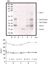
The IgE-binding intensities to major peanut allergens, such as Ara h 1, Ara h 2, and Ara h 3, were significantly reduced at pH 1.0 and 2.3, although there was no substantial change at pH 3.0 or 5.0 when compared with raw peanuts (Fig. 2). The IgE-binding intensities to Ara h 1 were also reduced by exposure to pH 1.0 and commercial vinegar, which corresponded to the reduced quantities of Ara h 1. IgE-binding to Ara h 2 disappeared at pH 1.0 and after treatment with commercial vinegar. The IgE-binding intensities to Ara h 3 were reduced in samples treated at pH 1.0 and 2.3, despite the increased quantity of this protein (Fig. 1).
DISCUSSION
The prevalence of peanut allergy in Korea is unknown; however, similar to other Asian countries, it appears to be low, which differs from the incidence in Western nations.11,12 Although the exact cause of this low prevalence remains unclear, one possible explanation is differences in eating habits, which can affect food allergenicity. The allergenicity of peanut proteins can be altered by different cooking methods.4,13 It is difficult to predict the physical and chemical changes which occur in cooked food, because food contains very complicated organic compounds. The most important finding of this study was that treatment of peanuts with vinegar affected their allergenicity, and that such changes in allergenicity differed with pH. These findings correspond with those of previous studies in which different cooking methods resulted in changes in the final allergenicity of peanuts,4,6,7 suggesting that treatment of peanuts with vinegar could lead to regional differences in the prevalence and severity of peanut allergy.
Nine allergens have been reported in peanuts, and Ara h 1, Ara h 2, and Ara h 3 are believed to be the major allergens. Of these, the importance of Ara h 2 in clinical peanut allergy has been suggested in previous studies.14-19 A correlation between clinical severity and the presence of low concentrations of Ara h 2 and higher levels of Ara h 1 and 3 was reported to indicate a greater potency of Ara h 2.16 Component-resolved diagnostics also revealed Ara h 2 as the most important component for accurate discrimination of subjects with peanut allergy vs. peanut-tolerant subjects.20 In this study, IgE reactivities toward Ara h 1, Ara h 2, and Ara h 3 were decreased after exposure of peanuts to acidic solutions, and the reduction in IgE reactivity to Ara h 2 was marked. While Ara h 2 has a greater resistance to proteolytic digestion and heat/chemical treatment, our data suggest that Ara h 2 was degraded in acidic conditions with a subsequent reduction in IgE binding.
In conclusion, the allergenicity of major peanut allergens is altered by acid treatment. Further research is needed to investigate changes in the allergenicity of major food allergens according to the various cooking methods used in each region.
 XML Download