

 PDF
PDF ePub
ePub Citation
 Print
Print


INTRODUCTION
Hemoptysis is the extra-oral release of blood emitted from the nose or mouth to the respiratory system, including the lungs. Pediatric cases of hemoptysis are uncommon, but some children experience life-threatening events.1 The common diagnostic evaluation usually consist s of a simple roentgenogram, computed tomography, bronchoscopy, and culture studies, although some of these are not easily available in clinical practice for children. A guideline or diagnostic algorithm based on common etiologies for pediatric hemoptysis in each community would be helpful.
Various etiologies of hemoptysis have been reported, depending on age and location. Adult cases of hemoptysis mainly occur as a result of infection, bronchial adenoma, or telangiectasia.2,3 However, in pediatric cases, major causative factors include infection, foreign body aspiration, and congenital heart diseases.4 In contrast, in Korea, pulmonary tuberculosis, lung cancer, and telangiectasia are causative factors for hemoptysis in adults.5 In Korean pediatric patients, despite the publication of some cases,6-8 major causative diseases for hemoptysis have not yet been reported. In this study, we analyzed the etiologies and clinical characteristics of hemoptysis in Korean children.
MATERIALS AND METHODS
The medical records of 40 patients aged 18 yr or younger who were admitted to the Department of Pediatrics, Samsung Seoul Hospital, Seoul, Korea between January 1996 and June 2008 with a chief complaint of hemoptysis were reviewed retrospectively, to assess gender, age distribution, amount of hemoptysis, causative diseases, treatment modalities, and prognosis. Patients with trauma, bleeding tendency, cancer, or hematologic disorders were excluded. According to the age distributions, patients were classified into an early childhood group (0-5 yr), late childhood group (6-10 yr), and adolescent group (≥11 yr). The amount of hemoptysis in 24 h was estimated to be minimal (≤20 mL), moderate (21-100 mL) or massive (>100 mL), based on the approximate amount of hemoptysis in one episode (<5, 50, or 100 mL) and the frequency of hemoptysis in 1 day.9
Treatment modalities were divided into medical treatments, including antibiotic administration, blood transfusion, and other conservative treatment, and surgical treatments, including bronchial artery embolization and surgery. Recurrence and prognosis were assessed based on a retrospective analysis of the medical records during the mean follow-up period, which was 27 months (range, 1-204 months).
RESULTS
Patient age and gender distribution
The 40 patients consisted of 18 boys and 22 girls, and the male-to-female ratio was 1:1.2. Patient age ranged from 10 months to 18 yr, with a median age of 6 yr and 3 months. Seventeen patients were included in the early childhood group; 8 in the late childhood; and 15 in the adolescent group.
Clinical characteristics
The median length of hospital stay was 6 days (range, 3-38 days). The diagnosis of the patient who was hospitalized for the longest period (38 days) was Heiner syndrome. The duration of hemoptysis ranged from 1 to 19 days, with a median period of 5 days. The longest duration (19 days) was in a patient with idiopathic pulmonary hemosiderosis. Of the 40 patients, there were 25 (62.5%) patients with a minimal amount of hemoptysis, whereas six children showed a massive amount of hemoptysis.
Causative diseases
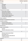
The main causative disease was respiratory tract infection (25%), followed by congenital heart diseases (17.5%). Additionally, there were four cases of Heiner syndrome (10%), three of respiratory neoplasm (7.5%), three of vasculitis syndrome (7.5%), three of bronchiectasis (7.5%), two of upper airway bleeding (5%), and four idiopathic cases (10%) (Table 1).
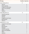
The etiology of hemoptysis was also assessed according to the age distribution. Infection, Heiner syndrome, and congenital heart disease were the major causative diseases in early childhood. During late childhood, various causative diseases such as vasculitis syndrome, bronchial tumor, and bronchiectasis were present. In the adolescent group, hemoptysis was mainly caused by infection and congenital heart disease, and bronchiectasis and bronchial neoplasms were causative diseases in this age group (Table 2).
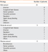
The patients with bronchiectasis, infection, congenital heart disease, or bronchial neoplasms showed a moderate or massive amount of bleeding, although no difference in the amount of hemoptysis was observed based on etiology (Table 3).
Treatment modalities
Hemoptysis disappeared in 35 patients (87.5%) with medical management alone, and surgical treatment was required in the remaining five patients. Three children with bronchial tumors received a lobectomy with tumor removal. A bronchial artery embolization was performed in one patient with a pulmonary arteriovenous fistula. A patient with a vascular malformation on the larynx was treated under general anesthesia with electrocauterization using rigid bronchoscopy.
Prognosis
Of the 40 patients, hemoptysis recurred in 12 (30%), whose causative diseases included Heiner syndrome and vasculitis syndrome. There were two cases of mortality (5%), one due to systemic lupus erythematosus and one due to lymphangiomatosis. The cause of death was persistent hemoptysis and subsequent respiratory failure.
DISCUSSION
Hemoptysis is not a common chief complaint in pediatric patients and is rarely life-threatening; therefore, a thorough investigation is necessary to identify the disease causing the hemoptysis. Infection is one of the most common etiologies for hemoptysis in children,4,10-12 however, a 10-yr retrospective study of pediatric hemoptysis in the US showed that cystic fibrosis was the most common cause, followed by congenital heart disease and infection.13 The etiology of hemoptysis in children may depend on race, region, and research methodology. In this study, infection and congenital heart disease were the common causes for pediatric hemoptysis in Korea. Although this is the first report on the etiology of hemoptysis in Korean children, selection bias should be considered, because the results were obtained from a single, tertiary medical institution. Therefore, multi-center studies are warranted to obtain more accurate information regarding the common etiology of hemoptysis in Korean children.
The hemoptysis pattern based on age distribution was examined. Coss-Bu et al13 reported in a US study that pediatric hemoptysis was most commonly noted in an adolescent group, 11 yr or older. However, we found it to be common in pediatric patients aged 5 yr or younger as well as those aged 11 yr or older. Our study showed that Heiner syndrome and congenital heart disease were the second most common causes for hemoptysis in the early childhood group. This is conceivable because food allergy and congenital diseases begin to manifest clinical symptoms during this period. Congenital heart disease was also a common cause of hemoptysis in the adolescent group of our study, which is consistent with a previous study in which hemoptysis was related to congenital heart disease and had a bimodal age distribution.13 Massive bleeding occurred in 15% of patients in our study, whereas in previous studies, approximately 5% had daily massive bleeding of >200 mL.9,10 These discrepancies may be attributable to differences in underlying diseases, the method used to estimate the amount of bleeding, or criteria for determining massive bleeding. For example, cystic fibrosis leads to a massive amount of hemoptysis in children,14,15 but it is a rare disease in the Korean population.16 In our series, bronchiectasis was the most common etiology in children with massive hemoptysis. None of the three patients with bronchiectasis had underlying causes such as cystic fibrosis or primary ciliary dyskinesia.
The amount of bleeding is minimal in most pediatric hemoptysis cases, and symptoms improve with conservative treatment.9,11 In the current study, 62.5% of cases had a minimal amount of hemoptysis, and in most cases (87.5%), hemoptysis improved with medical management alone. However, during the mean follow-up period of 27 months, hemoptysis recurred in 30% of patients. Mortality from hemoptysis is reported as 0-32% in children.11,14,15 It was 5% in the current study, and the cause of death was progression of respiratory failure due to pulmonary hemorrhage, rather than asphyxiation or shock by excessive bleeding. Considering that the common causes of hemoptysis in our study included chronic diseases such as Heiner syndrome and vasculitis syndrome, a long-term follow-up study is mandatory to determine the clinical course and recurrence of hemoptysis based on underlying diseases in pediatric patients.
In conclusion, the most common etiology in pediatric patients with hemoptysis was respiratory infection. In most cases, the amount of hemoptysis was minimal. These cases showed a good clinical course in which the symptoms were improved by conservative treatment alone; however, in some cases, a recurrence of hemoptysis was observed or surgical management was necessary. An accurate diagnosis of underlying diseases is essential for the treatment of hemoptysis.
 XML Download