

 PDF
PDF ePub
ePub Citation
 Print
Print


Endometrial polyps are a common gynecologic condition that affects up to 25% of the general population, most frequently peri- and postmenopausal women.1 These lesions are defined as localized overgrowths of endometrial tissue that contain a variable amount of glands, stroma, and blood vessels, covered by pseudostratified epithelium.2 Endometrial polyps are frequently associated with abnormal uterine bleeding and are often asymptomatic, incidentally detected during routine gynecologic examinations. Although the majority of polyps are benign, a small proportion may become malignant. The prevalence of endometrial cancer in polyps has been reported to range from 0-4.8%, depending on the selection of patients and resection methods used.1,3~14
With the expanding use of ultrasonography in outpatient clinics, the diagnosis of endometrial polyps has increased. Once polyps have been detected, operative hysteroscopy is considered the treatment of choice.10 Although surgical treatment is the only way to confirm the absence of malignancy, there is some controversy regarding the see-and-treat management of polyps due to their low malignancy potential. When clinicians have to face the choice between expectant management of polyps and a surgical procedure, they must balance the risk of malignancy against the healthcare costs and complications that accompany surgical intervention and unnecessary surgery.
The primary objective of our study was to evaluate the prevalence of premalignant and malignant lesions in endometrial polyps resected by surgical hysteroscopies in pre- and postmenopausal women. The secondary purpose was to investigate whether clinical parameters may predict the histopathologic features and attempt to help for optimal management of polyps.
Materials and Methods
From January 2010 to December 2011, all women who consecutively underwent hysteroscopic removal of endometrial polyps were analyzed, retrospectively. One thousand one hundred ninty-six women were found who matched the criteria. Women in whom resecting of lesions resulted in other histopathologic findings were excluded from the sample. A total of 1,070 women with pathologically confirmed endometrial polyps were included in this study. Patients were scheduled for operative hysteroscopy after a diagnosis of endometrial polyp was made either by hysteroscopy or by transvaginal sonography alone or combined with saline infusion sonography.
Demographic, clinical, and pathologic data were obtained using medical records, which included surgical notes and pathologic reports. A diagnosis of endometrial polyp by ultrasonography was made following a finding of focal endometrial thickening with a bright edge. When multiple polyps were present, the largest polyp diameter measured was recorded. Diagnostic hysteroscopy was performed using saline infusions to distend the uterine cavity, and an endoscope with a 2.8 mm diagnostic sheath was used. The endocervical canal, endometrial surface, vascularity, tubal ostia, endometrial polyp, myoma, and synechiae were evaluated.
Surgical hysteroscopy was performed by a gynecologist under general anesthesia, using a 5 or 10 mm resectoscope. Distension of the uterine cavity was obtained using normal saline solution at a pressure of 90 to 110 mmHg. When necessary, the cervix was dilated using Hegar dilators.
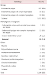
Tissue specimens from surgical hysteroscopy were placed in a container with 10% formaldehyde and sent to pathologists with specialized training in gynecologic pathology. Pathologic diagnosis distinguished between polyps that were recognized as benign (endometrial polyps and polyps with non-atypical simple hyperplasia and non-atypical complex hyperplasia), premalignant (polyps with atypical simple hyperplasia or atypical complex hyperplasia), malignant (harboring carcinoma), and non-polypoid lesions that were mistakenly diagnosed as polyps (myoma, polypoid adenomyoma, proliferative endometrium, secretary endometrium, disordered proliferative pattern, chronic inflammation, placental site nodules). The reason for this subdivision is the different rates of progression to endometrial adenocarcinoma: none or approximately 1% for benign, and variable, from 25 to 100%, for premalignant and malignant.15~17 The histopathologic definitions of endometrial hyperplasia and adenocarcinoma were described as follows: Endometrial simple hyperplasia was defined by moderate distortion of the endometrial architecture with crowding of glands, cystic dilatation, and non-complex budding. The lining epithelium of the gland is pseudo-stratified, showing mitotic activity with no atypia of cells. Atypical simple hyperplasia was defined by architecture similar to simple hyperplasia, but with more irregular glands; the glands are lined by atypical cells. Endometrial carcinoma was defined by crowded malignant tubular glands varying in size and invading the stroma.9
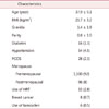
Clinical characteristics and known risk factors for endometrial cancer were evaluated, including age, body mass index (BMI), parity, diabetes (defined as fasting glucose > 126 mg/dL), hypertension (defined as diastolic blood pressure > 90 mmHg and/or systolic blood pressure > 140 mmHg), polycystic ovary syndrome (PCOS), menopausal status, use of hormonal replacement therapy (HRT), history of breast cancer, and use of tamoxifen. The diagnosis of PCOS was based on the Rotterdam criteria.18
The data are presented as mean ± standard deviations and number (percentage). Statistical analysis was carried out using IBM SPSS Statistics 16 software (SPSS Inc., Chicago, IL, USA). Frequency distributions were compared using Chi-square tests and Fisher's exact tests. Mean values were compared using the Student t-test. Logistic regression was performed as a multivariate analysis. The statistically significant level was established as: P values < 0.05.
Results
The mean age of the women in this study was 37.9 ± 9.1 years (range 16-81 years), and 96 women (8%) were postmenopausal. The clinical characteristics of the women who underwent hysteroscopic endometrial polypectomy are shown in Table 1.
Table 2 shows the final pathologic diagnosis of the resected lesions. One thousand one hundred fifty-seven women (96.7%) were diagnosed with benign endometrial pathologies, including 997 (83.4%) endometrial polyps, 15 (1.3%) polyps with non-atypical simple hyperplasia, and 19 (1.6%) polyps with non-atypical complex hyperplasia, as well as 126 (10.5%) other findings such as myoma, polypoid adenomyoma, proliferative endometrium, secreting endometrium, disordered proliferative pattern, chronic inflammation, and placental site nodule. Premalignant lesions consisted of 1 (0.1%) polyp with atypical simple hyperplasia and 12 (1.0%) polyps with atypical complex hyperplasia. Twenty-six malignant polyps (2.2%) were diagnosed.
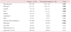
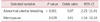
Aside from other pathologies, a total of 1,070 women with pathologically confirmed endometrial polyps were divided into two groups according to whether the polyps had benign or premalignant-malignant pathologies. The univariate analysis of factors associated with a risk for premalignant or malignant polyps is shown in Table 3. Among the clinical data considered, age, BMI, gravidity, parity, hypertension, abnormal uterine bleeding, and postmenopause were significantly higher in patients with premalignant and malignant pathologies (P < 0.05). The multivariate logistic regression for all significant variables at univariate analysis proved that abnormal uterine bleeding and postmenopause were the only factors that remained independently associated with a higher risk of malignancy, with odds ratios of 5.07 (95% CI, 2.25-11.41) and 3.41 (95% CI, 1.14-10.24), respectively (Table 4).
Discussion
Endometrial polyps are the most common cause of menorrhagia resistant to medical therapy in premenopausal women and can cause abnormal bleeding in postmenopausal patients.9,19 In the present study, with the large sample size of polyps resected by hysteroscopy, the prevalence of premalignancy and malignancy was 3.3% (1.1% hyperplasia with atypia and 2.2% endometrial adenocarcinoma). We included endometrial hyperplasia with atypia in the premalignant group since about 28% of untreated complex hyperplasia with atypia progress to malignancy, and the rate of concurrent endometrial carcinoma among hyperplasia with atypia is 42.6% at the time of hysterectomy.20,21
A varied incidence of malignancy associated with endometrial polyps has been reported in previous studies, ranging from 0% to 4.8%.1,3~14 A study involving one of the largest sample sizes (1,242 polyps resected by hysteroscopy) reported a high rate (4.8%) of malignant or premalignant lesions.9 In Korea, one study carried out by Cho et al.22 involving 449 endometrial polyps reported a prevalence of malignancy of 1.5% (0.2% hyperplasia with atypia and 1.3% endometrial adenocarcinoma). However, there are few reports regarding the malignant potential of endometrial polyps in Korea. A recent systemic review of 17 observational studies that included a total of 10,572 women noted that the prevalence of premalignancy and malignancy was 3.57%.10 Our results are in close agreement with these previous studies.
The differences in prevalence observed in the literature are due to differences in the study populations and methods used for the removal of polyps (for example, some based on specimens that were obtained by curettage, others via surgical hysteroscopy, etc.). In this study, we included a large unselected population of women with endometrial polyps, and all polyps were removed entirely by operative hysteroscopy. Hysteroscopy is effective and safe, as both the diagnosis and removal of endometrial polyps are outpatient procedures. This procedure can also remove polyps completely, inclusive of their stalks, so that the risk of malignancy can be estimated confidently.23,24 Hysteroscopic polypectomy is currently considered the gold standard for treatment.25
In the present study, we demonstrated that among women with endometrial polyps, postmenopause and abnormal uterine bleeding are both associated with an increased risk of endometrial premalignancy and malignancy. Numerous studies have reported that abnormal uterine bleeding increases the risk of malignancy. While Savelli et al.3 and Ben-Arie et al.4 reported that abnormal uterine bleeding was not a risk factor of malignancy in endometrial polyps, Ferrazzi et al.6 revealed that the risk of malignancy in women who were symptomatic was 10 times higher than those without symptoms. In addition, recent studies carried out by Wethington et al.26 and Costa-Paiva et al.27 reported that this risk is greatest in postmenopausal women with bleeding. In a meta-analysis, women with abnormal uterine bleeding had a 1.97-fold higher risk of developing premalignant or malignant polyps than asymptomatic women.10 In the present study we observed that the incidence of premalignant or malignant endometrial polyps was approximately 5 times greater in symptomatic women.
We also identified postmenopause as a significant factor associated with premalignancy and malignancy in endometrial polyps. Similar observations were reported by other authors.1,3,4,11,13,26~28 A systemic review reported that the relative risk for premalignancy and malignancy in postmenopausal women compared with premenopausal women was 3.86.10 In the present study, postmenopausal women had a 3.4 times greater prevalence of malignant polyps than their premenopausal counterparts. The progression from benign to malignant polyp is possibly caused by the accumulation of specific gene aberrations that relate to the proliferative process of the endometrium, which takes many years.29,30 This is why postmenopausal status and advanced age increase the risk of premalignant and malignant polyps. Some studies reported that advanced age was a risk factor for malignancy of polyps.1,3,27 However, we did not find an association between age and malignancy.This result may be related to the lower average age of our study population versus those of previous studies.
In this study, the risk of premalignant and malignant polyps was not significant associated with gravidity, parity, BMI, diabetes mellitus, hypertension, PCOS, HRT, history of breast cancer, use of tamoxifen, polyp size, or number of polyps. Tamoxifen and HRT are well-known risk factors for endometrial cancer.31,32 Nevertheless, unlike other studies, the results of the present study failed to find any association between the use of these therapies and increased malignancy of polyps. This lack of association may be due to the small number of women undergoing these therapies included in this study. In addition, we found no malignancies in women being treated with tamoxifen. A few studies have evaluated the association between polyp size and malignancy. Costa-Paiva et al.27 suggested that polyps larger than 15 mm were associated with increased malignancy, whereas other studies did not prove this association.8,12,28 We also found no significant association between polyp size and malignancy.
In the present study, we found that postmenopausal status and abnormal uterine bleeding are risk factors for premalignancy and malignancy in endometrial polyps. These results indirectly suggest that the hysteroscopic removal of polyps for histologic assessment should be done in postmenopausal women with abnormal uterine bleeding. This suggestion is in agreement with American Association of Gynecologic Laparoscopists (AAGL) practice guidelines.25 On the other hand, in women with no risk factors, conservative management is reasonable in order to minimize surgical risks and costs. Therefore, clinical decisions regarding the management of endometrial polyps should be individualized, assessing the risks and benefits of surgical procedures.
 XML Download