Abstract
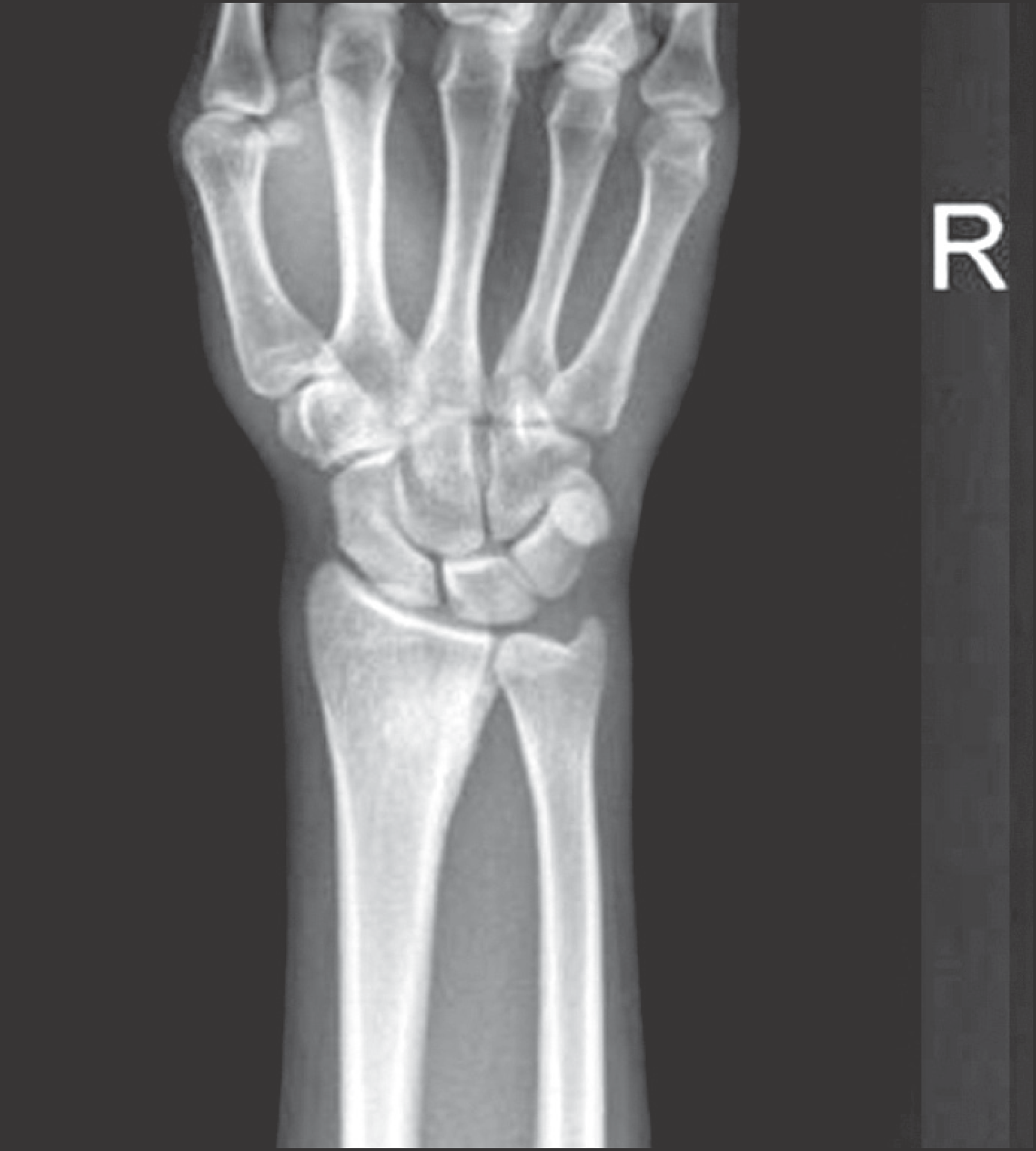
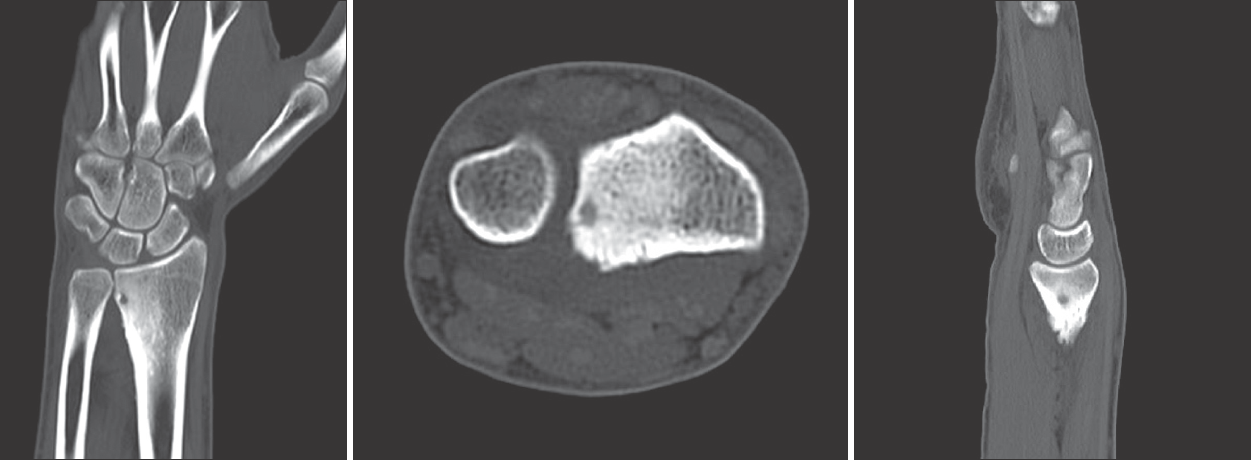
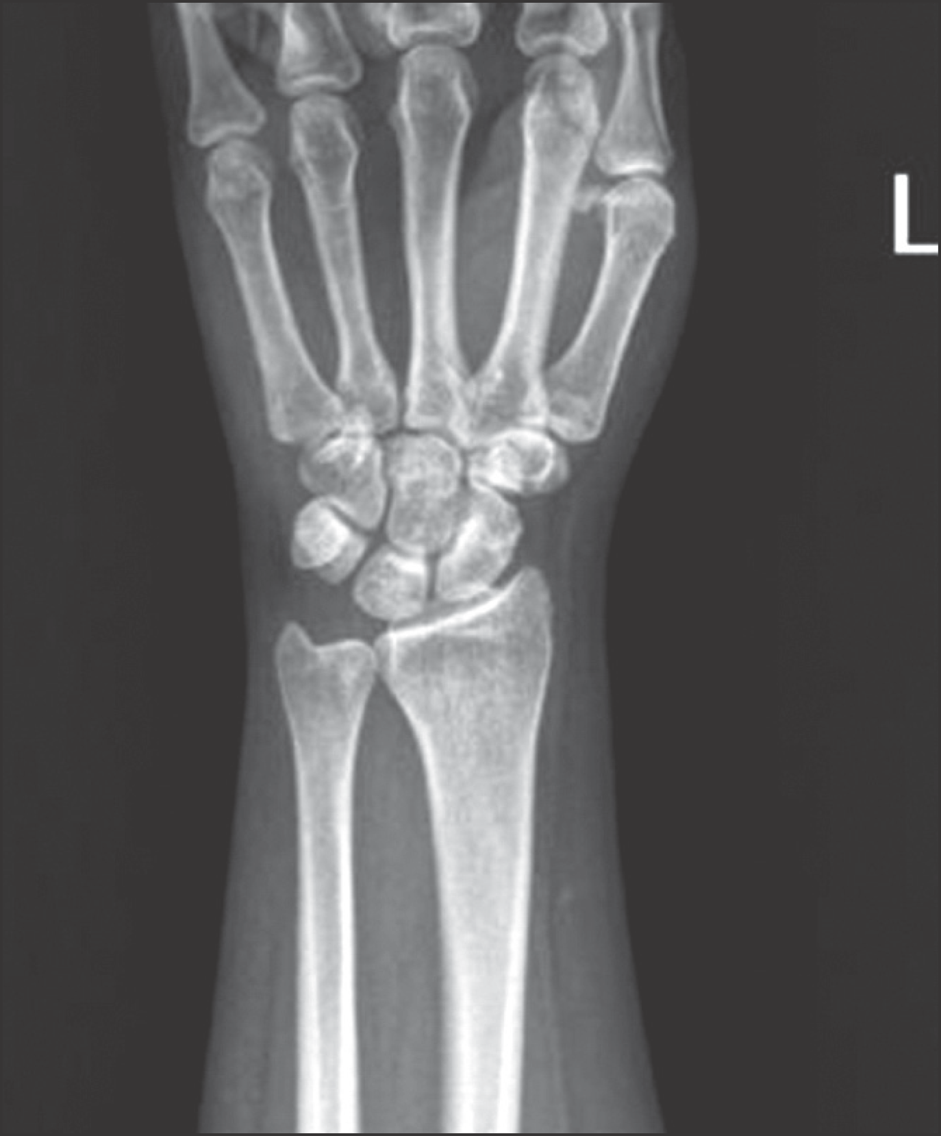
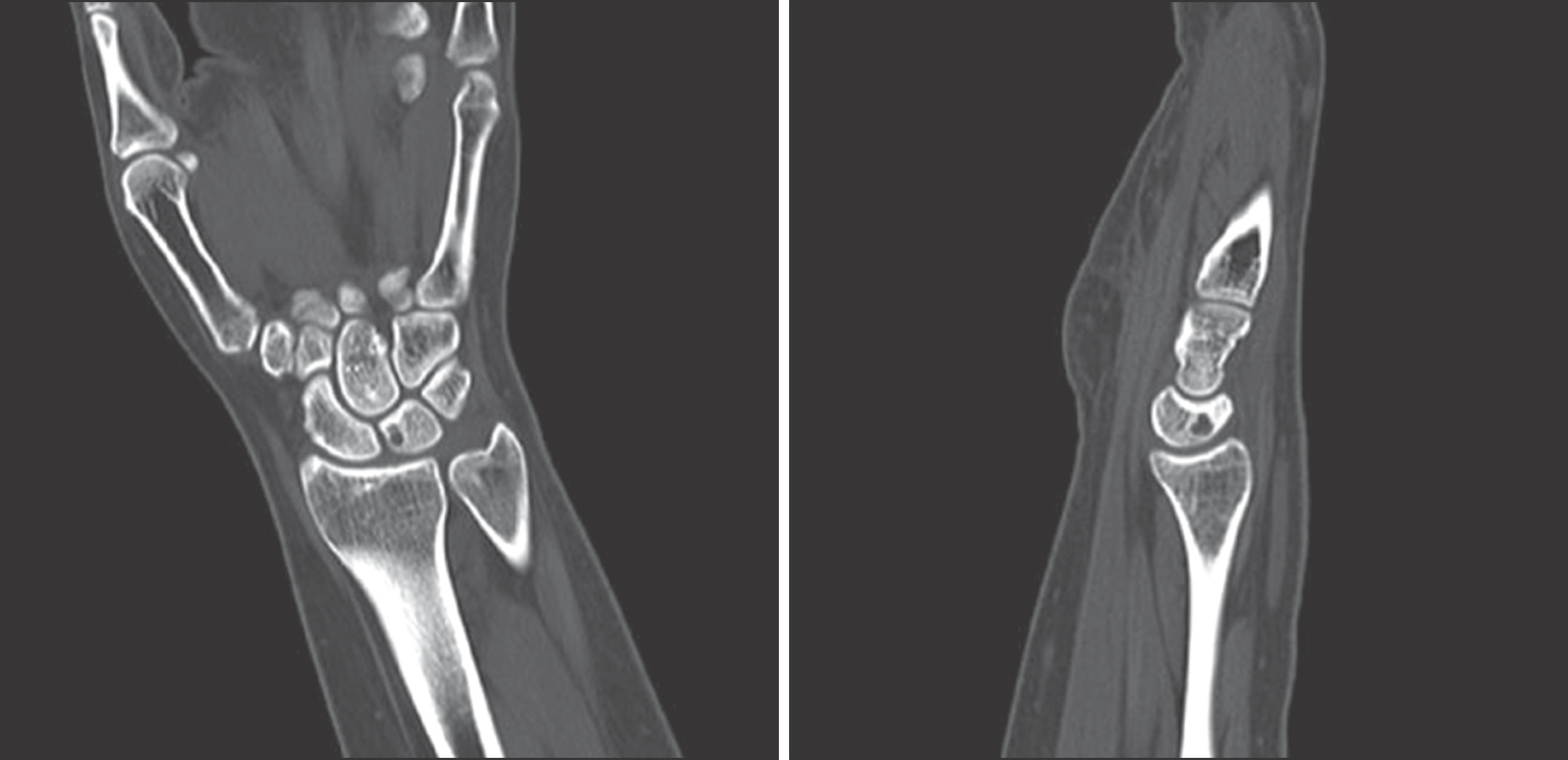
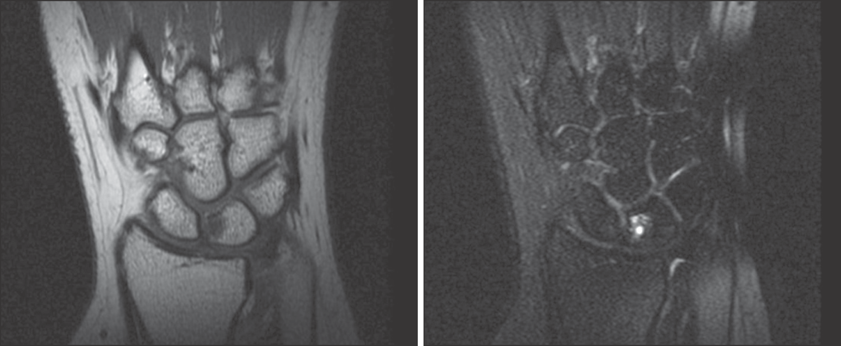
Osteoid osteoma can occur in all parts of the skeletal system. More than half occur in lower extremity and rare in wrist. Clinically pain is almost the only symptom worse at night and which is characterized by a rapid improvement by NSAID. We report the cases of osteoid osteoma which shows the characteristic symptoms and got a good results with appropriate imaging work up and surgical treatment.
References
1. Severo AL, Araújo R, Puentes R. Osteoid osteoma of the scaphoid bone: a case report. J Hand Surg Eur Vol. 2012; 37:797–9.

2. Jafari D, Shariatzade H, Mazhar FN, Abbasgholizadeh B, Dashtebozorgh A. Osteoid osteoma of the hand and wrist: a report of 25 cases. Med J Islam Repub Iran. 2013; 27:62–6.
3. Jafari D, Najd Mazhar F. Osteoid osteoma of the trapezoid bone. Arch Iran Med. 2012; 15:777–9.
4. Shin DB, Han SH, Soo RY, Ha DH, Ho LY. Osteoid osteoma of the distal phalanx of the thumb: a case report. J Korean Soc Surg Hand. 1998; 3:309–14.
5. Galdi B, Capo JT, Nourbakhsh A, Patterson F. Osteoid osteoma of the thumb: a case report. Hand (N Y). 2010; 5:423–6.

6. Goto T, Shinoda Y, Okuma T, et al. Administration of nonsteroidal anti-inflammatory drugs accelerates spontaneous healing of osteoid osteoma. Arch Orthop Trauma Surg. 2011; 131:619–25.

7. Sari S, Celikkanat S, Kara K, Akgun V, Karaman B. Osteoid osteoma radiofrequency ablation. JBR-BTR. 2014; 97:195.


 PDF
PDF ePub
ePub Citation
 Print
Print




 XML Download