Abstract
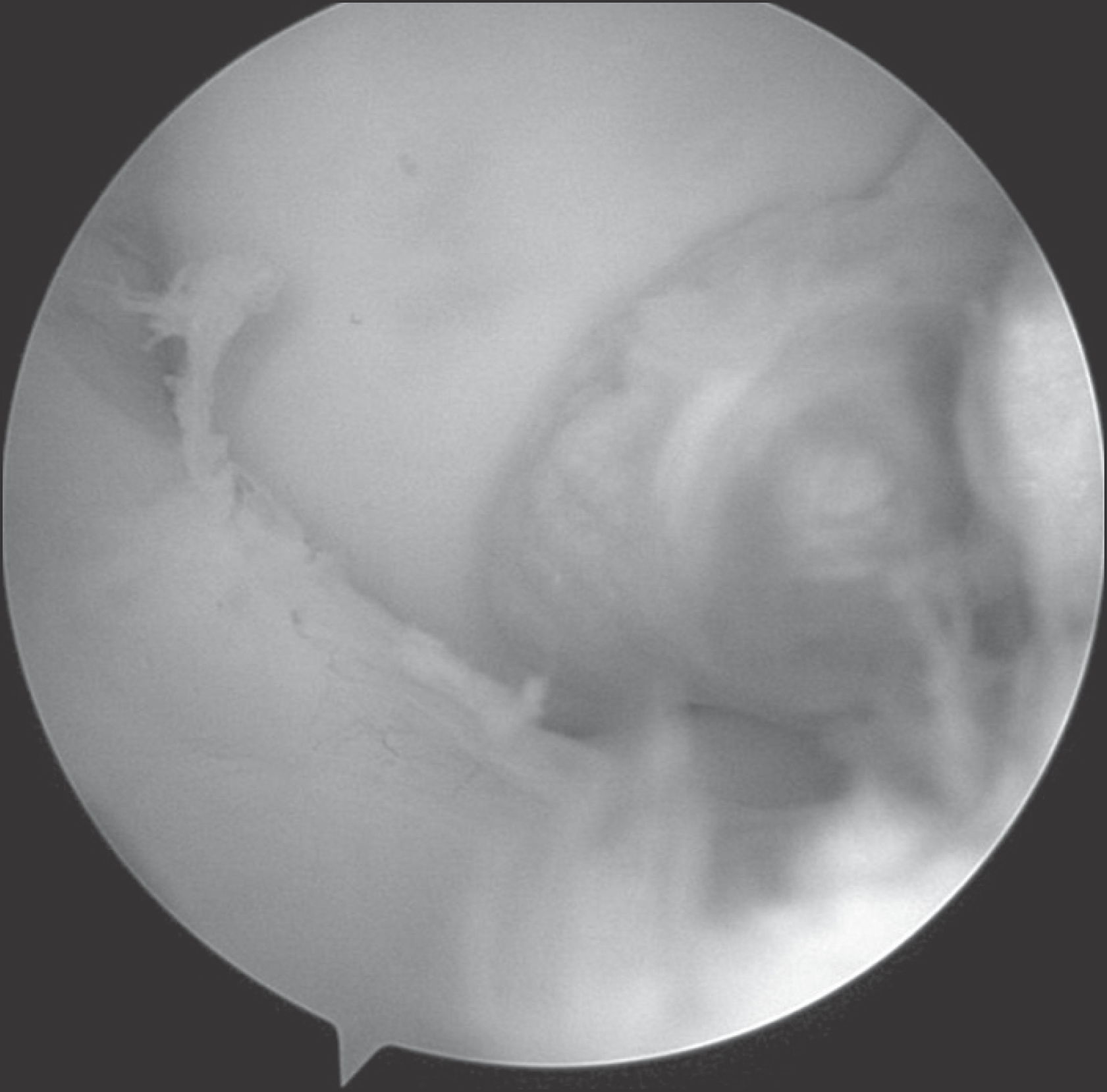
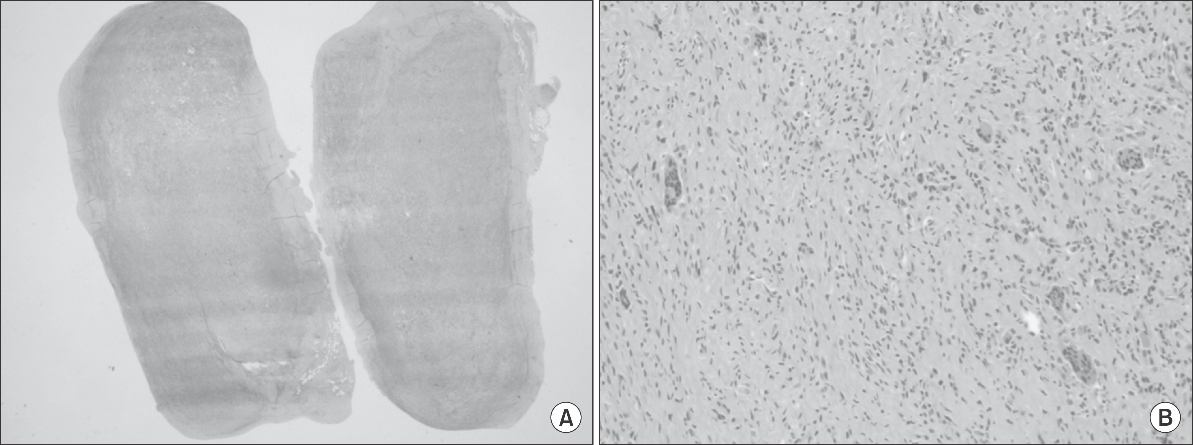
Localized forms of giant cell tumor are known to arise commonly in the synovial membrane of the finger joints. Multinucleated giant cells are its characteristic pathology finding, giant cell tumor shows a low rate of recurrence after complete excision. When occurring at the knee joints, giant cell tumor manifests a wide form of symptoms, from no symptom at all, to intermittent locking. Complete excision is possible by arthroscopy, but if done incompletely, it is reported to recur in 45% of cases. We present here a case of giant cell tumor that has arisen from the anterior portion of the posterior cruciate ligament, excised by arthroscopy and followed by pathologic confirmation.
References
1. Enzinger FM, Weiss SW. Benign tumors and tumor like lesions of synovial tissue. Enzinger FM, Weiss SW, editors. Soft tissue tumors. 3rd ed.St. Louis: Mosby;1995. p. 412–41.
2. Rosenberg AE. Soft tissue tumors and tumorlike lesions. Cotran RS, Kumar V, Robbins SL, editors. Robbins pathologic basis of disease. 5th ed.Philadelphia: Saunders;1994. p. 1260–1.
3. Chung WY, Kim YC, Jo SK. Localized form of tenosynovial giant cell tumor arising from the posterior Cruciate Ligament of the knee. 2 case report. J Korean Arthroscopy Soc. 2003; 7:87–91.
4. Kim KT, Kang MS. Localized tenosynovial giant cell tumor involving the posterior cruciate ligament of the Knee. 1 case report. J Korean Arthroscopy Soc. 2011; 15:113–6.
5. Insall JN, Scott WN. Surgery of the knee. 4th ed.Philadelphia: Elsevier;2006. p. 1026–9.
6. Koo BS, Kim KC, Lee HJ. Localized giant cell tumor of tendon sheath arising from the anterior cruciate ligament of the knee. 2 case report. J Korean Arthroscopy Soc. 1999; 3:146–9.
7. Lee GW, Lee KS, Song SH, Kim MK, Yun SH. Snow-man shaped nodular tenosynovitis in the knee. case report. J Korean Arthroscopy Soc. 1999; 3:44–7.
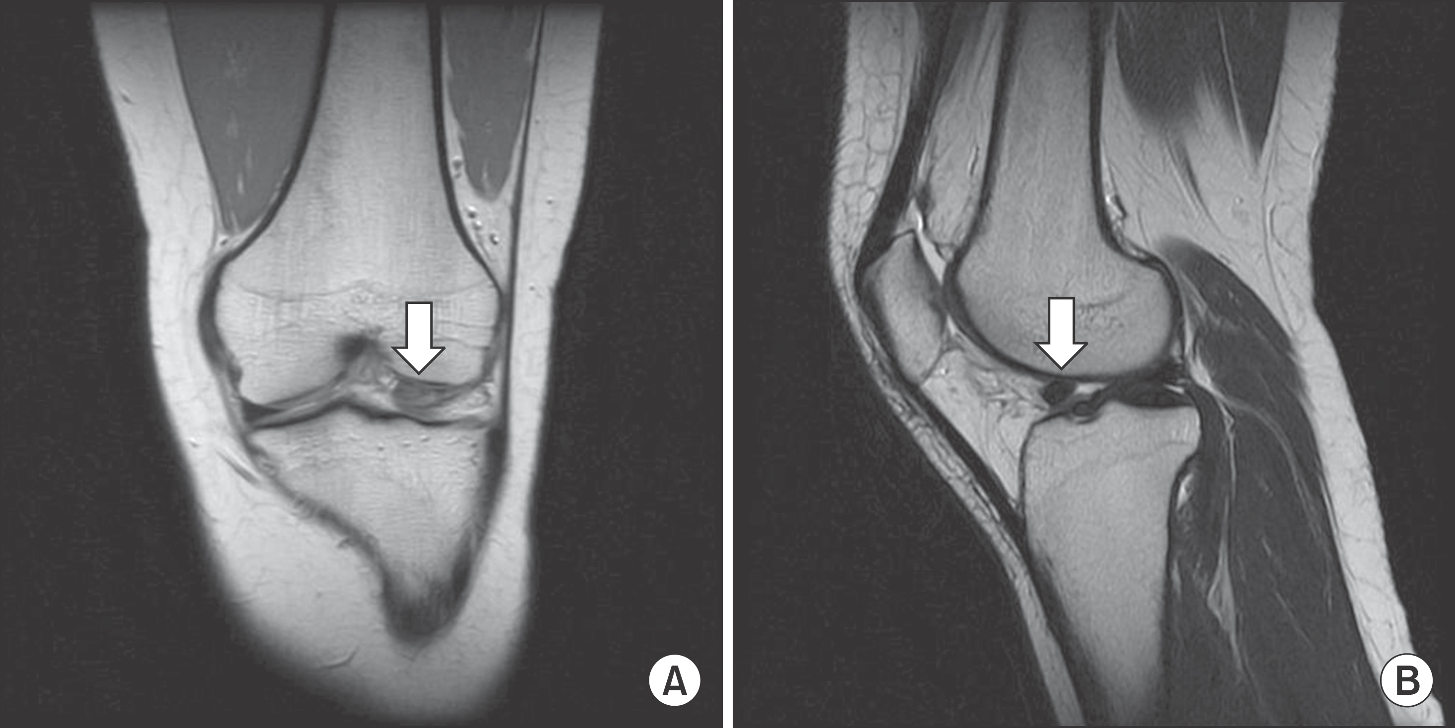
Figure 1.
(A) On proton density coronal image, a 9×6 mm sized mass (white arrow) with well circumscribed intermediate signal intensity was seen lateral side of knee joint. (B) On T2 sagittal image, a 9×6 mm sized mass (white arrow) with well circumscribed intermediate signal intensity was seen just anterior margin of PCL.


 PDF
PDF ePub
ePub Citation
 Print
Print


 XML Download