

 PDF
PDF ePub
ePub Citation
 Print
Print


Abstract
Inclusion body fibromatosis is a rare benign soft tissue neoplasm typically involving fingers and toes of children in mostly less than one year old. Histologic findings include spindle-shaped fibroblasts surrounded by dense stroma and small perinuclear eosinophilic inclusions in the cytoplasm. Although the tumor typically undergoes spontaneous regression, surgery is considered when functional impairment or deformity develops with the lesion. Unfortunately, recurrence rate was reported to be as high as 60 % following tumor excision. Authors would like to present our case where the tumor occurred in relatively older child and kissing lesion was found a few months after the surgery.
References
1. Reye RD. Recurring digital fibrous tumors of childhood. Arch Pathol. 1965; 80:228–31.
2. Netscher DT, Baumholtz MA, Popek E, Schneider AM. Nonmalignant fibrosing tumors in the pediatric hand: a clinicopathologic case review. Hand (N Y). 2009; 4:2–11.

3. Weiss SW, Goldblum JR. Enzinger and Weiss's soft tissue tumors. 5th ed.St. Louis: Mosby Elesevier;2008. p. 261–4.
4. Costa M, Venturino E, Rossello MI, Mazzotta P. La malattia di Reye: due nuove osservazioni associate a deformazione schele-trica falangea. Riv Chir Riab Mano Arto Sup. 1994; 31:211–5.
5. Drut R, Pedemonte L, Rositto A. Noninclusion-body infantile digital fibromatosis: a lesion heralding terminal osseous dysplasia and pigmentary defects syndrome. Int J Surg Pathol. 2005; 13:181–4.
6. Niamba P, Léauté-Labrèze C, Boralevi F, et al. Further documentation of spontaneous regression of infantile digital fibromatosis. Pediatr Dermatol. 2007; 24:280–4.
7. Grenier N, Liang C, Capaldi L, et al. A range of histologic findings in infantile digital fibromatosis. Pediatr Dermatol. 2008; 25:72–5.
8. Dey D, Nicol A, Singer S. Benign phyllodes tumor of the breast with intracytoplasmic inclusion bodies identical to infantile digital fibromatosis. Breast J. 2008; 14:198–9.
9. Spingardi O, Zoccolan A, Venturino E. Infantile digital fibromatosis: our experience and longterm results. Chir Main. 2011; 30:62–5.
Figure 1.
Photograph shows a round, soft tissue mass protruding in radial aspect of the left middle phalanx. (A) AP view (B) lateral view.
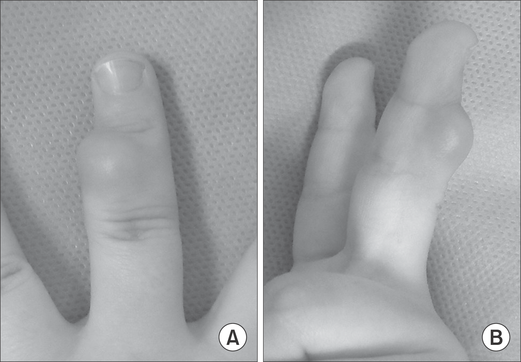
Figure 2.
Plain radiographs show that soft tissue density was increased in radial aspect of an affected finger. (A) AP view (B) lateral view.
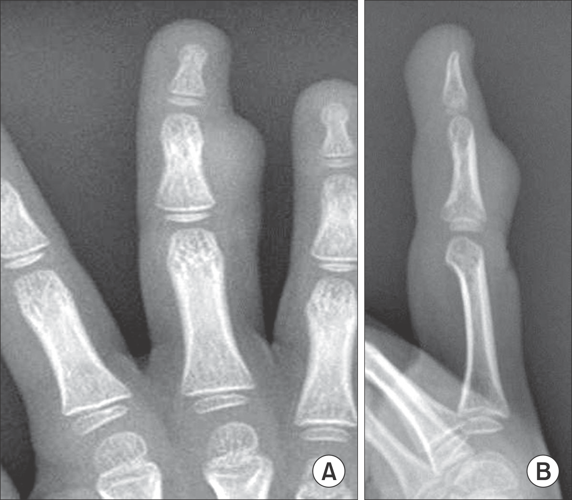
Figure 3.
MRI shows a well-delineated round mass of T1 low and T2 intermediate signal intensity. The tumor is located subcutaneously and abutting adjacent phalangeal bone and extensor tendon. There was marked diffuse homogeneous enhancement throughout the lesion following the administration of intravenous contrast.
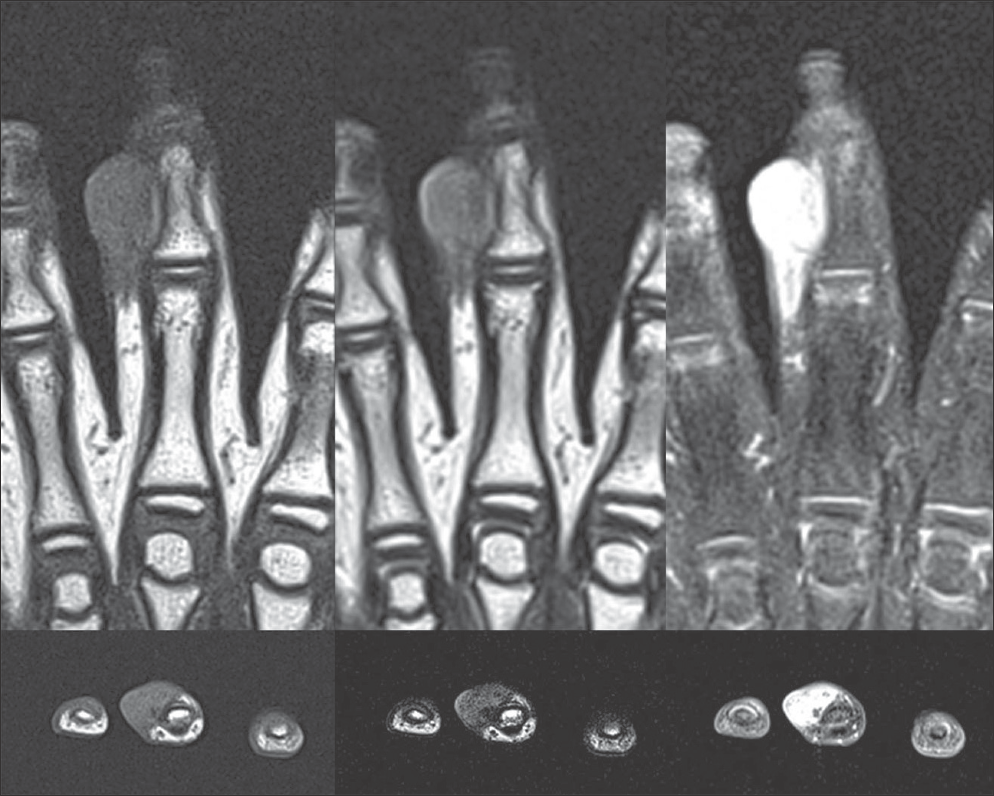
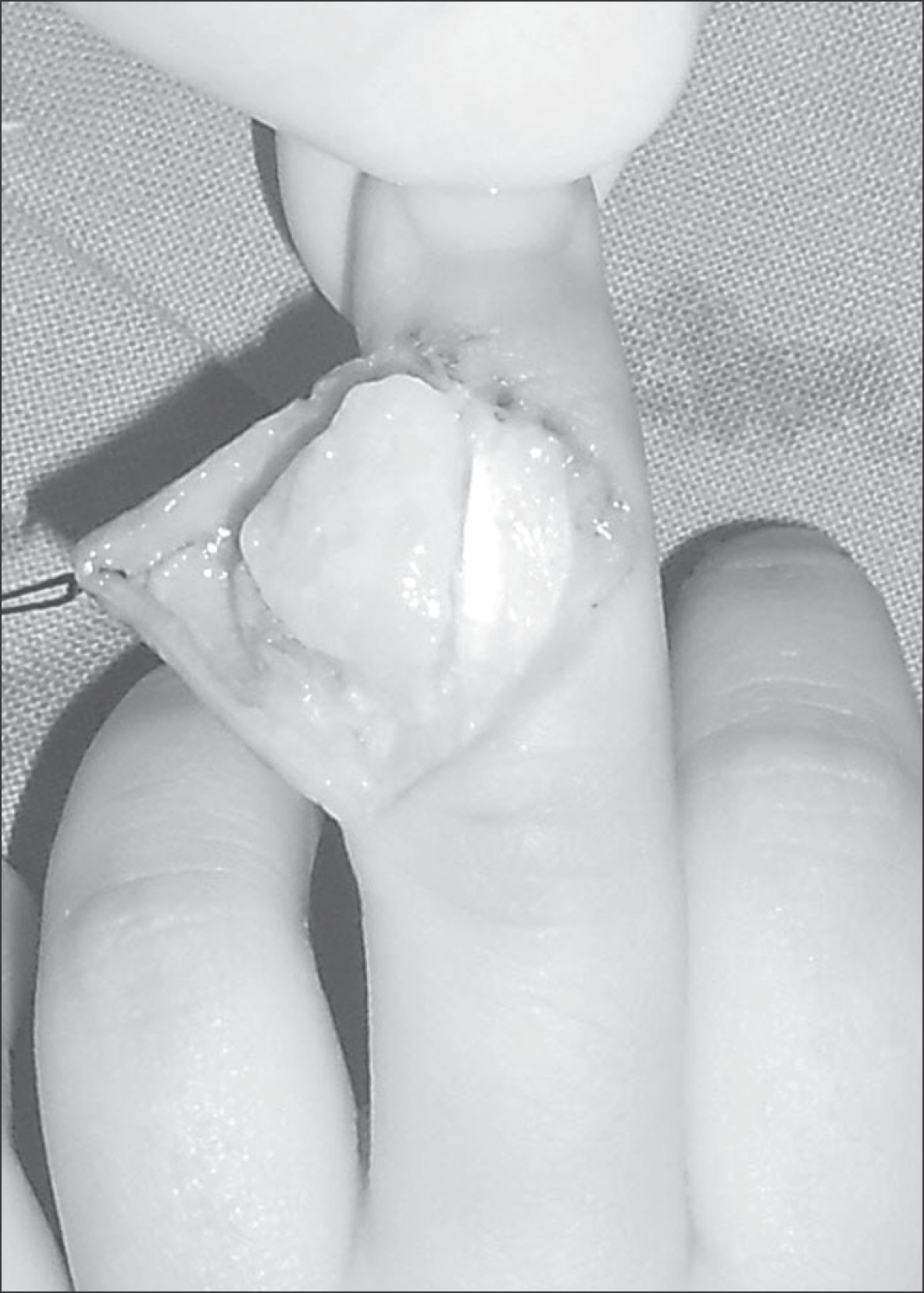
Figure 5.
Pathologic features. (A) The tumor consists of spindled cells with elongated, cytologically bland nuclei and rounded, pink, intracytoplasmic inclusions, some indenting the nucleus (Hematoxylin & Eosin, ×200). (B) Trichrome staining highlights intracytoplasmic inclusions (red dots, inclusion body) (trichrome ×200). (C) Immunostaining for smooth muscle actin (SMA) demonstrates peripheral tram-track pattern of muscle actins (SMA, ×200).

 XML Download