Abstract
Purpose
Fibrous dysplasia is related to the mutation of gene encoding the alpha-subunit of a signal-transducing G-protein and has variable clinical course. Operation can be performed to prevent functional disorder or structural deformity. After curettage, autologous bone graft were used to fill the defects after curettage. The aim of this study is to compare the result of autogenous cancellous bone grafting and allogenic bone grafting for fibrous dysplasia.
Materials and Methods
Among the patients who visit our hospital during the period of April, 1997 to October, 2013, we selected 34 patients who diagnosed fibrous dysplasia and visited our clinic over 1 year. There were 13 males and 21 females. Average age was 26.4 (range 2 to 57) years old. Autogenous bone graft (group I) in 5 cases, Non-autogenous bone graft (group II) in 30 cases. Iliac bone is used in all cases of autogenous bone graft. There were no significant difference in age, follow-up period, preoperational laboratory finding between two groups. Radiographic image was done to evaluate the recurrence of fibrous dysplasia or secondary degeneration.
Results
There were four cases in recurrence (group I: 1 case, group II: 3 cases, p=0.554). In all recurrent cases, reoperations were done using curettage and autogenous iliac bone graft. There was no re-recurrence after reoperation. One case of secondary aneurysmal bone cyst was confirmed (group II) and 1 cases of pathologic fractures had developed (group I: 0 case, group II: 1 cases, p=0.559). No malignant change occurred.
Go to : 
References
1. Stephenson RB, London MD, Hankin FM, Kaufer H. Fibrous dysplasia. An analysis of options for treatment. J Bone Joint Surg Am. 1987; 69:400–9.
2. DiCaprio MR, Enneking WF. Fibrous dysplasia. Pathophysiology, evaluation, and treatment. J Bone Joint Surg Am. 2005; 87:1848–64.
4. Jaffe H. Tumors and Tumorous Conditions of the Bones and Joints. Philadelphia: Lea and Febiger;1958. p. 117–42.
7. Fowler BL, Dall BE, Rowe DE. Complications associated with harvesting autogenous iliac bone graft. Am J Orthop (Belle Mead NJ). 1995; 24:895–903.
9. Schlumberger HG. Fibrous dysplasia of single bones (monostotic fibrous dysplasia). Mil Surg. 1946; 99:504–27.

10. Harris WH, Dudley HR Jr, Barry RJ. The natural history of fibrous dysplasia. An orthopaedic, pathological, and roentgenographic study. J Bone Joint Surg Am. 1962; 44-A:207–33.
Go to : 
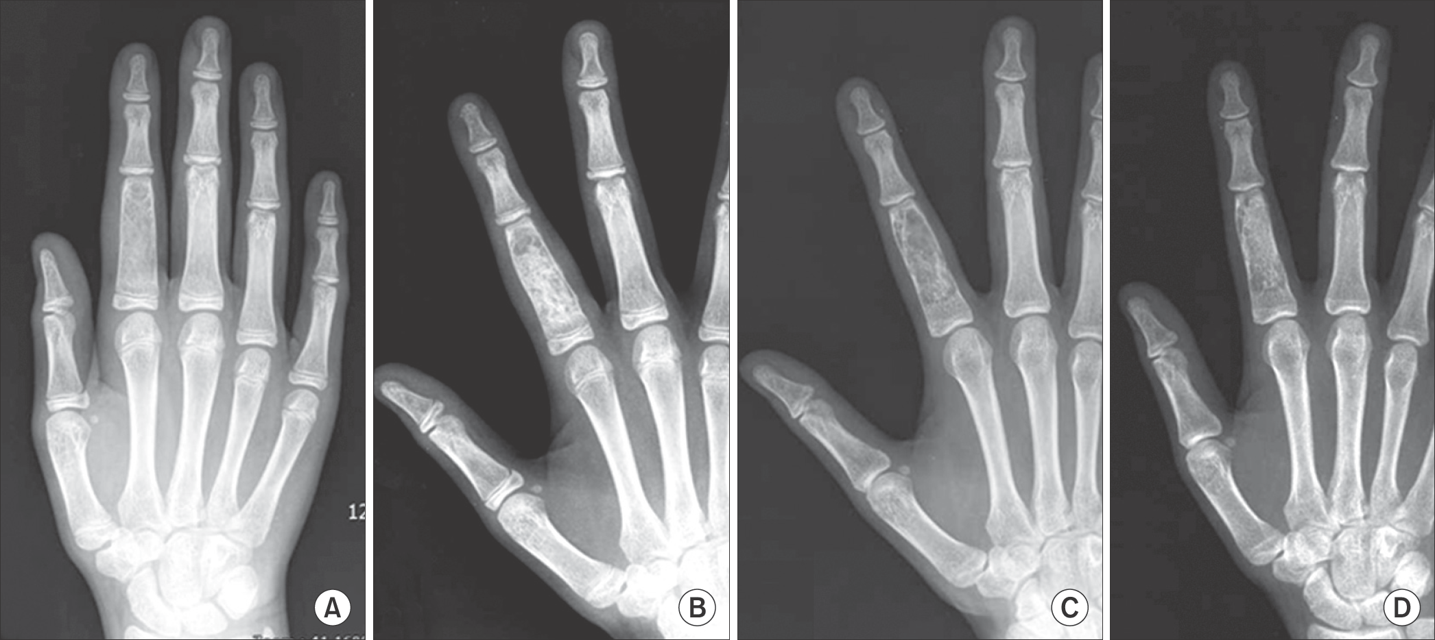
 | Figure 1.Fibrous dysplasia of the proximal phalanx in a 10-year-old girl. (A) The radiogram show a radiolucent area on proximal phalanx of index finger. (B) The postoperative radiogram at 1month show internal filling with autogenous bone graft on proximal phalanx of index finger. (C) The postoperative radiogram at 4 years after surgery show a radiolucent area on proximal phalanx of index finger suggesting recurrence. (D)The Radiogram at 4 years after re-operation shows healing with new bone formation. |
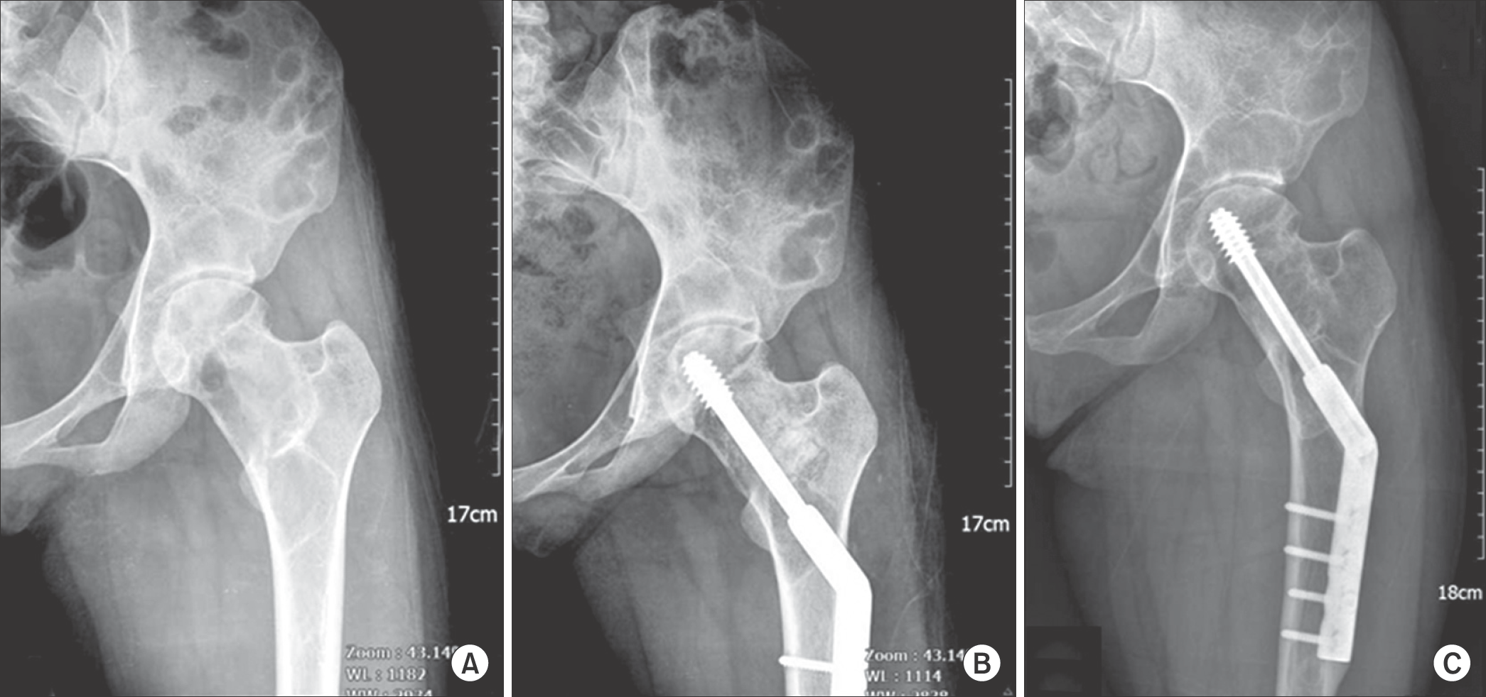
 | Figure 2.Fibrous dysplasia of the proximal femur in 18-years-old girl. (A) The radiogram shows a large radiolucent area with marginal sclerosis on left proximal femur. (B) The radiogram made 1 month after surgery shows allogenic bone graft and internal fixation with dynamic hip screw on left proximal femur. (C) The postoperative radiogram at 9 years shows healing with new bone formation. |
Table 1.
Patients treated with bone grafting on fibrous dysplasia


 PDF
PDF ePub
ePub Citation
 Print
Print


 XML Download