

 PDF
PDF ePub
ePub Citation
 Print
Print


Abstract
Purpose
To diagnose soft tissue tumor, such as lipoma and Schwannoma, magnetic resonance imaging (MRI) is sufficient in most cases. However, various characteristics are found in MRI images of Schwannoma, thus other type of tumors are often misdiagnosed as Schwannoma with MRI images. In this study, we evaluate the diagnostic value of specific MRI findings of Schwannoma.
Materials and Methods
From January 2002 to May 2013, 104 patients who are suspected as Schwannoma rith MRI images are included in data, and the final diagnosis is confirmed with biopsy. Patients are divided into group 1 and group 2 who are confirmed as Schwannoma and other disease with biopsy, respectively.
Results
92 patients were diagnosed as Schwannoma (group 1) and 12 patients were diagnosed as other disease (group 2). We investigate the diagnostic value of specific MRI findings of Schwannoma. 41 patients of group 1 (45%) and 0 patients of group 2 (0%) showed target sign, 47 patients of group 1 (51%) and 2 patients of group 2 (17%) showed fascicular sign, 44 patients of group 1 (48%) and 5 patients of group 2 (42%) showed fat split sign, 28 patients of group 1 (30%) and 1 patients of group 2(9%) showed nerve entering and exiting sign, and 8 patients of group 1 (9%) and 6 patients of group 2 (50%) showed none of four specific findings on their MRI images. 52 patients of group 1 (57%) and 5 patients of group 2 (42%) have tumors on the pathway of nerve. Target sign could be considered as the best diagnostic value of the sign we investigate (p<0.05).
Conclusion
Although specific MRI findings have powerful diagnostic value, patients are often misdiagnosed as Schwannoma with MRI findings. Therefore, if patients who are suspected as Schwannoma based on MRI findings have no target sign on their MRI images, we should consider the possibility of other disease.
Go to : 
References
1. Pilavaki M, Chourmouzi D, Kiziridou A, Skordalaki A, Zarampoukas T, Drevelengas A. Imaging of peripheral nerve sheath tumors with pathologic correlation: pictorial review. Eur J Radiol. 2004; 52:229–39.
2. Kransdorf M, Murphey MD. Imaging of soft tissue tumors. Philadelphia: Saunders;1997. p. 235–73.
3. Stout AP. The peripheral manifestations of thespecific nerve sheath tumor (neurilemmoma). Am J Cancer. 1935; 24:751–96.
4. Stull MA, Moser RP Jr, Kransdorf MJ, Bogumill GP, Nelson MC. Magnetic resonance appearance of peripheral nerve sheath tumors. Skeletal Radiol. 1991; 20:9–14.

5. Cerofolini E, Landi A, DeSantis G, Maiorana A, Canossi G, Romagnoli R. MR of benign peripheral nerve sheath tumors. J Comput Assist Tomogr. 1991; 15:593–7.
6. Suh JS, Abenoza P, Galloway HR, Everson LI, Griffiths HJ. Peripheral (extracranial) nerve tumors: correlation of MR imaging and histologic findings. Radiology. 1992; 183:341–6.
7. Woodruff JM, Selig AM, Crowley K, Allen PW. Schwannoma (neurilemoma) with malignant transformation. A rare, distinctive peripheral nerve tumor. Am J Surg Pathol. 1994; 18:882–95.
8. Abernathey CD, Onofrio BM, Scheithauer B, Pairolero PC, Shives TC. Surgical management of giant sacral schwannomas. J Neurosurg. 1986; 65:286–95.
9. Hennigan TW, Branfoot AC, Theodorou NA. Ancient neurilemmoma of the pelvis. J R Soc Med. 1992; 85:416–7.
10. Hems TE, Burge PD, Wilson DJ. The role of magnetic resonance imaging in the management of peripheral nerve tumours. J Hand Surg Br. 1997; 22:57–60.
Go to :
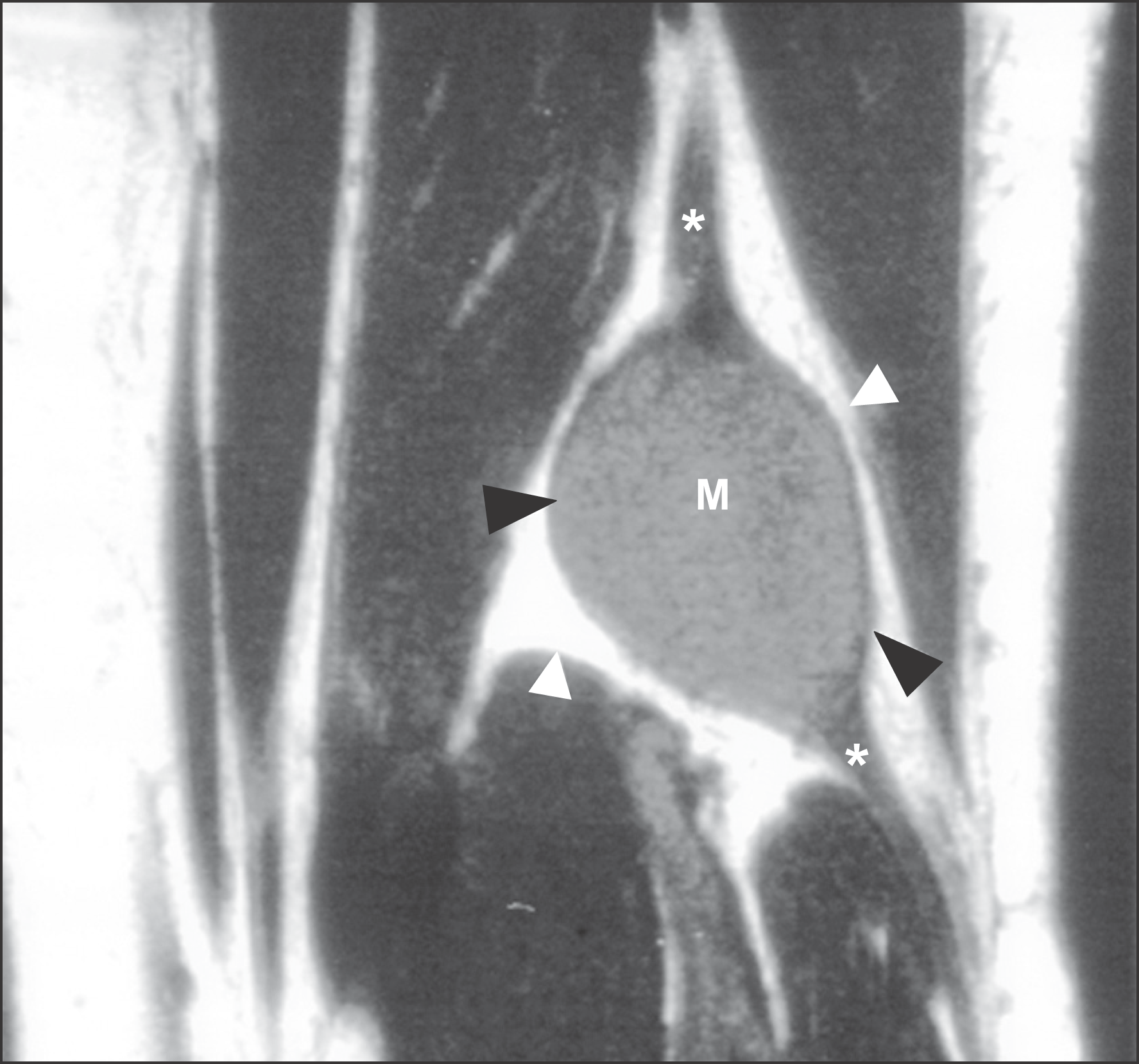 | Figure 1.A well-defined fusiform mass with entering and exiting nerve. The mass is sharply marginated (black arrow) which is seen along the nerve (white arrow). |
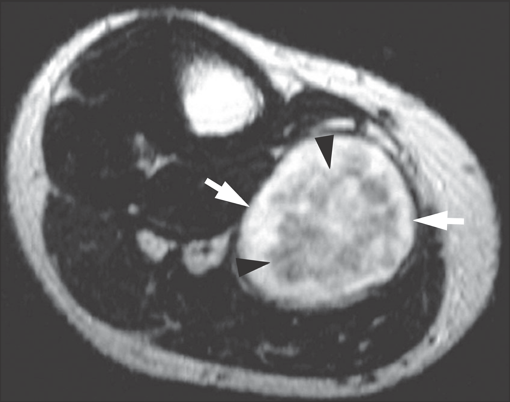 | Figure 2.Axial T2-weighted image in a different patient demonstrating peripheral hyperintense (arrows) and central hypointense (arrowheads) signal (the “target” sign). |
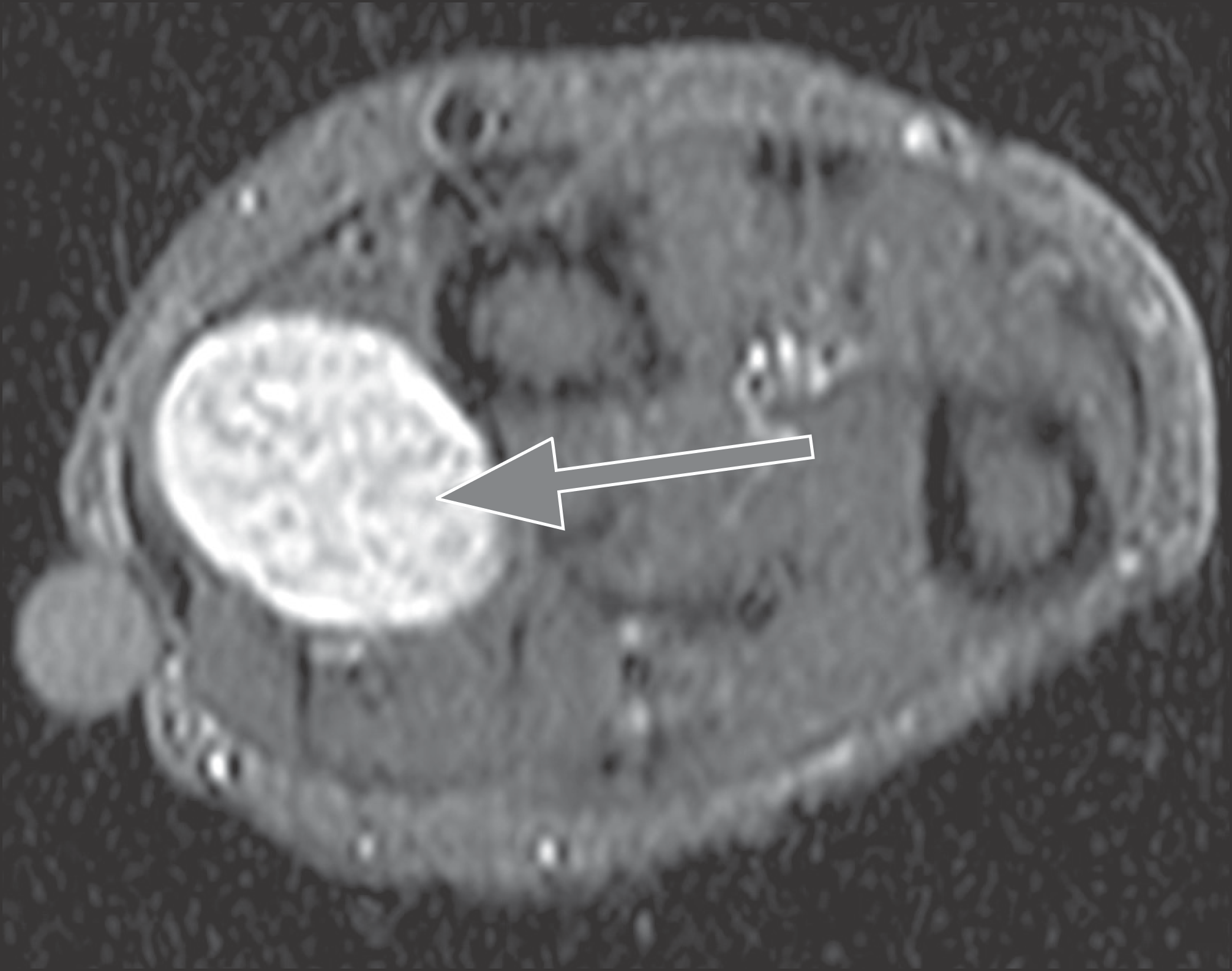 | Figure 3.Fascicular sign. Multiple ring-like structures are seen throughout the lesion (arrow). |
 | Figure 4.Sagittal T1 (A)/T2 (B)-weighted image demonstrating well-marginated mass hypointense (A)/hyperintense (B) signal (Fascicular sign (+)). |
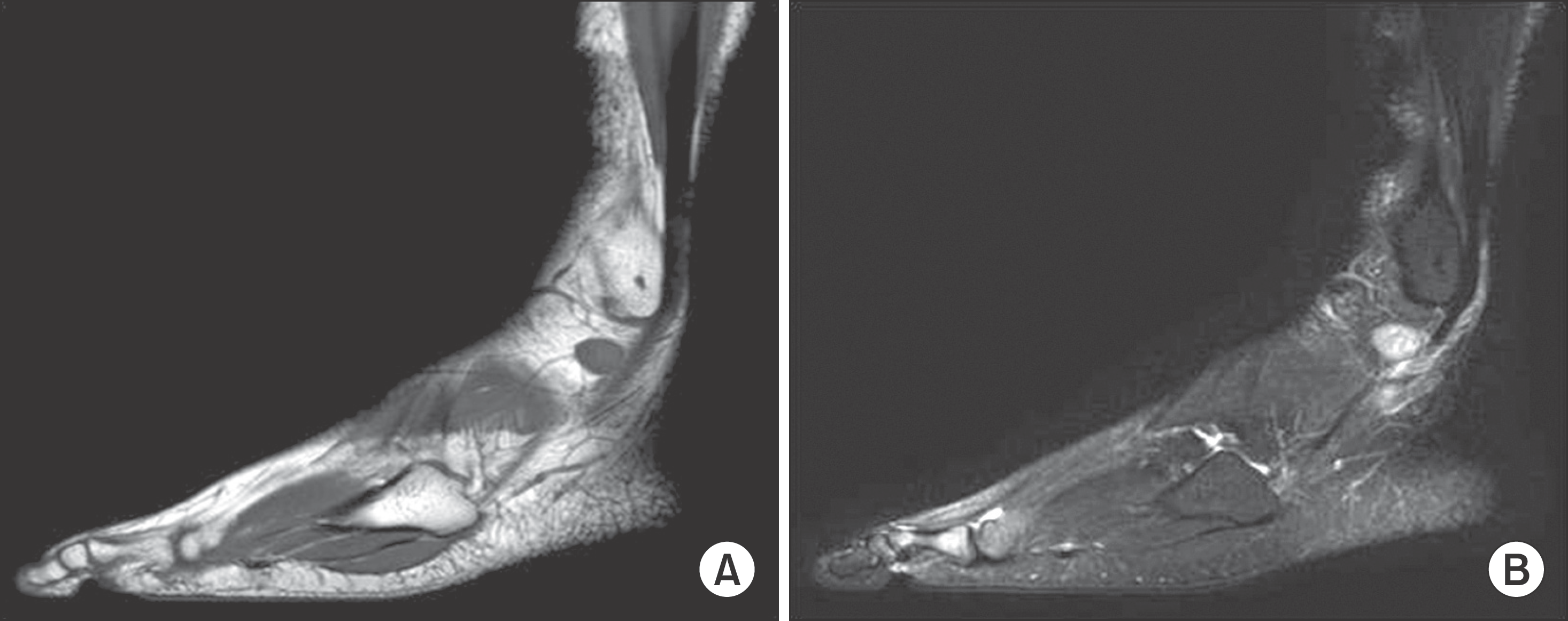
 | Figure 5.Coronal T1 (A)/T2 (B)-weighted image demonstrating well-marginated mass hyporintense (A)/hyperintense (B) signal (Split fat sign/nerve entering sign (+/+)). |
 XML Download