

 PDF
PDF ePub
ePub Citation
 Print
Print


Abstract
A femoral bone tumor causing a valgus deformity by affecting the growth plate was found. Long intramedullary diaphyseal tumor was separated by septum at the metapysis. Low grade chondrosarcoma was confirmed diagnosed by pathologists. Progressive limb deformity can be a sign of bone tumor in growing period.
References
1. Kusuzaki K, Murata H, Takeshita H, et al. Usefulness of cy-tofluorometric DNA ploidy analysis in distinguishing benign cartilaginous tumors from chondrosarcomas. Mod Pathol. 1999; 12:863–72.
2. Aprin H, Riseborough EJ, Hall JE. Chondrosarcoma in children and adolescents. Clin Orthop Relat Res. 1982; 166:226–32.

3. Haygood TM, Teot L, Ward WG, Allen A, Monu JU. Low-grade chondrosarcoma in a 12-year-old boy. Skeletal Radiol. 1995; 24:466–8.
4. Young CL, Sim FH, Unni KK, McLeod RA. Chondrosarcoma of bone in children. Cancer. 1990; 66:1641–8.
5. Huvos AG, Marcove RC. Chondrosarcoma in the young. A clinicopathologic analysis of 79 patients younger than 21 years of age. Am J Surg Pathol. 1987; 11:930–42.
6. Ishida T, Iijima T, Goto T, Kawano H, Machinami R. Concurrent enchondroma and periosteal chondroma of the humerus mimicking chondrosarcoma. Skeletal Radiol. 1998; 27:337–40.
7. Yamamoto Y, Washimi O, Yamada H, Washimi Y, Itoh M, Kuroda M. Concurrent periosteal chondroma and enchondroma of the fibula mimicking chondrosarcoma. Skeletal Radiol. 2006; 35:302–5.
8. Babinet A, de Pinieux G, Tomeno B, Forest M, Anract P. Intracortical chondrosarcoma. A case report. J Bone Joint Surg Am. 2003; 85-A:533–5.
9. Leerapun T, Hugate RR, Inwards CY, Scully SP, Sim FH. Surgical management of conventional grade I chondrosarcoma of long bones. Clin Orthop Relat Res. 2007; 463:166–72.
10. Greenspan A, Jundt G, Remagen W. Differential diagnosis in orthopaedic oncology. 2nd ed.Philadelphia: Lippincott Williams & Wilkins;2007. p. 212–7.
Figure 1.
A 15-year-old man has progressive valgus deformity of left lower limb. (A) Photograph shows valgus deformed lower limb. (B) Radiograph demonstrate 27 degree valgus deformed femur at the meta-diaphysis area.
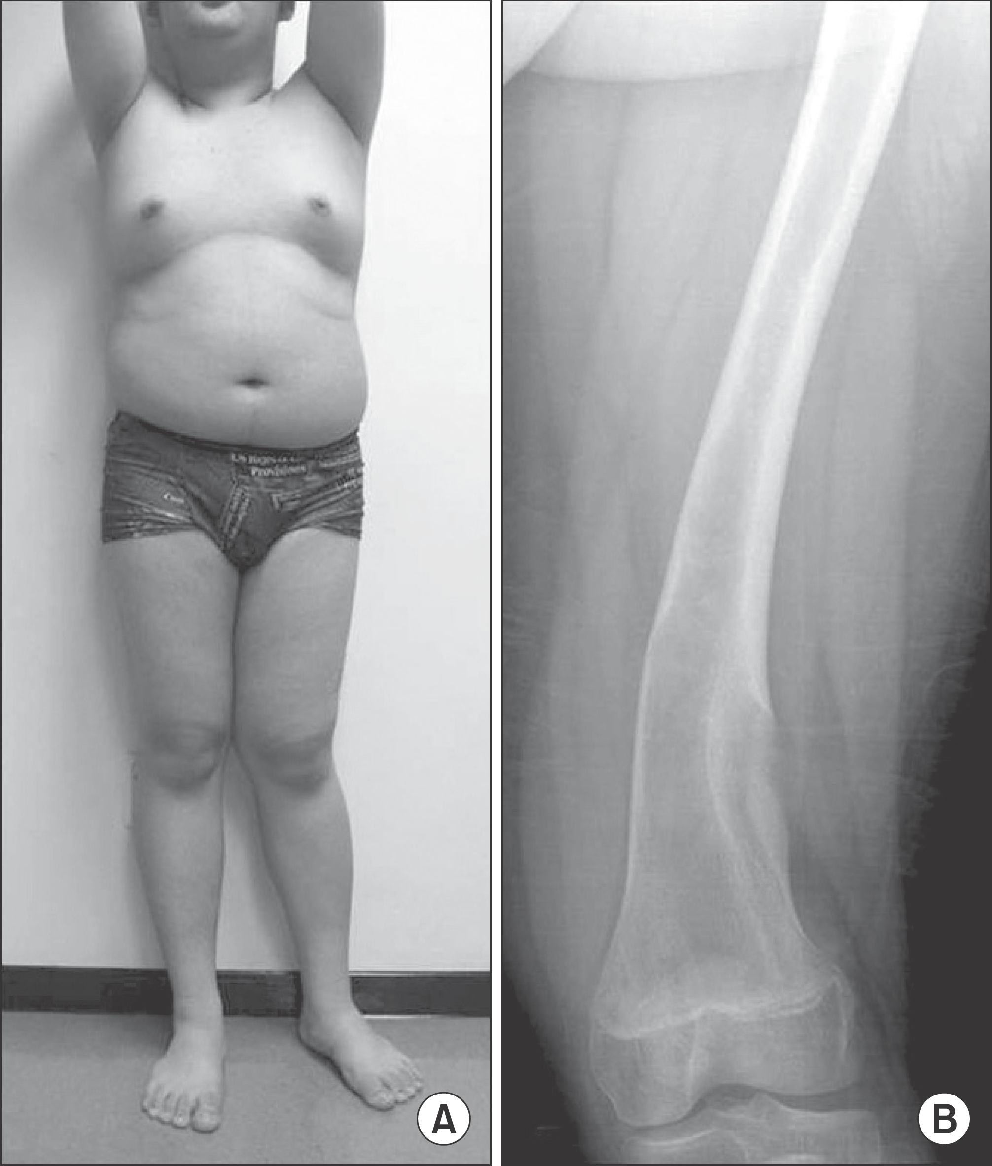
Figure 2.
(A) T1-weighted coronal images show the meta-diaphysial intramedullary and intracortical lesion. (B) Coronal computed tomography obtained after open biopsy showing valgus curved femur may caused close connecting growth plate of the intracortical lesion (arrow). (C) Axial computed tomography sections showing the intramedullary calcific stippling (superior), distinguished intracortical osteolytic lesion (middle), and elliptical tailed of lateral intracortical lesion toward growth plate (inferior).
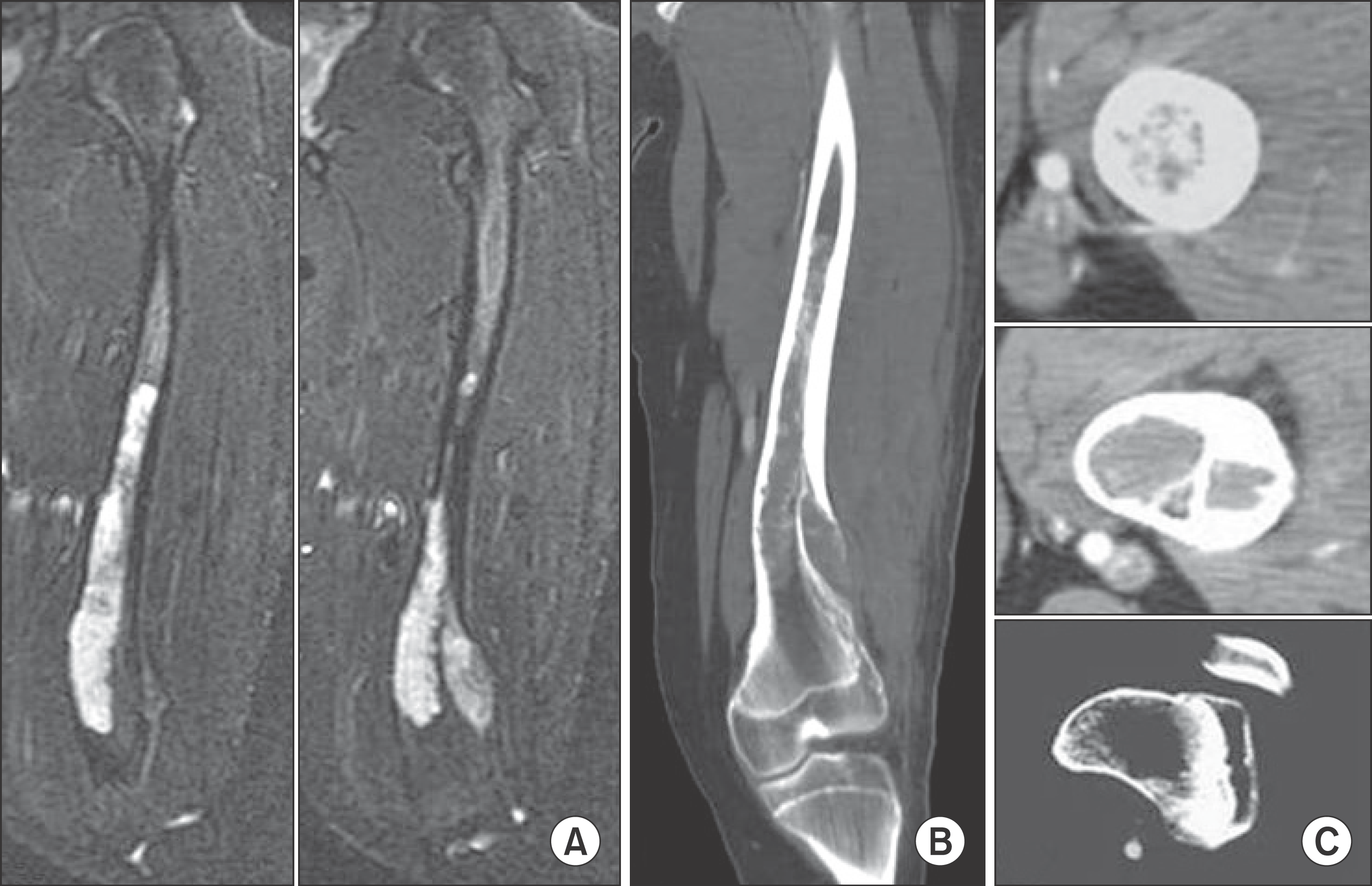
Figure 3.
(A) and (B) Coronal MRI showed mountain peak shape of the lateral growth plate to the distal tail of the intracortical lesion.
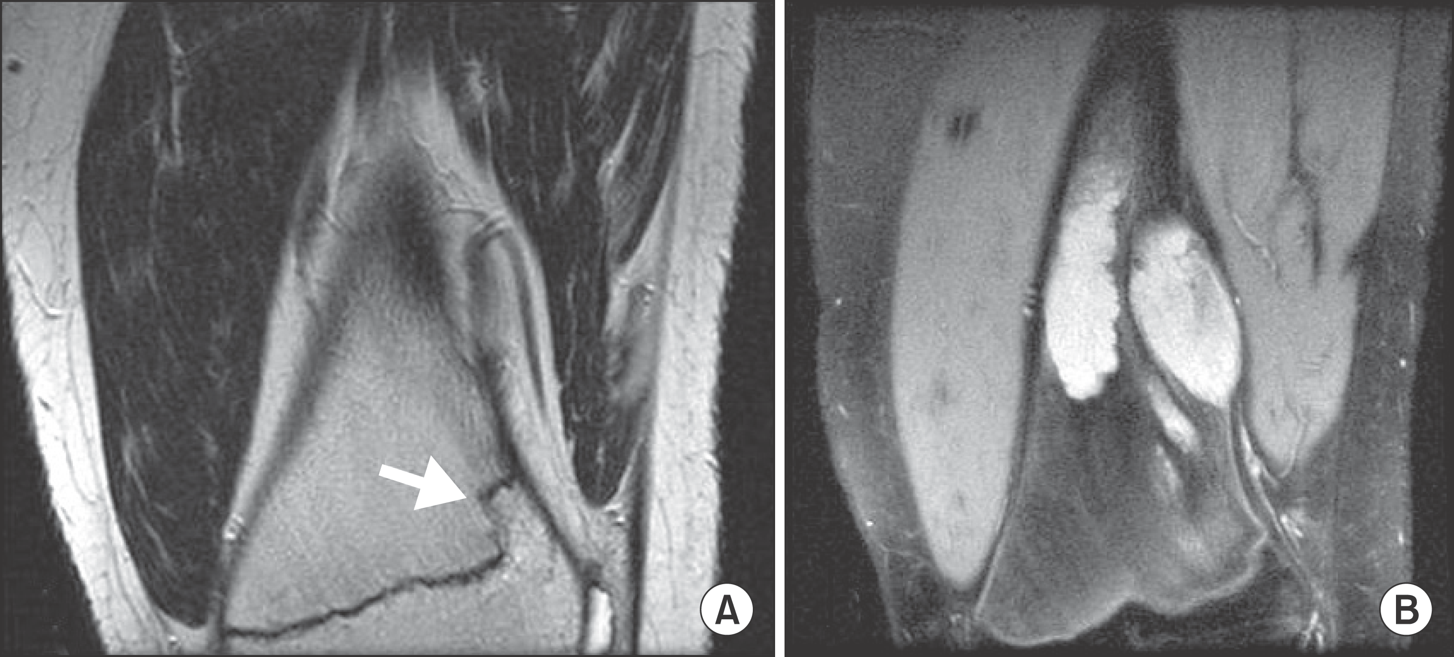
Figure 4.
(A) The photograph of coronal cut specimen reveal the distinguished intramedullary and intracortical lesion. (B) Photomicrograph of intramedullary and (C) intracortical lesion showing a low grade chondrosarcoma with intrusion of surrounding mature lamellar bone, (Hematoxylin and eosin, ×100). (D) The obvious atypia of chondrocytes was not revealed on the high-magnification photomicrograph (Hematoxylin and eosin, ×400).
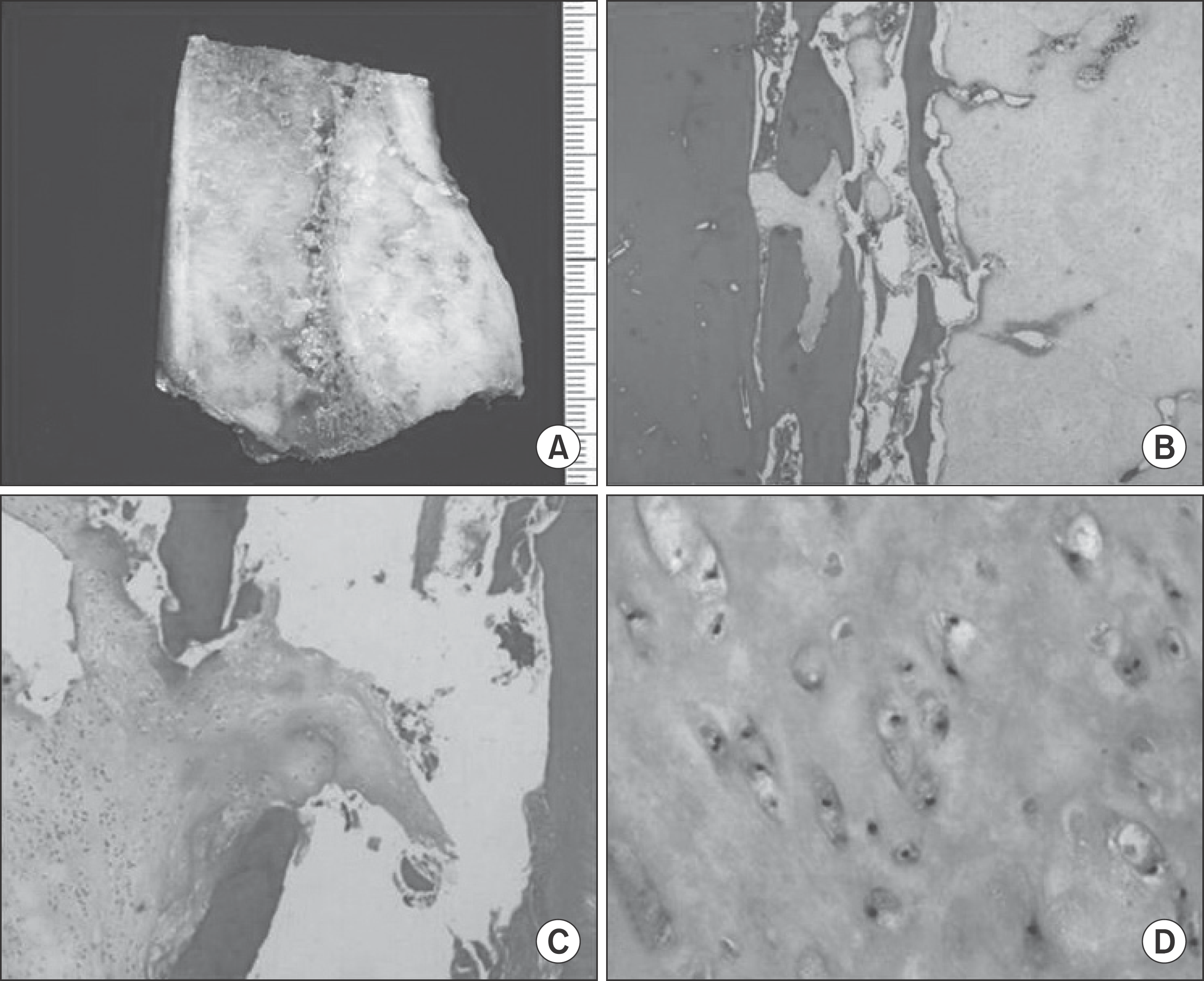
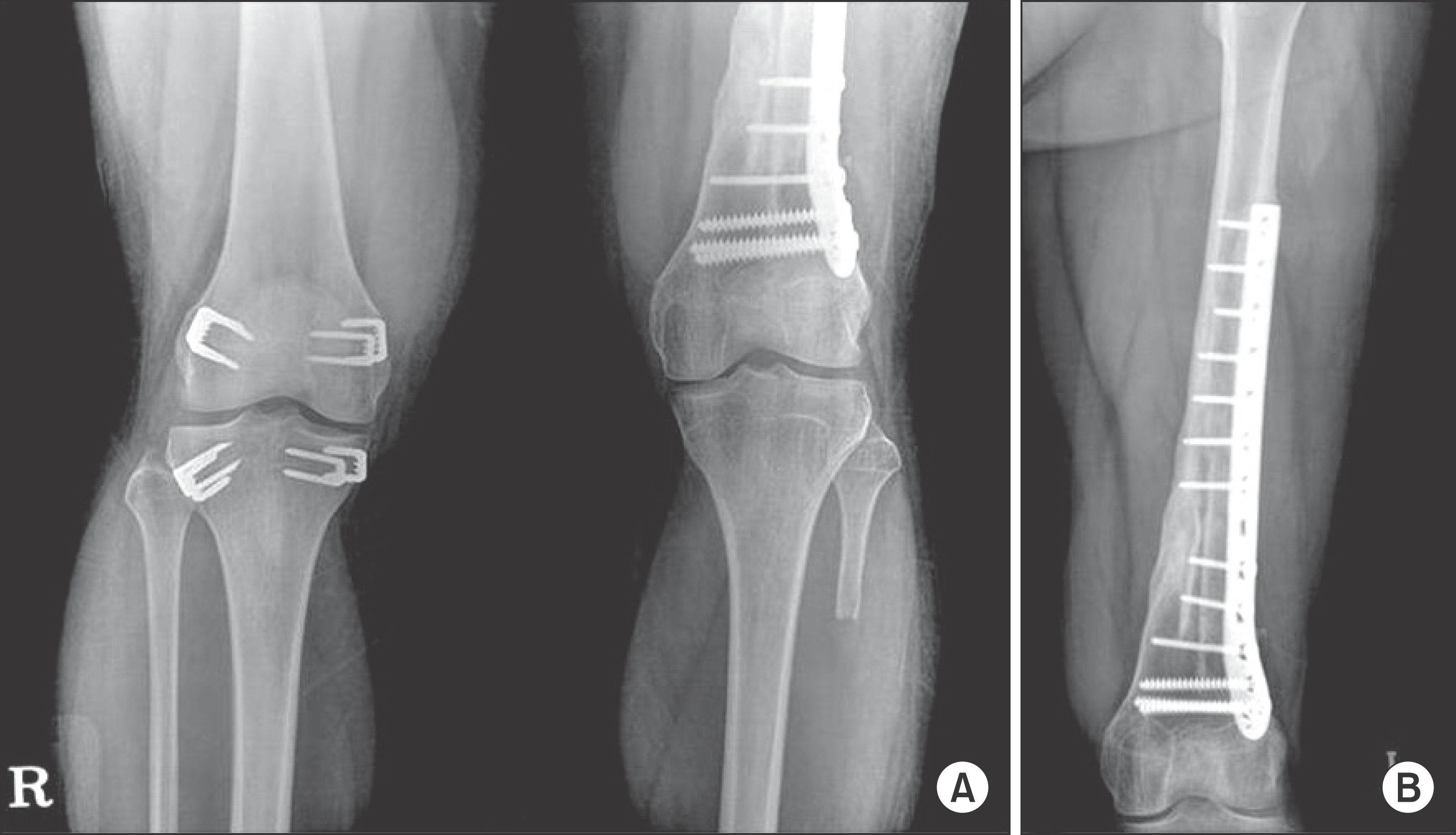
Figure 5.
Post operative six-year follow up (A) both knee anteroposterior radiograph shows the epiphysiodesis around right knee and different knee joint level by shortening of left femur. (B) Left femur anteroposterior radiograph demonstrates well incorporation of allograft and intramedullary loaded autogenous fibular graft with correction of deformity.
 XML Download