

 PDF
PDF ePub
ePub Citation
 Print
Print


Abstract
Rosai-Dorfman disease (RDD) is an idiopathic histioproliferative disorder of lymph node and extranodal site. Bone involvement is very rare. We report a case of extranodal RDD of the tibia in 32-year old male. The patient presented with pain with no evidence of lymphadenopathy. Clinico-radiologic diagnosis was metastatic carcinoma or Langerhans cell histiocytosis, but, histopathologic examination confirmed the diagnosis with RDD. We performed curettage on the osteolytic lesion of tibia. In South Korea, there was no report about RDD of the extremity and we want to report this case with review of the literature.
References
1. Demicco EG, Rosenberg AE, Björnsson J, Rybak LD, Unni KK, Nielsen GP. Primary Rosai-Dorfman disease of bone: a clinicopathologic study of 15 cases. Am J Surg Pathol. 2010; 34:1324–33.
2. Lai KL, Abdullah V, Ng KS, Fung NS, van Hasselt CA. Rosai-Dorfman disease: presentation, diagnosis, and treatment. Head Neck. 2013; 35:E85–8.

3. George J, Stacy G, Peabody T, Montag A. Rosai-Dorfman disease manifesting as a solitary lesion of the radius in a 41-year-old woman. Skeletal Radiol. 2003; 32:236–9.
4. Orvets ND, Mayerson JL, Wakely PE Jr. Extranodal Rosai-Dorfman disease as solitary lesion of the tibia in a 56-year-old woman. Am J Orthop (Belle Mead NJ). 2013; 42:420–2.
5. Shin BJ, Lee JC, Nam JS, et al. Rosai-Dorfman disease occurred in epidural space of the thoracic spinal canal. J Korean Soc Spine Surg. 2006; 13:64–8.
6. Tripathy K, Misra A, Sahu AK, Patnaik K. Extranodal Rosai-Dorfman disease in a carpal bone. Indian J Orthop. 2012; 46:487–9.
7. Rittner RE, Baumann U, Laenger F, Hartung D, Rosenthal H, Hueper K. Whole-body diffusion-weighted MRI in a case of Rosai-Dorfman disease with exclusive multifocal skeletal involvement. Skeletal Radiol. 2012; 41:709–13.
Figure 1.
Anteroposterior view (A) & lateral view (B) of plain radiographs of left tibia show osteolytic bony lesion with endosteal scalloping.
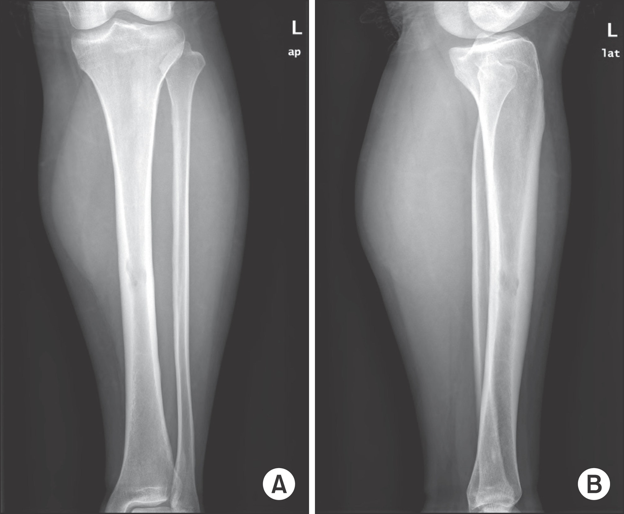
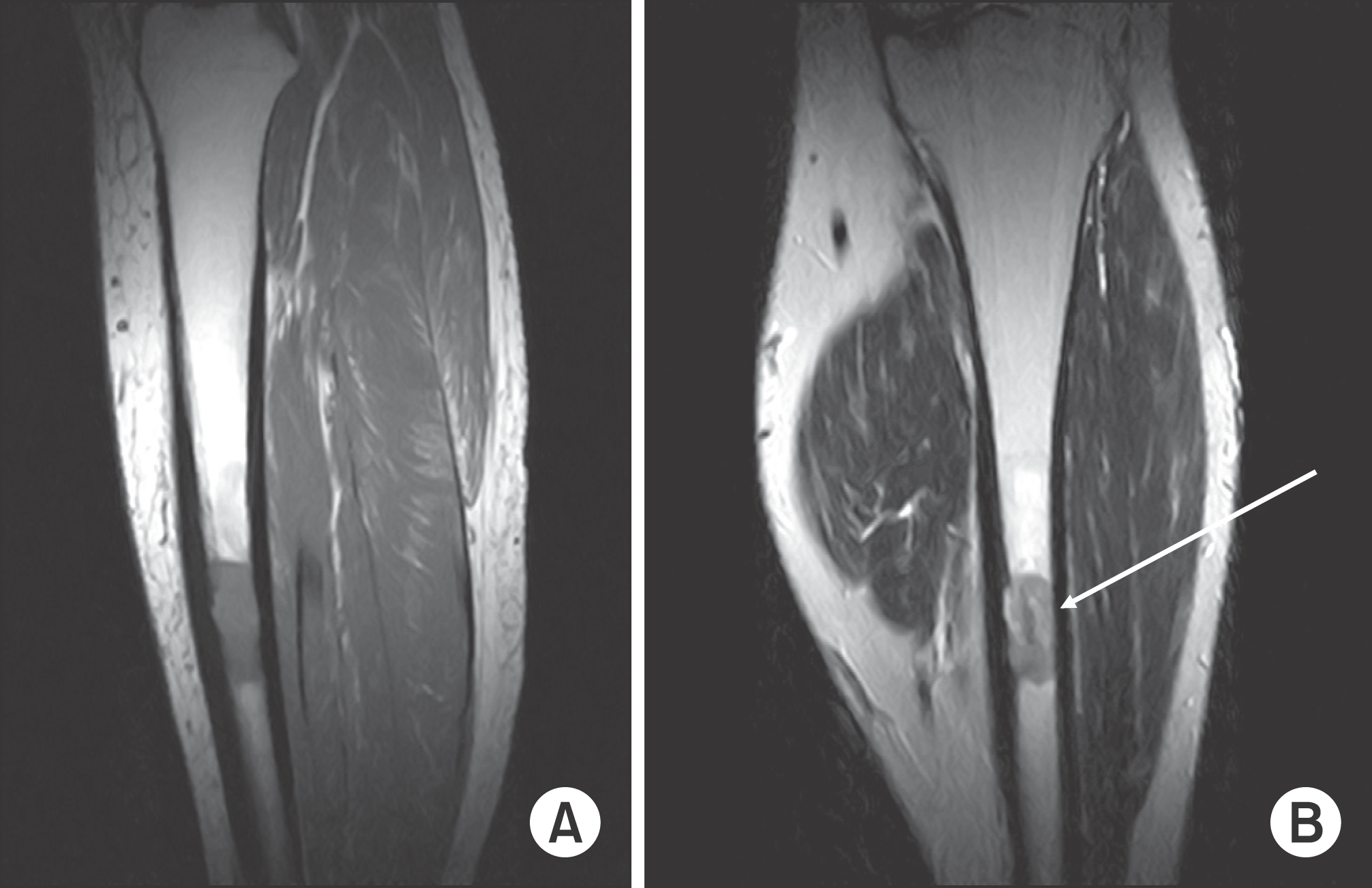
Figure 2.
(A) Sagittal T1-weigthed MR image of lower leg shows bony lesion with low signal intensity. (B) Coronal T2-weighted MR image shows bony lesion with low to intermediate signal intensity.

 XML Download