

 PDF
PDF ePub
ePub Citation
 Print
Print


Abstract
Osteochondromadevelop most commonly at distal femur, proximal humerus and proximal tibia, but the rib osteochondroma was reported less commonly. In this report, scapular snapping syndrome complicated by adventitious bursa and soft tissue pseudotumor surrounding the osteochondroma of the 6th rib body was treated successfully by surgical excision of them. We report this rare case with reviewing the relevant literature.
Go to : 
References
1. Milch H. Snapping scapula. Clin Orthop. 1961; 20:139–50.
2. McCluskey GM III, Bigliani LU. Surgical management of refractoryscapulothoracic bursitis. Otrhop Trans. 1991; 15:801.
3. Milch H. Partial scapulectomy for snapping of the scapula. J Bone Joint Surg Am. 1950; 32-A:561–6.

4. Milch H, Burman MS. Snapping scapula and humerusvarus Report of six cases. Arch Surg. 1933; 26:570–88.
5. Parsons TA. The snapping scapula and subscapular exostoses. J Bone Joint Surg Br. 1973; 55:345–9.
6. Cuomo F, Blank K, Zuckerman JD, Present DA. Scapular osteochondroma presenting with exostosis bursata. Bull Hosp Jt Dis. 1993; 52:55–8.
7. Shogry ME, Armstrong P. Case report 630: Reactive bursa formation surrounding an osteochondroma. Skeletal Radiol. 1990; 19:465–7.
8. Michelle AA, Davies JJ, Krueger FJ, Lichtor JM. Scientific articles. N Y State J Med. 1950; 50:1353–6.
9. Merolla G, Cerciello S, Paladini P, Porcellini G. Snapping scapula syndrome: current concepts review in conservative and surgical treatment. Muscles Ligaments Tendons J. 2013. 380–90.
10. Kuhn JE, Plancher KD, Hawkins RJ. Symptomatic scapulothoracic crepitus and bursitis. J Am Acad Orthop Surg. 1998; 6:267–73.
Go to :
 | Figure 1.On preoperative x-ray about 1.2 cm size exophyticbony mass (white arrow) is noted on the middle part ofthe left 6th rib (A). On MRI about 1.2×1 cm sized exophytic bony lesion (white arrow) in posterolateral arc of the left 6th rib with surrounding loculated fluid-signal like collection (white arrow head) is seen (B). |
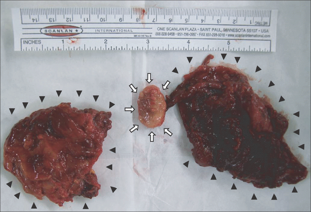 | Figure 2.Excised osteochondroma (white arrow) with surrounding soft tissue (dark arrow head) is shown. The surrounding soft tissue has soft and amorphous shape bursal sac and relatively hard pseudotumor, which cannot be demarcated well. |
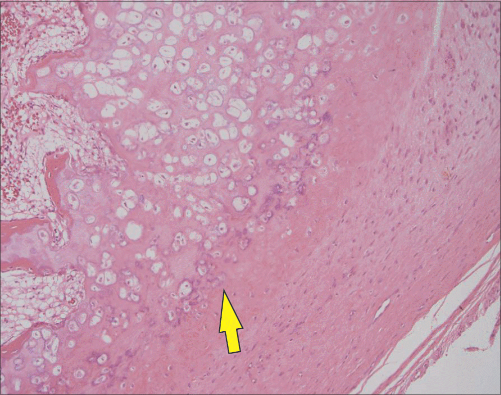 | Figure 3.Histologic section shows thickened bone and cartilage fragment. The mature bone stalk is covered with well differentiated cartilagenuous cap with irregular tidemark (H&E, ×40). |
Table 1.
Review of the Cases Reported in the International Literature with Rib Osteochondroma
 XML Download