Abstract
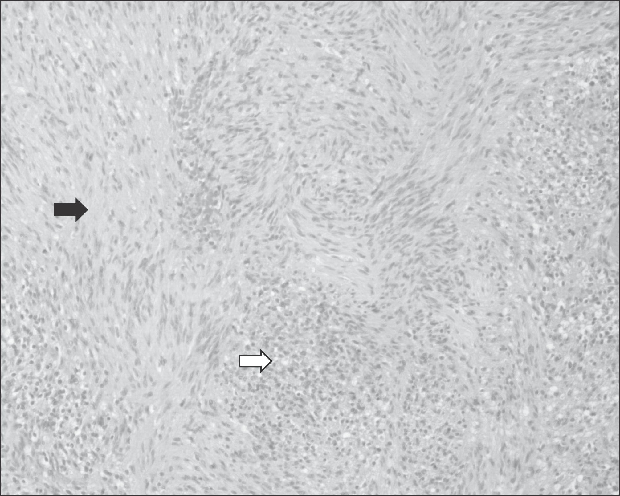
We experienced a case of 63 years old male patient who had synchronous rectus abdominis intramuscular schwannoma and chest wall lipoma. Schwannoma is rare benign tumor which derived from nerve sheath and mainly peripheral nerve of flexor part. The authors report rare synchronous schwannoma and lipoma development.
References
1. Das Gupta TK, Brasfield RD. Tumors of peripheral nerve origin: benign and malignant solitary schwannomas. CA Cancer J Clin. 1970; 20:228–33.
2. Adani R, Baccarani A, Guidi E, Tarallo L. Schwannomas of the upper extremity: diagnosis and treatment. Chir Organi Mov. 2008; 92:85–8.

3. Kehoe NJ, Reid RP, Semple JC. Solitary benign peripheral-nerve tumours. Review of 32 years' experience. J Bone Joint Surg Br. 1995; 77:497–500.

4. Schultz E, Sapan MR, McHeffey-Atkinson B, Naidich JB, Arlen M. Case report 872. "Ancient" schwannoma (degenerated neurilemoma). Skeletal Radiol. 1994; 23:593–5.
6. Lee SH, Jung HG, Lee HK. Neurilemoma of trunk and extremities. J Korean Bone Joint Tumor Soc. 1996; 4:88–93.

7. Bahk WJ RS, Kang YK, Lee AH. Schwannoma of the extremities. J Korean Bone Joint Tumor Soc. 2003; 9:148–54.
8. Pyun YS, Kim SR, Joh YR. Surgical treatment of the neurilemoma in extremities. J Korean Bone Joint Tumor Soc. 1998; 4:88–93.
Figure 2.
(A) Transverse CT image shows well-defined low density soft tissue mass on Rt. anterior chest wall. (B) Transverse CT image shows well-defined mass in rectus abdominis muscle.
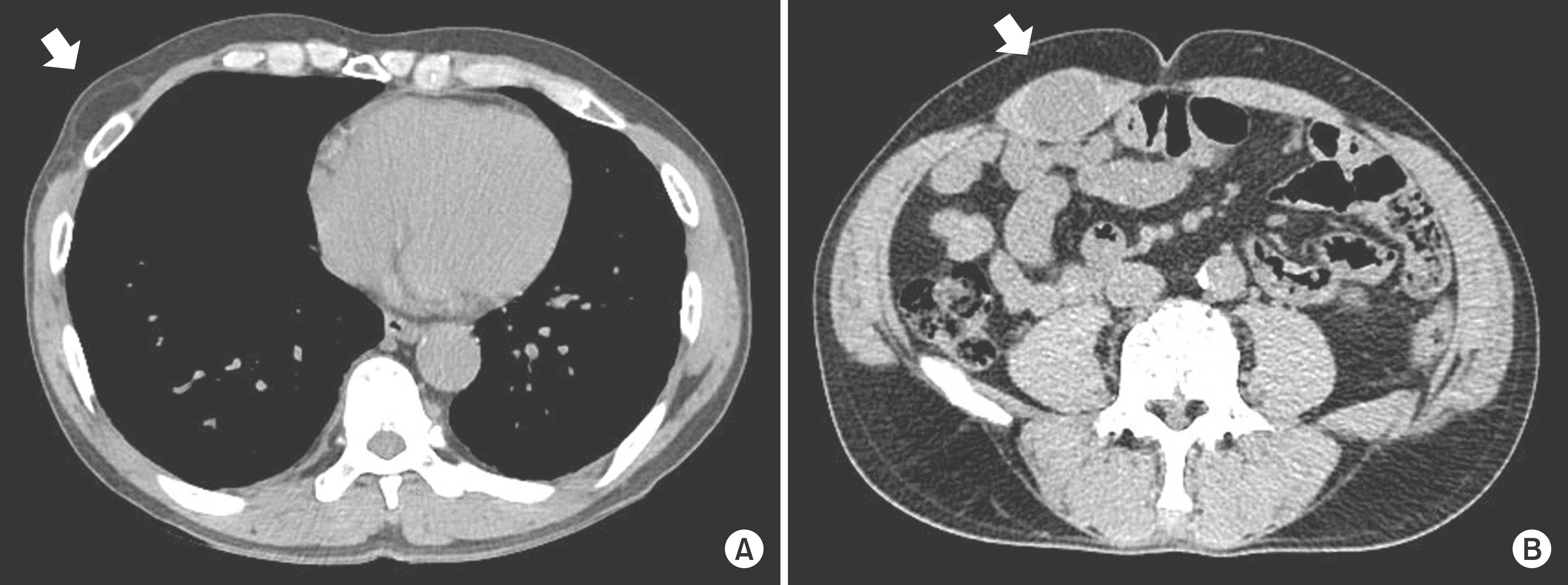
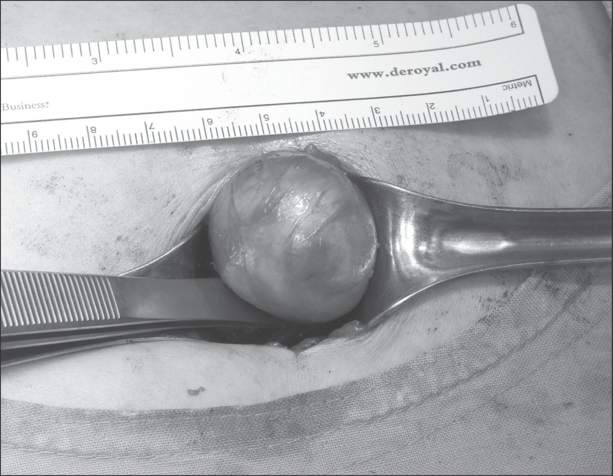
Figure 3.
In the gross finding of the intra-muscular mass in rectus abdominis, a round, pink and well-capsulated mass is shown.


 PDF
PDF ePub
ePub Citation
 Print
Print


 XML Download