

 PDF
PDF ePub
ePub Citation
 Print
Print


Abstract
Bizarre parosteal osteochondromatous proliferation (Nora's lesion) is a rare benign tumor and known to be primarily occur in the small tubular bone of the hands and feet. However, it is very unusual to be reported that it occurs in metatarsal bone in Korea. Thus, we report this tumor of metatarsal bone including the literature review because we have experienced this example.
Go to : 
References
1. Nora FE, Dahlin DC, Beabout JW. Bizarre parosteal osteochondromatous proliferations of the hands and feet. Am J Surg Pathol. 1983; 7:245–50.

2. Meneses MF, Unni KK, Swee RG. Bizarre parosteal osteochondromatous proliferation of bone (Nora's lesion). Am J Surg Pathol. 1993; 17:691–7.
3. Torreggiani WC, Munk PL, Al-Ismail K, et al. MR imaging features of bizarre parosteal osteochondromatous proliferation of bone (Nora's lesion). Eur J Radiol. 2001; 40:224–31.
4. Kim BH, Park YK, Kim YW, Yang MH. Bizarre Parosteal Osteochondromatous Proliferation: A report of five cases. Korean J Pathol. 1996; 30:733–8.
5. van der Walt JD, Ryan JF. Parosteal osteogenic sarcoma of the hand. Histopathology. 1990; 16:75–8.
6. Chung YK, Shin SI, Kim SW, Kim KW. Bizarre parosteal osteochondromatous proliferation of hand – a case report-. J Korean Bone and Joint Tumor Soc. 1998; 4:166–9.
7. Gruber G, Giessauf C, Leithner A, et al. Bizarre parosteal osteochondromatous proliferation (Nora lesion): a report of 3 cases and a review of the literature. Can J Surg. 2008; 51:486–9.
8. Choi JH, Gu MJ, Kim MJ, Choi WH, Shin DS, Cho KH. Fibrosarcoma in bizarre parosteal osteochondromatous proliferation. Skeletal Radiol. 2001; 30:44–7.
Go to :
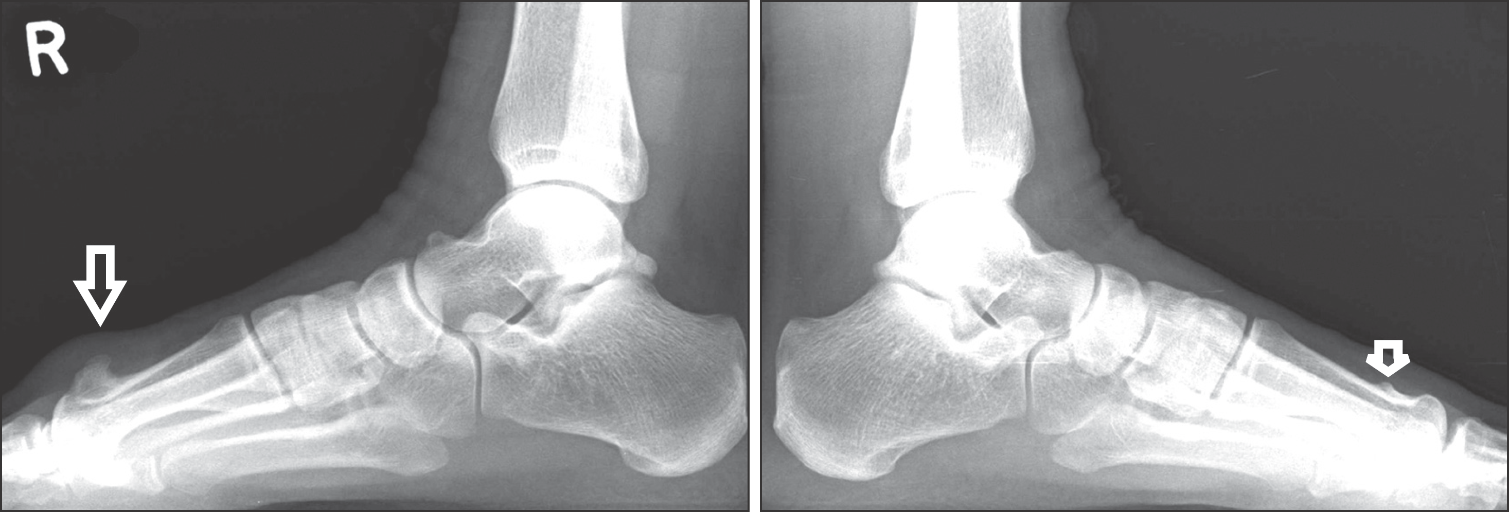 | Figure 1.Arrow at plain radiograph shows a bony mass in the both 1st metatarsal head dorsal area. |
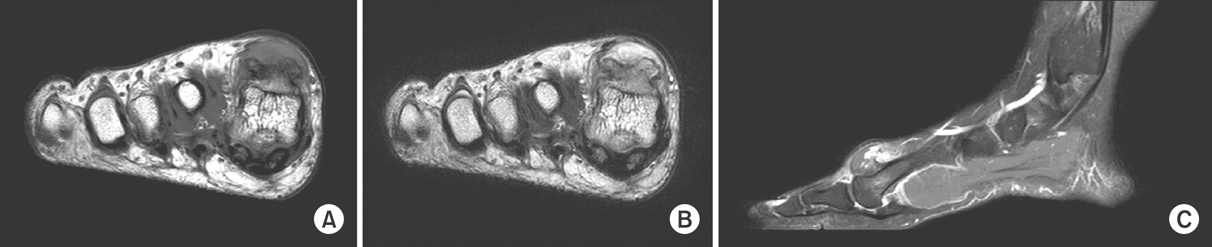 | Figure 2.The MRI shows that the tumor is homogenous appearance and there is no bony deconstruction, medulla abnormality, continuity to bone marrow, that suggests bizarre parosteal osteochondromatous proliferation. (A) T1 enhanced image (B) T2 enhanced image (C) Enhancement of soft tissue and bursa around the tumor in the MRI with intravenous gadolinium contrast suggests inflammation of soft tissue due to irritation from tumor. |

 XML Download