Abstract
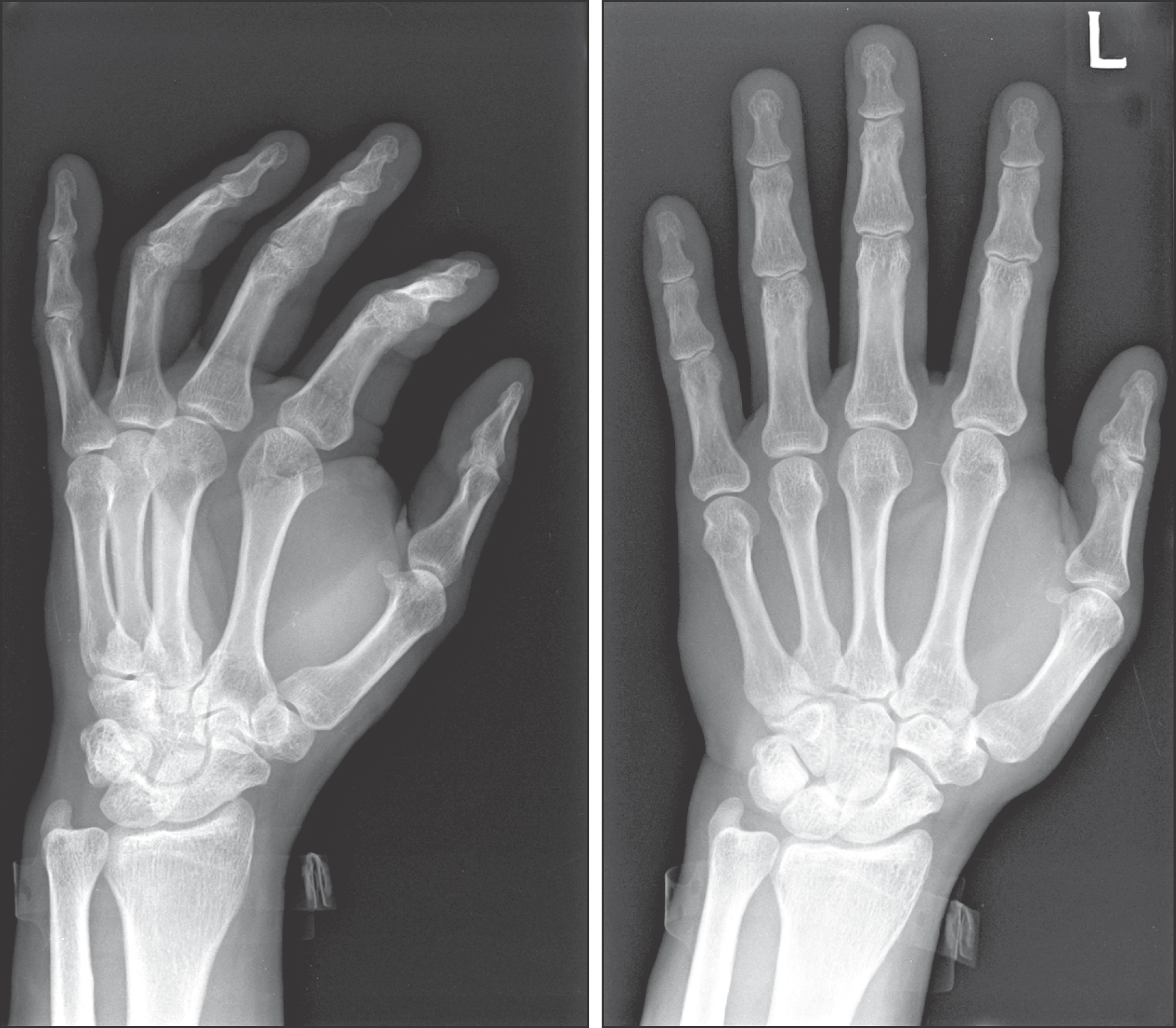
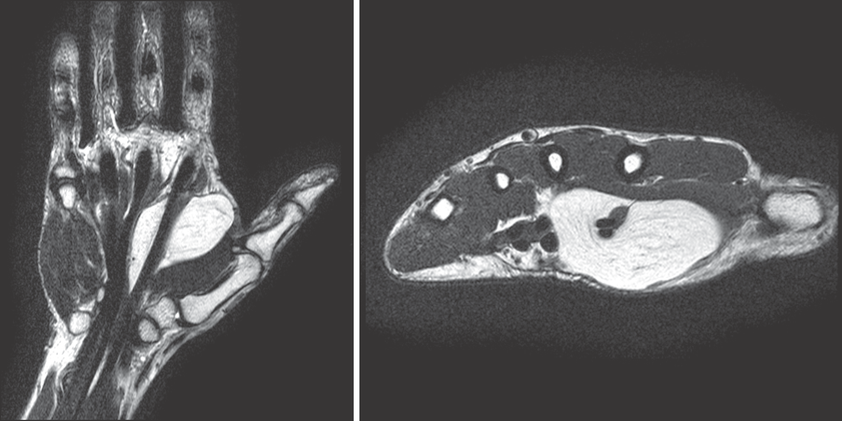
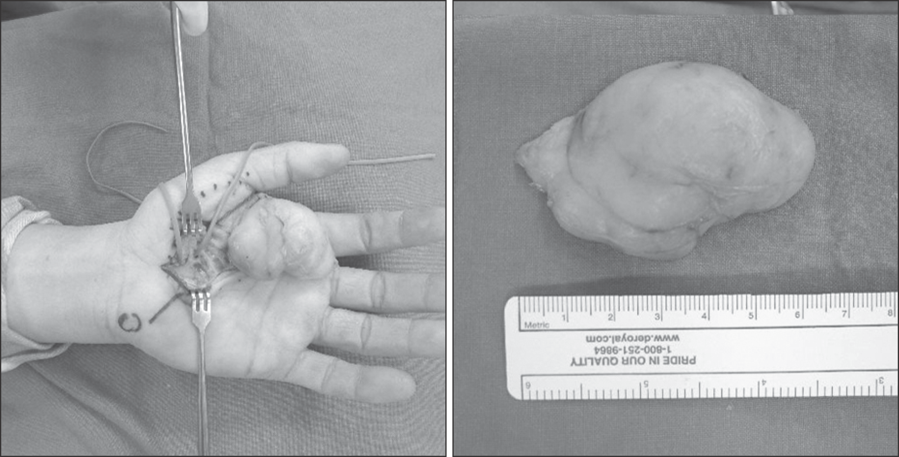
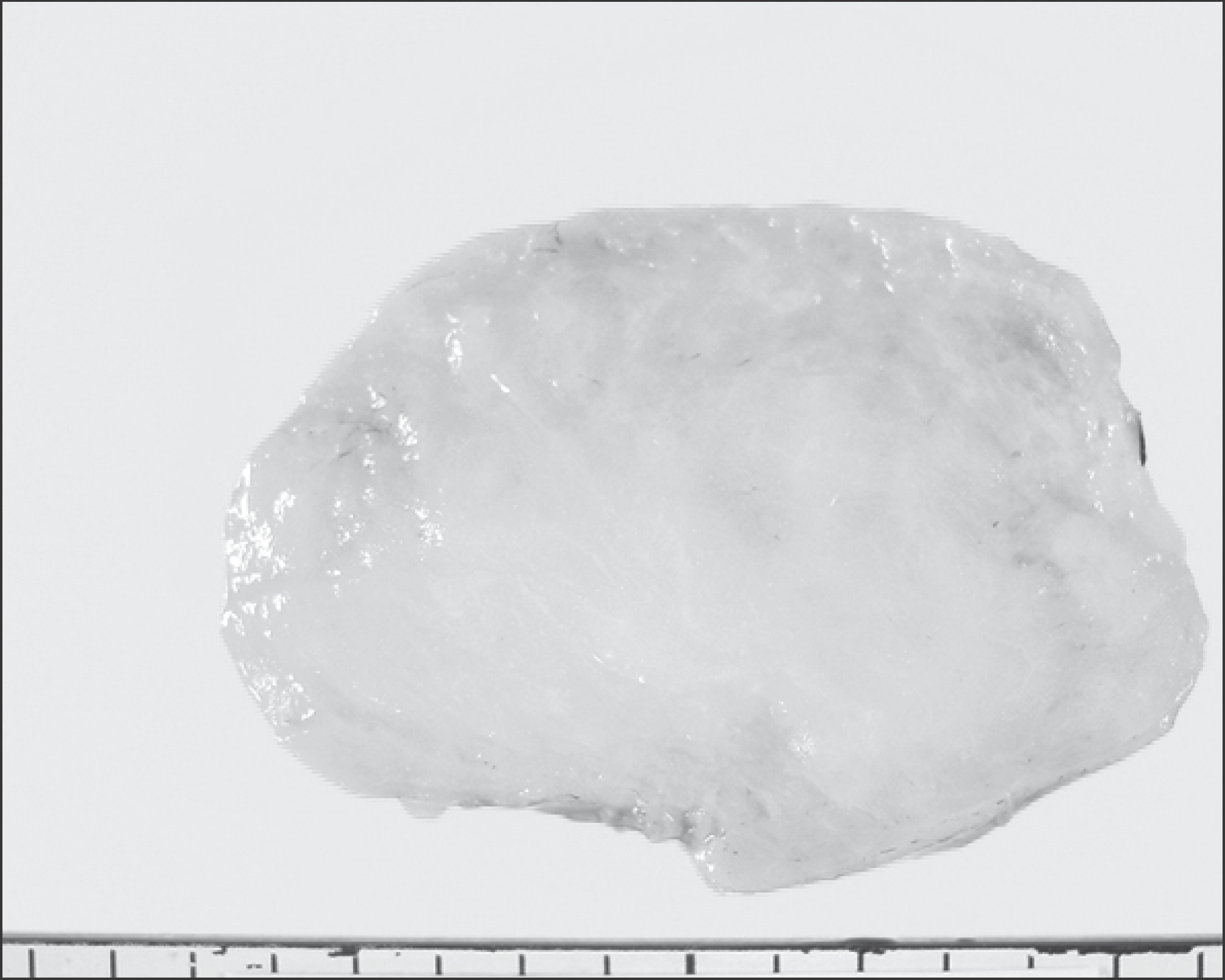
Lipomas are the commonest soft tissue tumor. However, those arsing in the hand are infrequent. Lipomas in the hand that exhibit a size of more than 5 cm call giant lipoma, these are very rare only case reports and small series of this entity have been described. We could experience a case about giant lipoma of the hand which cannot easily contact, we report a case including review of literatures.
References
1. Kamath BJ, Kamath KR, Bhardwaj P, Shridhar , Sharma C. A giant lipoma in the hand – report of a rare case. Online J Health Allied Scs. 2006; 5:1–6.
2. Cribb GL, Cool WP, Ford DJ, Mangham DC. Giant lipomatous tumours of the hand and forearm. J Hand Surg Br. 2005; 30:509–12.

3. Higgs PE, Young VL, Schuster R, Weeks PM. Giant lipomas of the hand and forearm. South Med J. 1993; 86:887–90.

6. Paarlberg D, Linscheid RL, Soule EH. Lipomas of the hand. Including a case of lipoblastomatosis in a child. Mayo Clin Proc. 1972; 47:121–4.
7. Johnson CJ, Pynsent PB, Grimer RJ. Clinical features of soft tissue sarcomas. Ann R Coll Surg Engl. 2001; 83:203–5.
8. Capelastegui A, Astigarraga E, Fernandez-Canton G, Saralegui I, Larena JA, Merino A. Masses and pseudomasses of the hand and wrist: MR findings in 134 cases. Skeletal Radiol. 1999; 28:498–507.

9. Regan JM, Bickel WH, Broders AC. Infiltrating benign lipomas of the extremities. West J Surg Obstet Gynecol. 1946; 54:87–93.


 PDF
PDF ePub
ePub Citation
 Print
Print


 XML Download