

 PDF
PDF ePub
ePub Citation
 Print
Print


Abstract
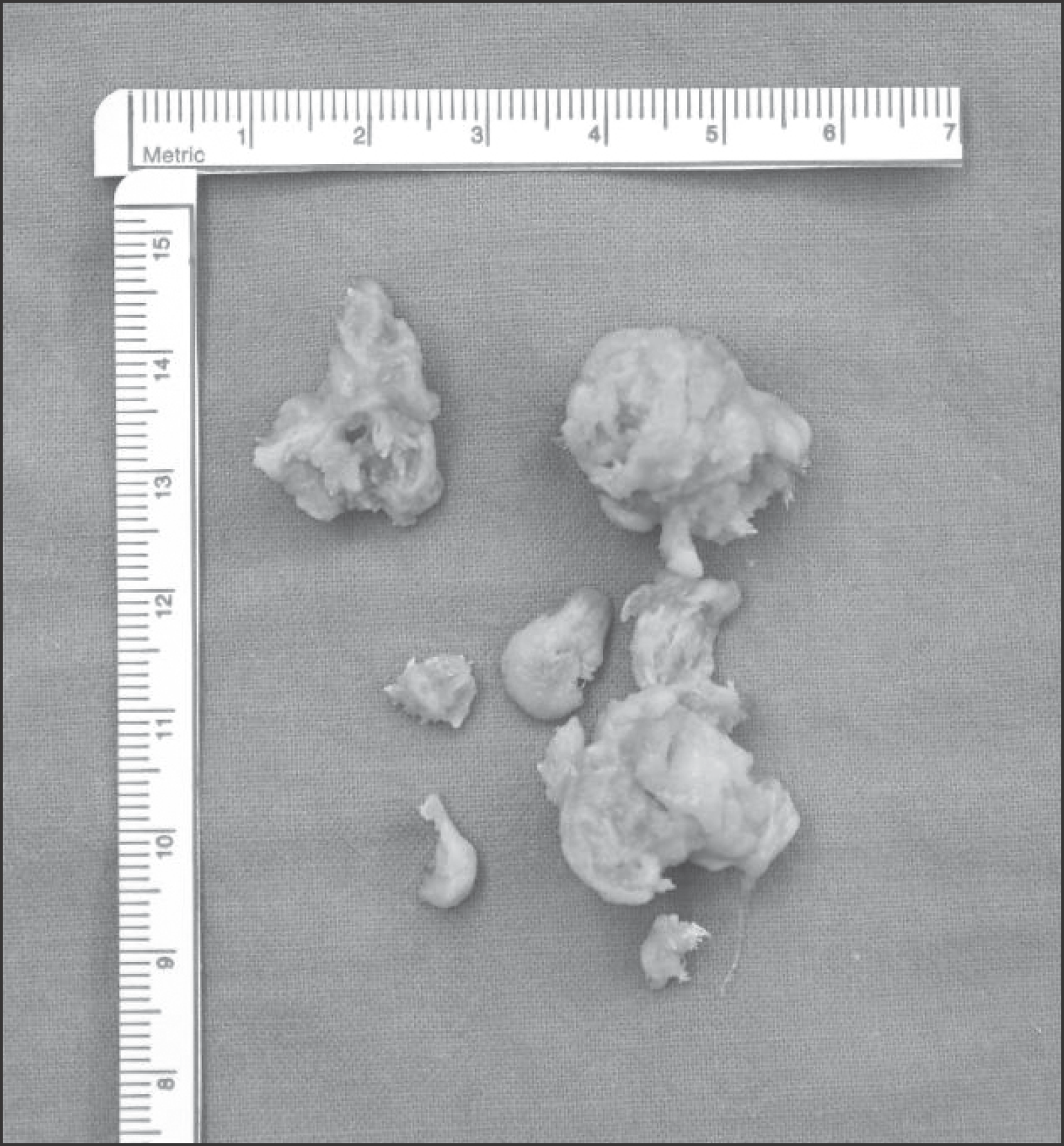
Heterotopic ossification is an abnormal bone formation after surgery or without any reason. Large joint, such as hip and knee joint, is a known most common site. Operation itself and postoperative early range of motion exercise are risk factors. We present a case of heterotopic ossification mimics neurogenic tumor after high tibial osteotomy.
References
1. Harwin SF, Stein AJ, Stern RE, Kulick RG. Heterotopic ossification following primary total knee arthroplasty. J Arthroplasty. 1993; 8:113–6.

2. Cook J, Scott RD. Bony ankylosis following total knee arthroplasty: a case report. J Arthroplasty. 2005; 20:122–4.
3. Furia JP, Pellegrini VD Jr. Heterotopic ossification following primary total knee arthroplasty. J Arthroplasty. 1995; 10:413–9.
4. Hasegawa M, Ohashi T, Uchida A. Heterotopic ossification around distal femur after total knee arthroplasty. Arch Orthop Trauma Surg. 2002; 122:274–8.
5. Figgie HE 3rd, Goldberg VM, Heiple KG, Moller HS 3rd, Figgie MP. The incidence and significance of heterotopic ossification following total knee arthroplasty. Adv Orthop Surg. 1986; 10:12–7.
6. Mollan RA. Serum alkaline phosphatase in heterotopic para-articular ossification after total hip replacement. J Bone Joint Surg Br. 1979; 61B:432–4.
7. Isobe K, Shimizu T, Akahane T, Kato H. Imaging of ancient schwannoma. AJR Am J Roentgenol. 2004; 183:331–6.
8. Pape HC, Marsh S, Morley JR, Krettek C, Giannoudis PV. Current concepts in the development of heterotopic ossification. J Bone Joint Surg Br. 2004; 86:783–7.
Figure 1.
Plain radiograph of the right knee showing 2×2 cm sized calcified mass in the posterior knee joint (arrow).
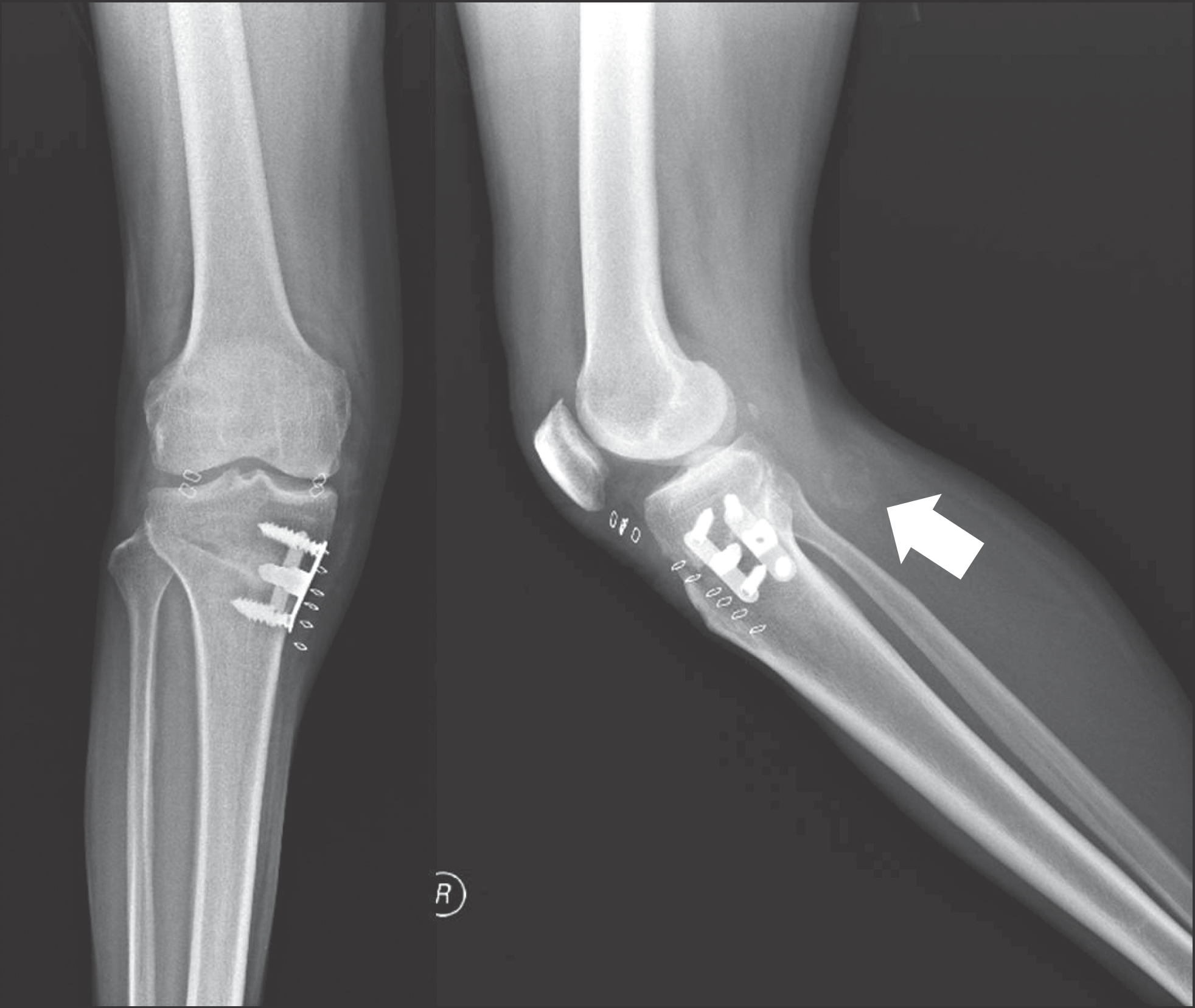
Figure 3.
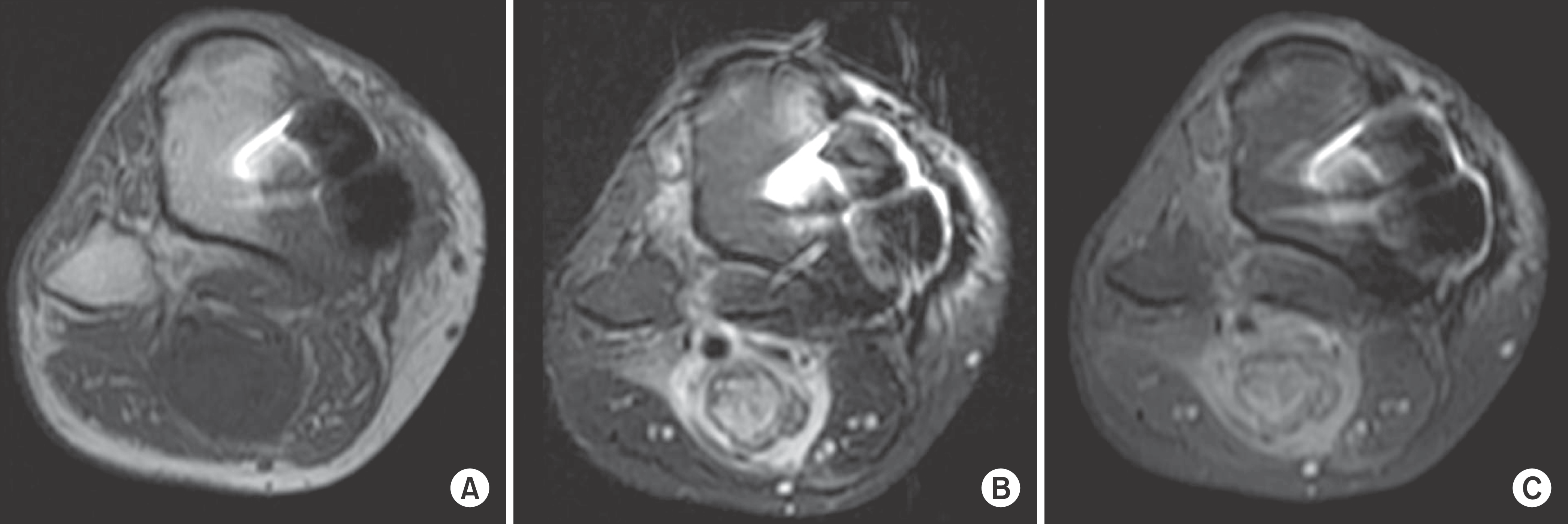
Preoperative magnetic resonance image shows 3×2.5×2 cm sized tumor arising from tibial nerve in popliteal region. (A) Axial T1weighed MR image shows inhomogeneous low signal intensity. (B) Axial T2 weighted MR image shows heterogeneous high signal intensity. (C) Axial gadolinum enhanced MR image shows heterogeneous enhanced mass.

 XML Download