

 PDF
PDF ePub
ePub Citation
 Print
Print


Abstract
Ganglion cysts that occurred within sheaths of peripheral nerves have been documented in literatures, but it is relatively rare condition. The peroneal nerve is the most common site of involvement. Other reported sites of involvement are the radial, ulnar, median, sciatic, tibial and posterior interosseous nerves. We report a case of the intraneual ganglion cyst within peroneal nerve and another case of the intraneural ganglion cyst within a medial plantar nerve that were successfully excised without neurologic complication.
Go to : 
References
1. Coleman SH, Beredjeklian PK, Weiland AJ. Intraneural ganglion cyst of the peroneal nerve accompanied by complete foot drop. A case report. Am J Sports Med. 2001; 29:238–41.
2. Patel P, Schucany WG. A rare case of intraneural ganglion cyst involving the tibial nerve. Proc (Bayl Univ Med Cent). 2012; 25:132–5.

3. Nucci F, Artico M, Santoro A, et al. Intraneural synovial cyst of the peroneal nerve: report of two cases and review of the literature. Neurosurgery. 1990; 26:339–44.
4. Yamazaki H, Saitoh S, Seki H, Murakami N, Misawa T, Takaoka K. Peroneal nerve palsy caused by intraneural ganglion. Skeletal Radiol. 1999; 28:52–6.
5. Coakley FV, Finlay DB, Harper WM, Allen MJ. Direct and indirect MRI findings in ganglion cysts of the common peroneal nerve. Clin Radiol. 1995; 50:168–9.
6. Fansa H, Plogmeier K, Gonschorek A, Feistner H. Common peroneal nerve palsy caused by a ganglion. Case report. Scand J Plast Reconstr Surg Hand Surg. 1998; 32:425–7.
7. Lowenstein J, Towers J, Tomaino MM. Intraneural ganglion of the peroneal nerve: importance of timely diagnosis. Am J Orthop (Belle Mead NJ). 2001; 30:816–9.
8. Spinner RJ, Atkinson JL, Scheithauer BW, et al. Peroneal intraneural ganglia: the importance of the articular branch. Clinical series. J Neurosurg. 2003; 99:319–29.
9. Miskovsky S, Kaeding C, Weis L. Proximal tibiofibular joint ganglion cysts: excision, recurrence, and joint arthrodesis. Am J Sports Med. 2004; 32:1022–8.
Go to :
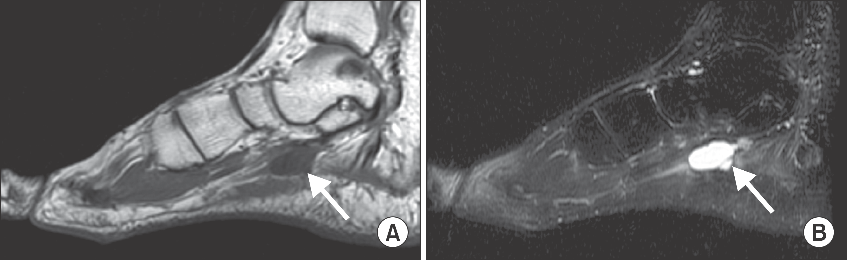 | Figure 1.Sagittal T1 weighted (A), Sagittal T2 weighted (B) MR images show intraneural ganglion within medial plantar nerve (arrow). |
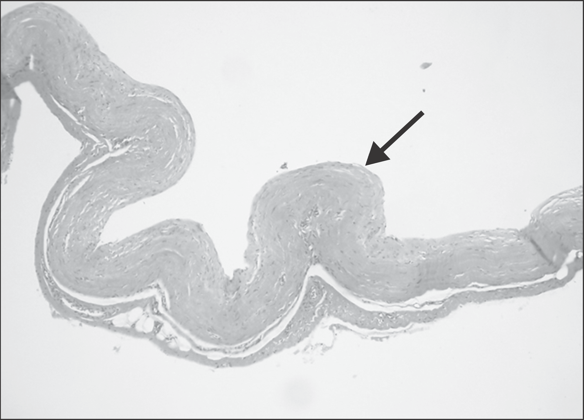 | Figure 2.Light micrographs shows a fibrous connective tissue that is a portion of cyst wall (arrow). The cystic wall is not lined by synovial cells (H & E stain, ×100). |

 XML Download