

 PDF
PDF ePub
ePub Citation
 Print
Print


Abstract
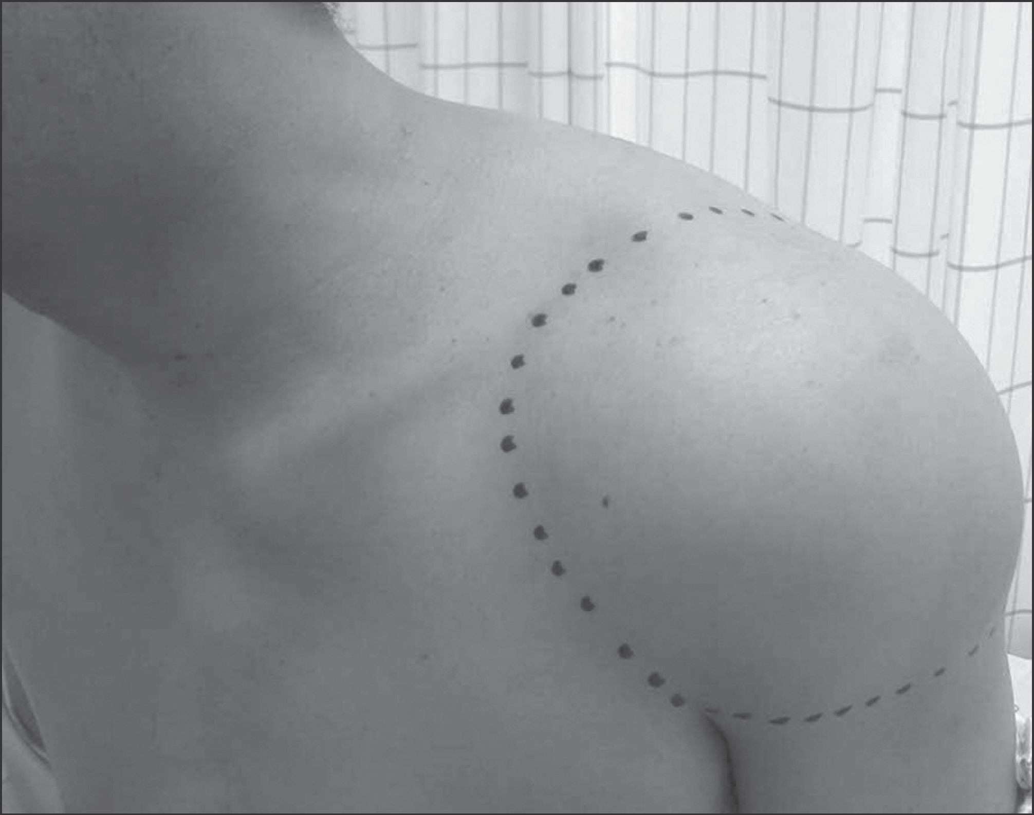
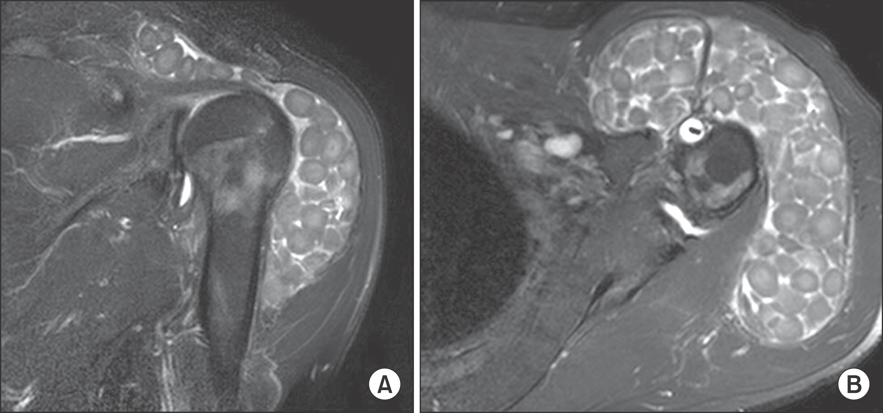
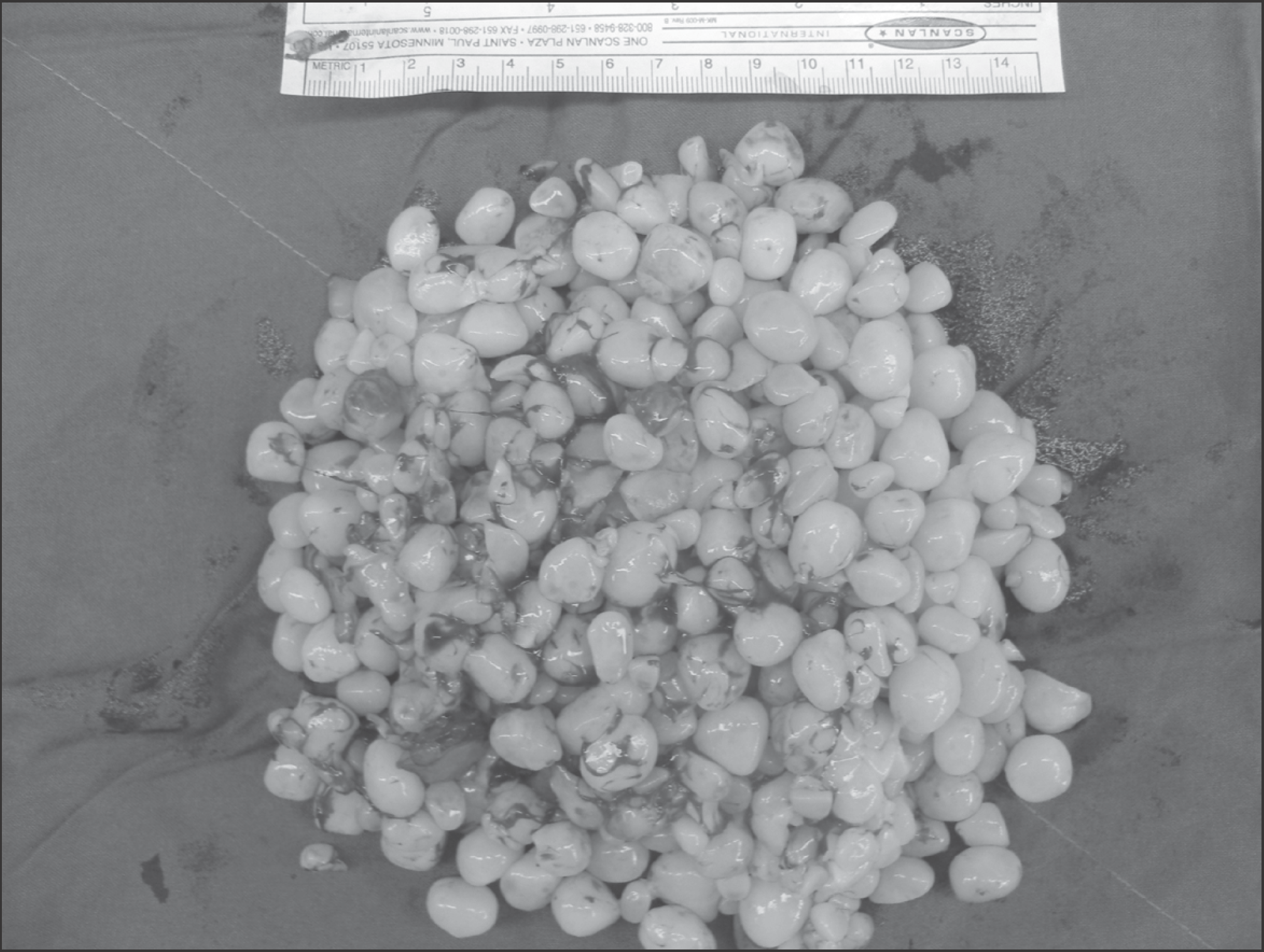
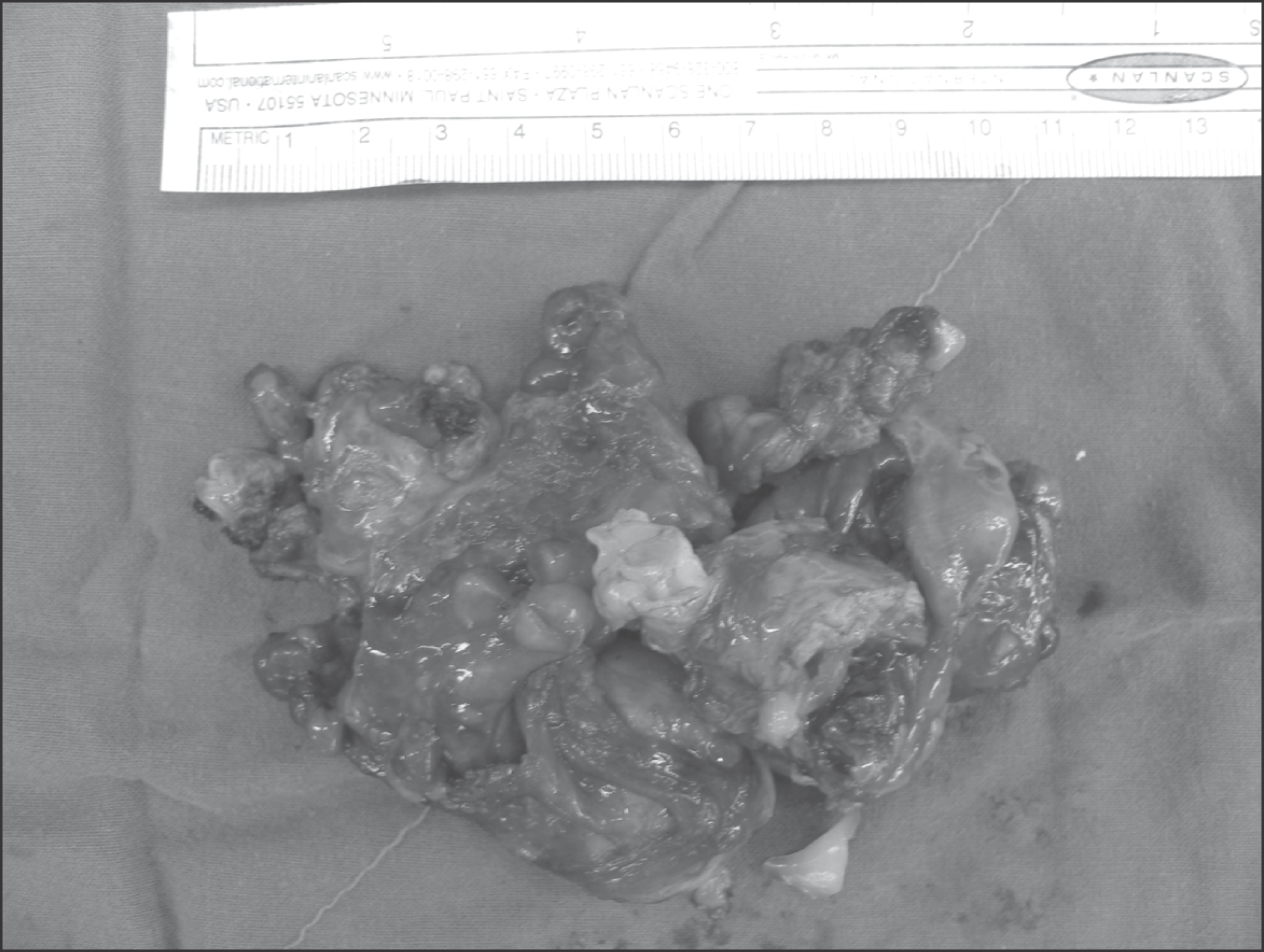
Multiple rice body formation is a complication of chronic bursitis frequently associated with seronegative rheumatoid arthritis or tuberculosis. It resembles synovial chondromatosis on imaging and clinically. We report on a pathologically diagnosed multiple rice body formation in subacromial and subdeltoid bursitis in a 44-year-old man who was treated by surgical removal and bursectomy. At 16 months after the removal, range of motion of affected shoulder was normal. No evidence of recurrence of rice body in plain X-ray and ultrasonography. Multiple rice body formed in chronic subacromial and subdeltoid bursitis could be treated with surgical removal and bursectomy successfully.
Go to : 
References
1. Chen A, Wong LY, Sheu CY, Chen BF. Distinguishing multiple rice body formation in chronic subacromial-subdeltoid bursitis from synovial chondromatosis. Skeletal Radiol. 2002; 31:119–21.

2. Mutlu H, Silit E, Pekkafali Z, et al. Multiple rice body formation in the subacromial-subdeltoid bursa and knee joint. Skeletal Radiol. 2004; 33:531–3.
3. Kim RS, Lee JY, Jung SR, Lee KY. Tuberculous subdeltoid bursitis with rice bodies. Yonsei Med J. 2002; 43:539–42.
4. Königshausen M, Seybold D, Heyer CM, Muhr G, Gekle C. Tuberculous rice body synovitis of the shoulder joint. Orthopade. 2009; 38:1106–12.
5. Cheung HS, Ryan LM, Kozin F, McCarty DJ. Synovial origins of Rice bodies in joint fluid. Arthritis Rheum. 1980; 23:72–6.
6. Popert AJ, Scott DL, Wainwright AC, Walton KW, Williamson N, Chapman JH. Frequency of occurrence, mode of development, and significance or rice bodies in rheumatoid joints. Ann Rheum Dis. 1982; 41:109–17.
7. Bruggeman NB, Sperling JW, Shives TC. Arthroscopic technique for treatment of synovial chondromatosis of the glenohumeral joint. Arthroscopy. 2005; 21:633.
8. Fowble VA, Levy HJ. Arthroscopic treatment for synovial chondromatosis of the shoulder. Arthroscopy. 2003; 19:E2.
9. Tokis AV, Andrikoula SI, Chouliaras VT, Vasiliadis HS, Georgoulis AD. Diagnosis and arthroscopic treatment of primary synovial chondromatosis of the shoulder. Arthroscopy. 2007; 23:1023. .e1–5.
10. Moosikasuwan JB, Miller TT, Burke BJ. Rotator cuff tears: clinical, radiographic, and US findings. Radiographics. 2005; 25:1591–607.
Go to :

 XML Download