

 PDF
PDF ePub
ePub Citation
 Print
Print


Abstract
Extraskeletal chondroma is a benign soft tissue tumor which is composed of hyaline cartilage but arises from the fibrous stroma rather than from mature cartilaginous or osseous tissue. Extraskeletal chondroma is relatively rare and occurs most frequently in the soft tissue around the joints of hands and feet. We present one case of extraskeletal chondroma in a finger of a young woman.
References
1. Kransdorf MJ, Meis JM. From the archives of the AFIP. Extraskeletal osseous and cartilaginous tumors of the extremities. Radiographics. 1993; 13:853–84.

2. Le Corroller T, Bouvier-Labit C, Champsaur P. Diffuse mineralization of forearm extraskeletal chondroma. Joint Bone Spine. 2008; 75:479–81.
3. De Riu G, Meloni SM, Gobbi R, Contini M, Tullio A. Soft-tissue chondroma of the masticatory space. Int J Oral Maxillofac Surg. 2007; 36:174–6.
4. Kudawara I, Ueda T, Araki N. Extraskeletal chondroma around the knee. Clin Radiol. 2001; 56:779–82.
5. Adaletli I, Laor T, Yin H, Podberesky DJ. Extraskeletal chondroma: another diagnostic possibility for a soft tissue axillary mass in an adolescent. Case Rep Orthop. 2011; 2011:309328.
6. Papagelopoulos PJ, Savvidou OD, Mavrogenis AF, Chloros GD, Papaparaskeva KT, Soucacos PN. Extraskeletal chondroma of the foot. Joint Bone Spine. 2007; 74:285–8.
7. Singh R, Sharma AK, Magu NK, Kaur KP, Sen R, Magu S. Extraskeletal osteochondroma in the nape of the neck: a case report. J Orthop Surg (Hong Kong). 2006; 14:192–5.
Figure 1.
Photograph shows a protruding mass in radial aspect of the right 5th middle phalanx. (A) AP view (B) lateral view.
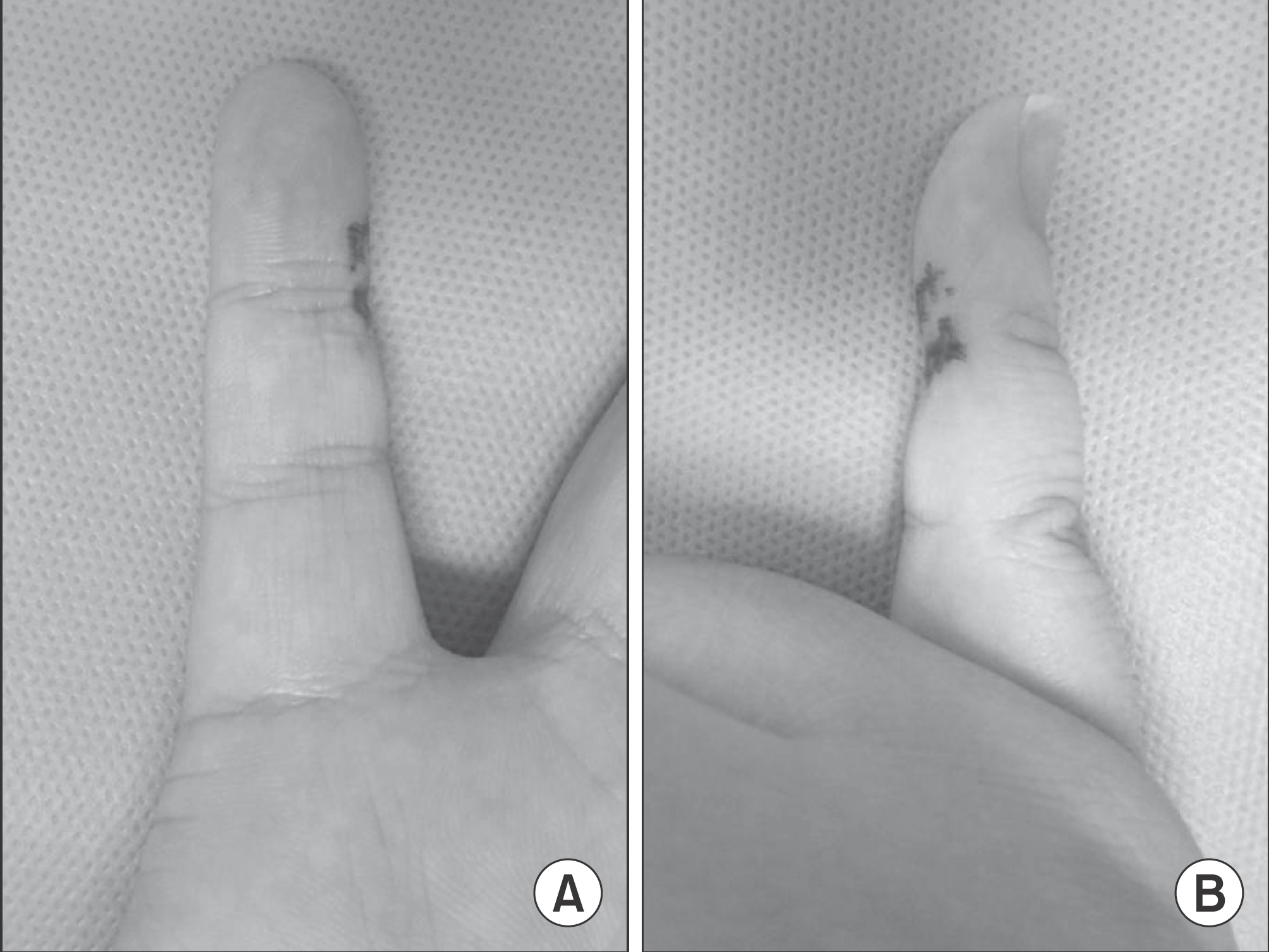
Figure 3.
Ultrasonograph shows an ovoid-shaped non-echoic lesion in right 5th finger middle phalanx, which measures 0.3×0.3×1.0 cm. (A) Axial view, (B) Saggital view.
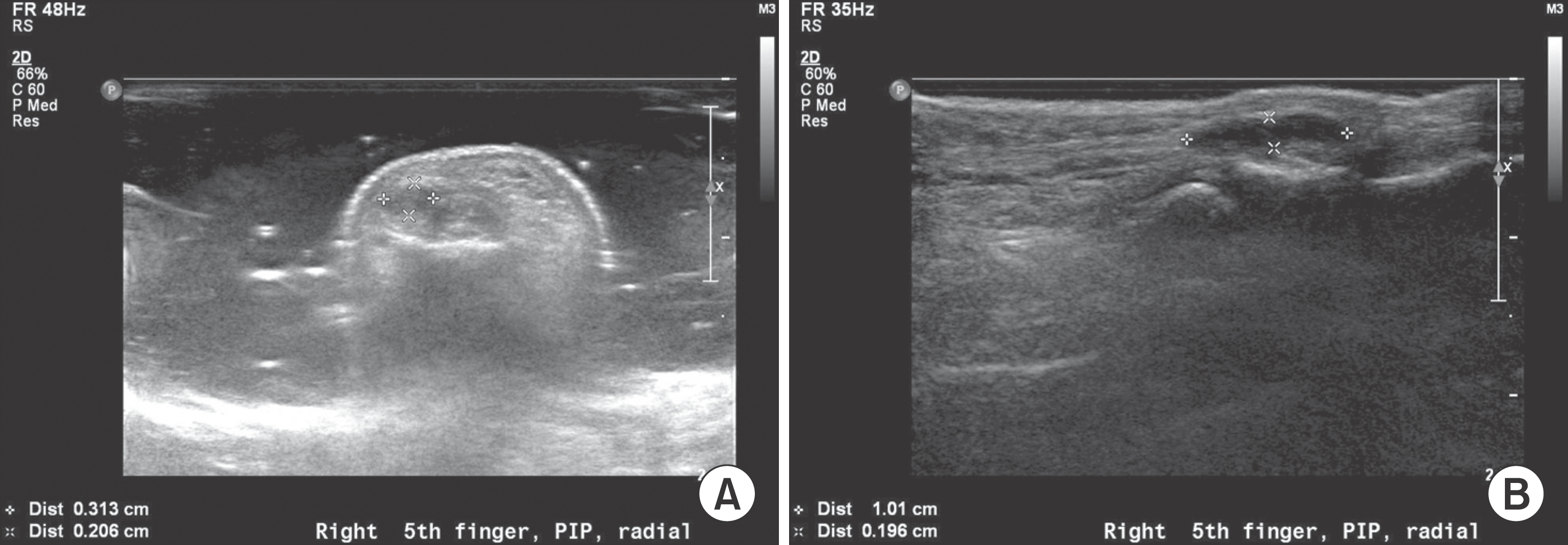
Figure 4.
MRI shows a well demarcated ovoid mass of T1 low to intermediate and T2 intermediate signal intensity. There was marked diffuse heterogeneous enhancement throughout the lesion following the administration of intravenous contrast.
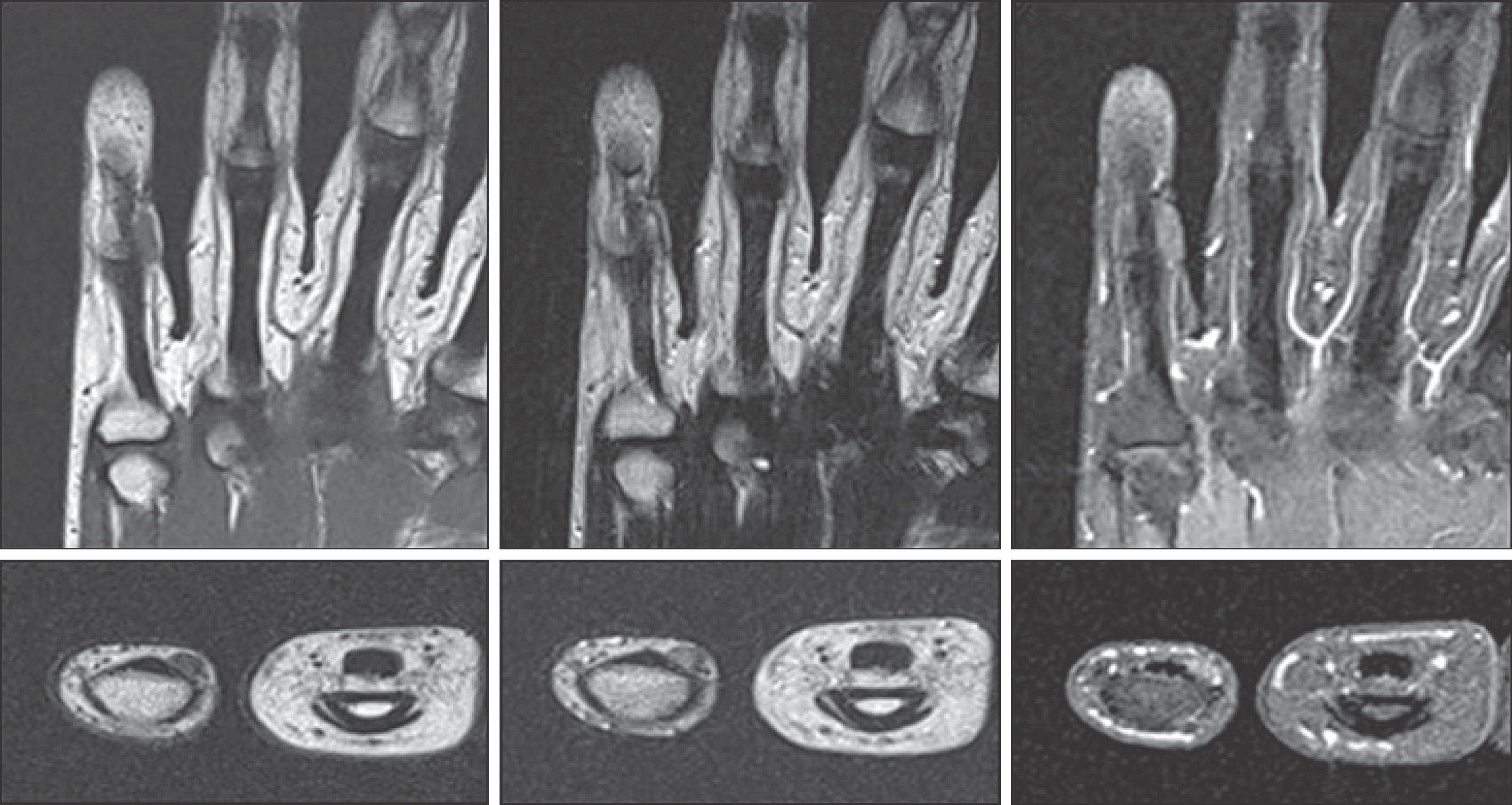
Figure 5.
Photographs show a gray-whitish, nodular soft tissue mass. The tumor rested upon the digital nerve.
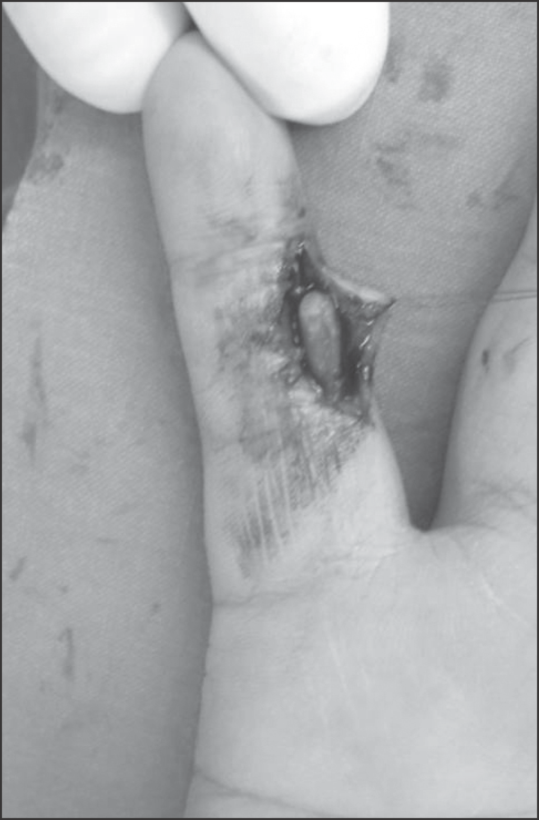
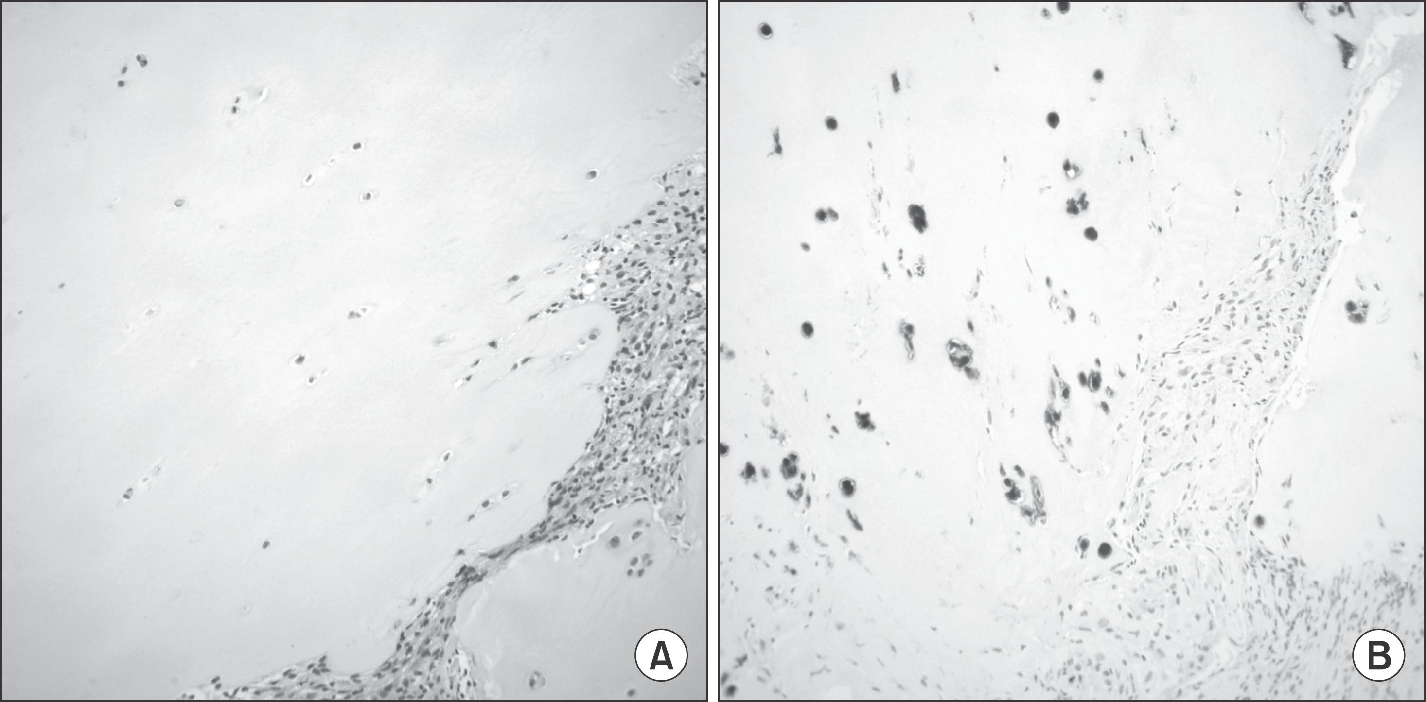
Figure 6.
Pathologic features of extra-skeletal chondroma. (A) The tumor consists of the circumscribed lobules of mature hyaline cartilage. The chondrocytic tumor cells are identified in lacunae (Hematoxylin & Eosin, ×200). (B) The tumor cells of the extraskeletal chondroma are positive with S100 protein as with normal chondrocytes (Immunohistochemistry, ×200).
 XML Download