

 PDF
PDF ePub
ePub Citation
 Print
Print


Abstract
Materials and Methods
We retrospectively reviewed 7 patients with giant cell tumor of the sacrum who were treated at out institution between 1990 and 2012.
Results
There were 2 men and 5 women with mean age of 23.6 years. The average follow up was 52.3 months (range, 15–73 months). Six patients received surgical treatment. Intralesional curettage was performed for the 5 patients and marginal resection for another one patient. The remaining one patient was received radiation only. The patients who received radiation therapy and marginal excision had no residual or recurrent tumors. Of 5 patients with intra-lesional excision, one patient needs one more operation; two patients need two more operation for local control of the giant cell tumor. The remaining two patients failed to gain local control in spite of additional treatments.
References
1. Sung HW, Shu WP, Wang HM, Yuai SY, Tsai YB. Surgical treatment of primary tumors of the sacrum. Clin Orthop Relat Res. 1987; 215:91–8.

2. Turcotte RE. Giant cell tumor of bone. Orthop Clin North Am. 2006; 37:35–51.
3. Campanacci M, Baldini N, Boriani S, Sudanese A. Giant-cell tumor of bone. J Bone Joint Surg Am. 1987; 69:106–14.
4. Feigenberg SJ, Marcus Jr RB, Zlotecki RA, Scarborough MT, Berrey BH, Enneking WF. Radiation therapy for giant cell tumors of bone. Clin Orthop Relat Res. 2003; 411:207–16.
5. McDonald DJ, Sim FH, McLeod RA, Dahlin DC. Giant-cell tumor of bone. J Bone Joint Surg Am. 1986; 68:235–42.
6. Sung HW, Kuo DP, Shu WP, Chai YB, Liu CC, Li SM. Giantcell tumor of bone: analysis of two hundred and eight cases in Chinese patients. J Bone Joint Surg Am. 1982; 64:755–61.
7. Guo W, Ji T, Tang X, Yang Y. Outcome of conservative surgery for giant cell tumor of the sacrum. Spine (Phila Pa 1976). 2009; 34:1025–31.
8. Leggon RE, Zlotecki R, Reith J, Scarborough MT. Giant cell tumor of the pelvis and sacrum: 17 cases and analysis of the literature. Clin Orthop Relat Res. 2004; 423:196–207.
9. Thangaraj R, Grimer RJ, Carter SR, Stirling AJ, Spilsbury J, Spooner D. Giant cell tumour of the sacrum: a suggested algorithm for treatment. Eur Spine J. 2010; 19:1189–94.
10. Martin C, McCarthy EF. Giant cell tumor of the sacrum and spine: series of 23 cases and a review of the literature. Iowa Orthop J. 2010; 30:69–75.
11. Rock MG, Sim FH, Unni KK, et al. Secondary malignant giantcell tumor of bone. Clinicopathological assessment of nineteen patients. J Bone Joint Surg Am. 1986; 68:1073–9.
12. Laskin WB, Silverman TA, Enzinger FM. Postradiation soft tissue sarcomas. An analysis of 53 cases. Cancer. 1988; 62:2330–40.
13. Dray MS, Miller MV. Paget's osteosarcoma and post-radiation osteosarcoma: secondary osteosarcoma at Middlemore Hospital, New Zealand. Pathology. 2008; 40:604–10.
14. Kong CB, Hong YS, Lee KY, et al. Malignant transformation of benign giant cell tumor. J Korean Bone Joint Tumor Soc. 2012; 18:14–9.
15. Lackman RD, Khoury LD, Esmail A, Donthineni-Rao R. The treatment of sacral giantcell tumours by serial arterial embolisation. J Bone Joint Surg Br. 2002; 84:873–7.
16. Hosalkar HS, Jones KJ, King JJ, Lackman RD. Serial arterial embolization for large sacral giantcell tumors: mid- to longterm results. Spine (Phila Pa 1976). 2007; 32:1107–15.
17. Bertoni F, Present D, Sudanese A, Baldini N, Bacchini P, Campanacci M. Giant-cell tumor of bone with pulmonary metastases. Six case reports and a review of the literature. Clin Orthop Relat Res. 1988; 237:275–85.
18. Thomas D, Henshaw R, Skubitz K, et al. Denosumab in patients with giantcell tumour of bone: an open-label, phase 2 study. Lancet Oncol. 2010; 11:275–80.
19. Branstetter DG, Nelson SD, Manivel JC, et al. Denosumab induces tumor reduction and bone formation in patients with giantcell tumor of bone. Clin Cancer Res. 2012; 18:4415–24.
Figure 1.
Fourteen-year-old woman suffered from a perianal numbness and defecation difficulty. (A, B) Initial simple lateral X-ray and T2 weighted sagittal MRI shows a giant cell tumor at S2-S3. (C, D) 5 years later after the radiation treatment, Simple lateral X-ray and T2 weighted sagittal MRI shows sclerotic change and stationary lesion.
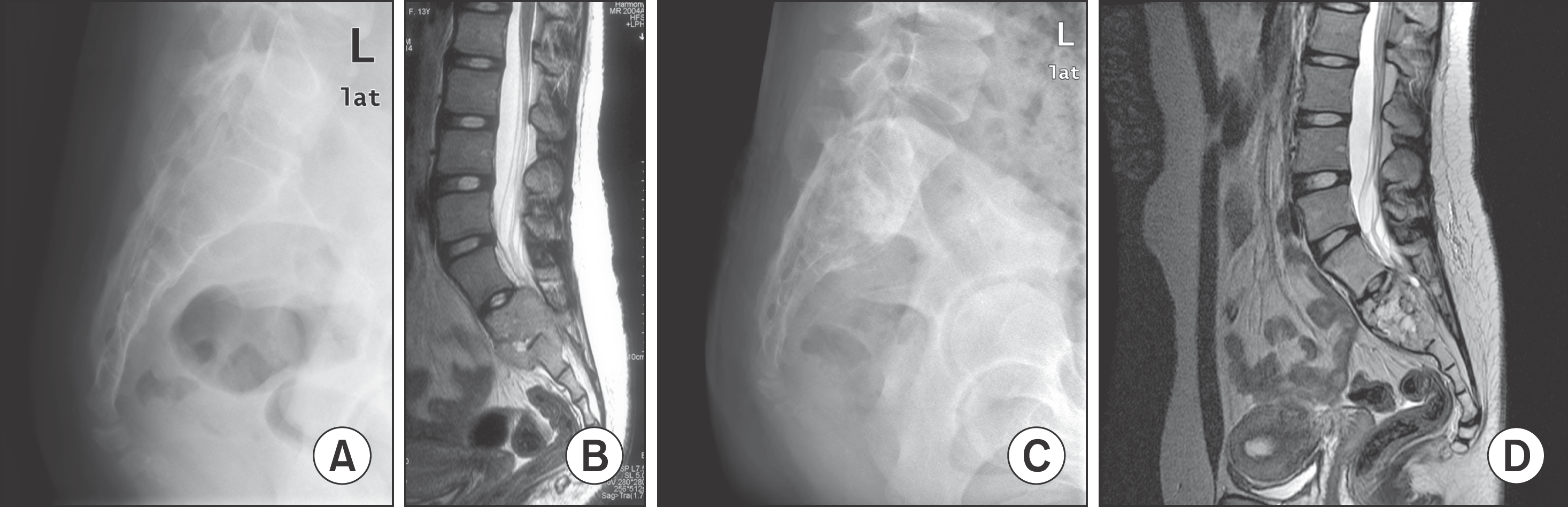
Figure 2.
Twenty five-year-old woman complained about her right hip pain for two years. (A) Pre-operative plain radiograph, (B) CT, (C) Pre-operative T2 weighted MRI, and (D) Post-operative X-ray. There was no recurrence of giant cell tumor until last follow up.

Figure 3.
Thirty one-year-old woman suffered from radiating pain for four months. (A) Pre-operative lateral X-ray and (B) pre-operative T2 weighted MRI show giant cell tumor at S2-S4 level. (C) Immediate post-operative X-ray shows packed gauzes due to massive intra-operative bleeding. (D) 4 years later after the operation, MRI shows excised coccyx at first operation and remnant tumors. So, re-curettage was performed. (E) 5 years after the first operation, T2 weighted MRI shows no residual or recurrent tumor.
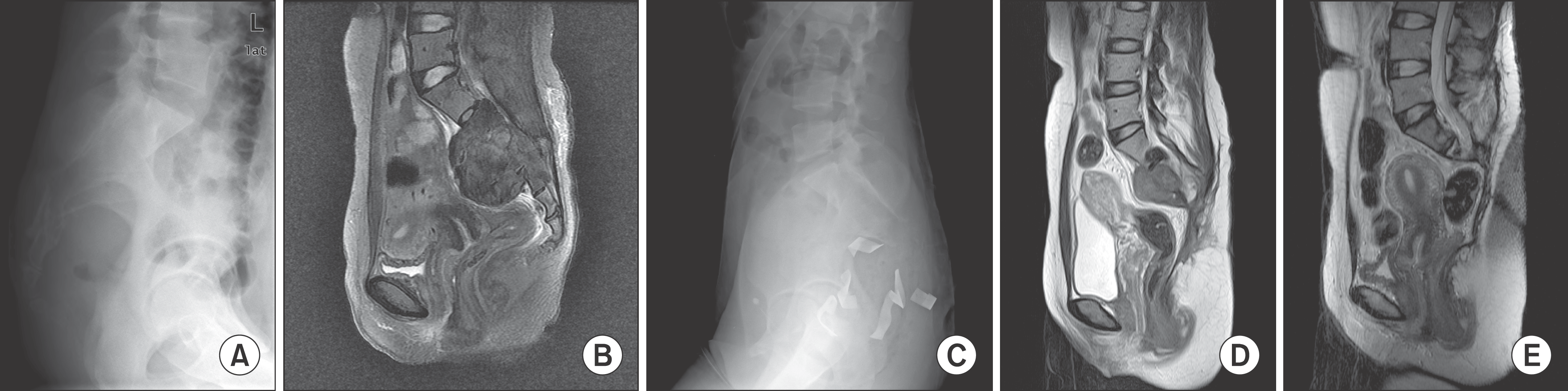
Figure 4.
Eighteen year-old women developed pelvic pain about two years ago. Three months ago, weakness of lower extremity was developed. (A) Pre-operative T2 weighted MRI shows giant cell tumor at S2-S3 level. (B) 3 months later after the operation, T2 weighted MRI shows recurrent lesions. She received radiation therapy twice. (C) T1 fat suppression enhanced MRI after the radiation treatment shows aggravated lesions. Re-curettage was performed. (D) 6 months later the reoperation, follow-up T2 weighted MRI shows aggravated lesions. (E) The lesion aggravated gradually and last follow-up pelvis CT shows huge pre-sacral mass. She has difficulties in the urination and the defecation.
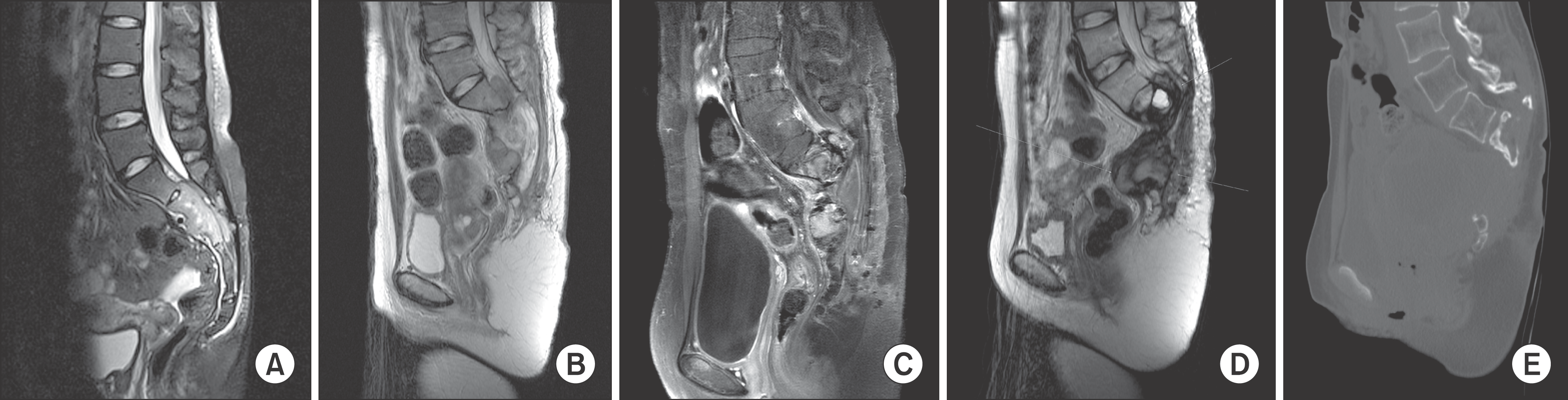
Table 1.
Demographic and Clinicopathologic Data
Table 2.
Data of Arterial Embolization and Blood Loss During Operation
 XML Download