

 PDF
PDF ePub
ePub Citation
 Print
Print


Abstract
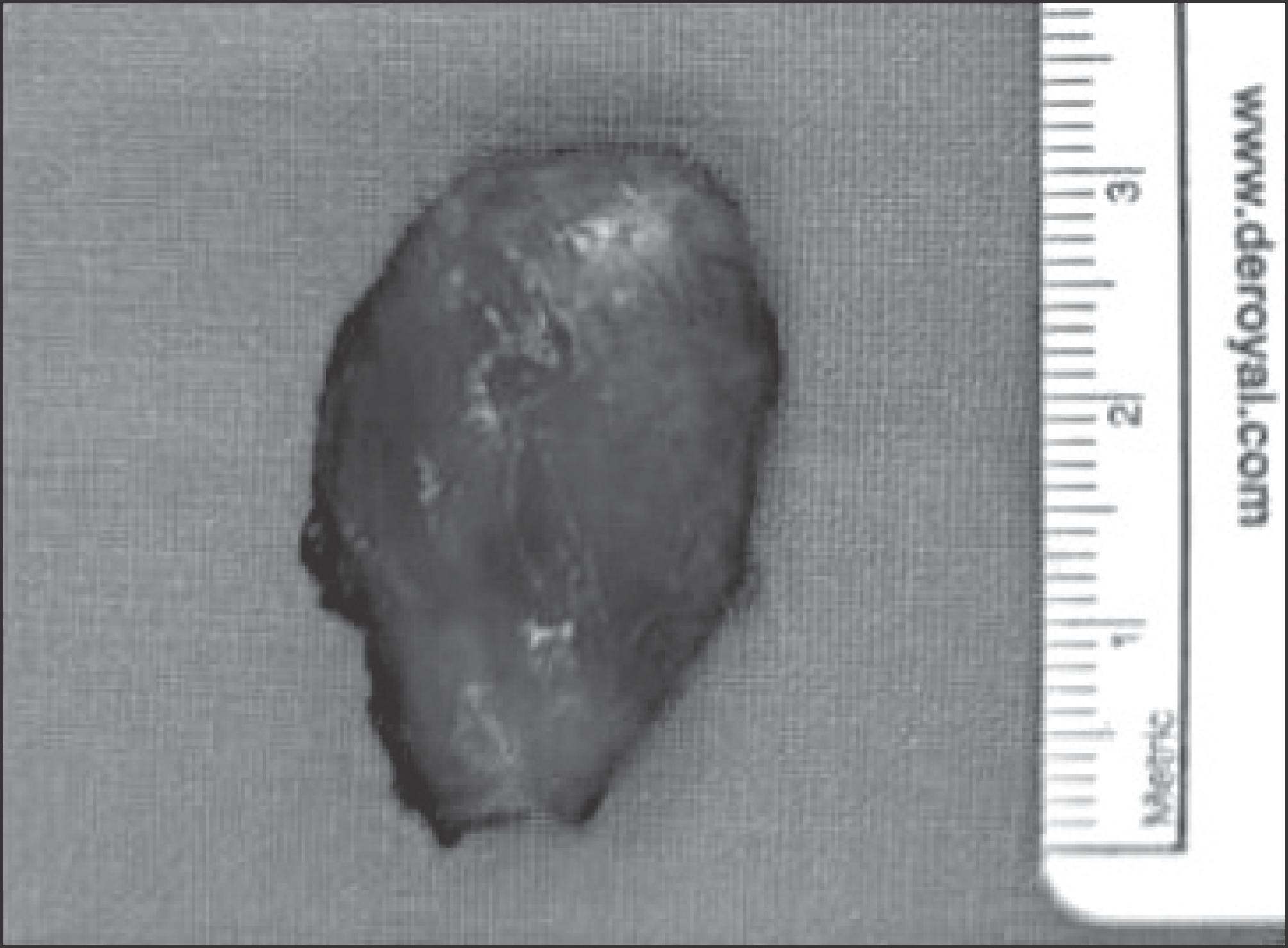
Ancient schwannoma is a variant of schwannoma and is characterized slowly growing tumor with degenerative change. And it is reported that schwannoma is relatively rare in extensor area. As a rare cause of solitary ancient schwannoma in extensor area of upper arm, we report it.
References
1. Adani R, Baccarani A, Guidi E, Tarallo L. Schwannomas of the upper extremity: diagnosis and treatment. Chir Organi Mov. 2008; 92:85–8.

2. Louis DS. Peripheral nerve tumors in the upper extremity. Hand Clin. 1987; 3:311–8.
3. Kehoe NJ, Reid RP, Semple JC. Solitary benign peripheral-nerve tumours. Review of 32 years' experience. J Bone Joint Surg Br. 1995; 77:497–500.
4. Schultz E, Sapan MR, McHeffey-Atkinson B, Naidich JB, Arlen M. Case report 872. “Ancient” schwannoma (degenerated neurilemoma). Skeletal Radiol. 1994; 23:593–5.
5. Graviet S, Sinclair G, Kajani N. Ancient schwannoma of the foot. J Foot Ankle Surg. 1995; 34:46–50.
6. Kim SM, Seo SW, Lee JY, Sung KS. Surgical outcome of schwannomas arising from major peripheral nerves in the lower limb. Int Orthop. 2012; 36:1721–5.
7. Isobe K, Shimizu T, Akahane T, Kato H. Imaging of ancient schwannoma. AJR Am J Roentgenol. 2004; 183:331–6.
Figure 1.
Plain elbow lateral x-ray shows soft tissue tumor density in extensor area without calcification.
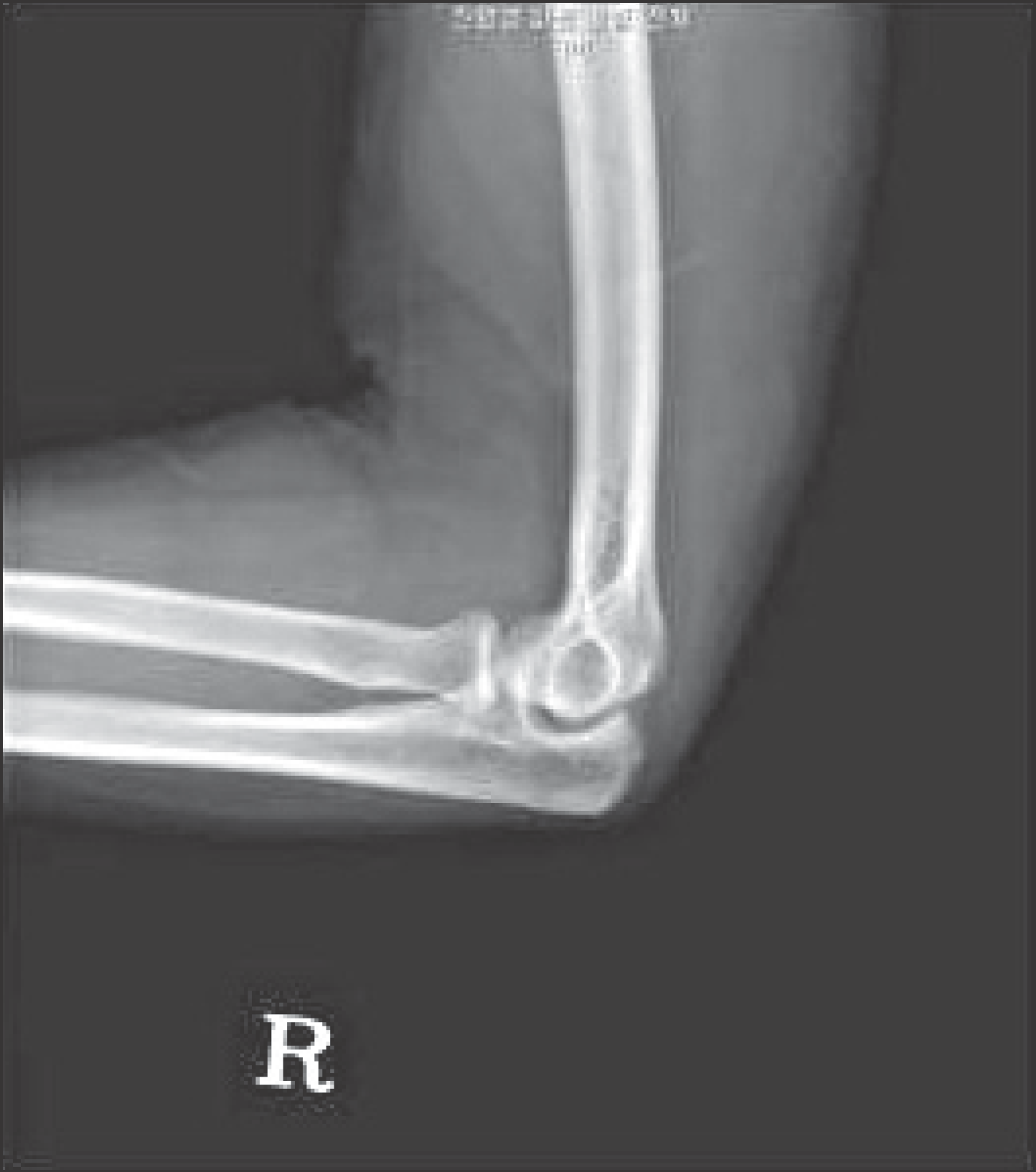
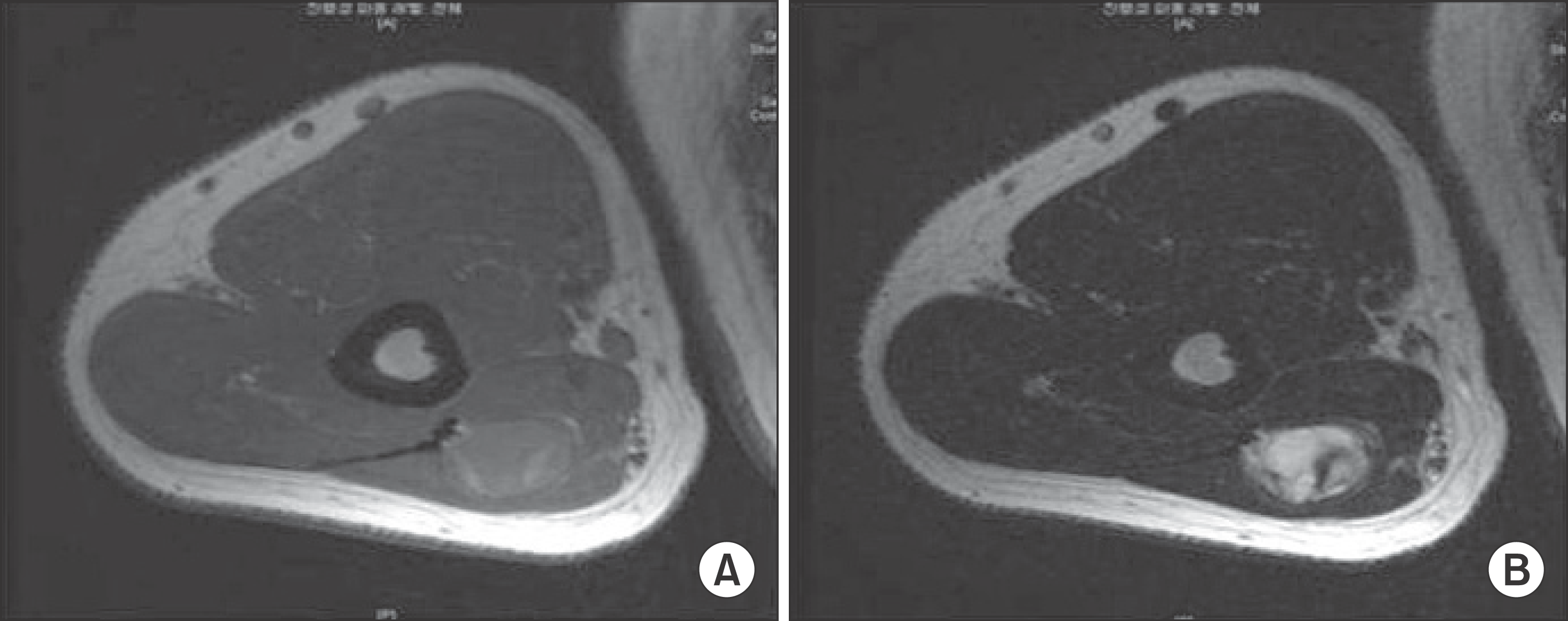
Figure 2.
These are the magnetic resonance image findings of mass. (A) Axial T1-weighted MR image shows inhomogeneous low signal intensity. (B) Axial T-2 weighted MR image shows heterogenous high signal intensity.

 XML Download