Abstract
Erdheim Chester disease (ECD) is very rare non-Langerhans cell histiocytosis (LCH) which occurs in the skeletal system and multiple organs. As it is progressive, sometimes it causes fatal results. However, it is often misdiagnosed as LCH or multiple bone metastasis and, thus, is very difficult to diagnose. In Korea, only 10 cases were first reported in 1999. In particular, there have been a few orthopedic approaches or reports in English-speaking literatures, and no report has been issued in Korea. The authors performed bone biopsy in patients with knee and lower extremity pain who were referred for the integrated treatment. We attempts to report this diagnosis experience with literature review.
Go to : 
References
1. Chester W. Über Lipoidgranulomatose (Over lipoid granulomatosis). Virchows Arch Pathol Anat Physiol. 1930. 279. : 561–602.
2. Veyssier-Belot C, Cacoub P, Caparros-Lefebvre D, et al. Erdheim-Chester disease. Clinical and radiologic characteristics of 59 cases. Medicine (Baltimore). 1996; 75:157–69.

3. Schmidt HH, Gregg RE, Shamburek R, Brewer BH Jr, Zssssech LA. Erdheim-Chester disease: low low-density lipoprotein levels due to rapid catabolism. Metabolism. 1997; 46:1215–9.

4. Park YK, Ryu KN, Huh B, Kim JD. Erdheim-Chester disease: a case report. J Korean Med Sci. 1999; 14:323–6.

5. Resnick D, Greenway G, Genant H, Brower A, Haghighi P, Emmett M. Erdheim-Chester disease. Radiology. 1982; 142:289–95.

6. Atkins HL, Klopper JF, Ansari AN, Iwai J. Lipid (cholesterol) granulomatosis (Chester-Erdheim disease) and congenital megacalices. Clin Nucl Med. 1978; 3:324–7.

7. Haroche J, Amoura Z, Dion E, et al. Cardiovascular involvement, an overlooked feature of Erdheim-Chester disease: report of 6 new cases and a literature review. Medicine (Baltimore). 2004; 83:371–92.
Go to : 
 | Figure 2.On histologic finding, pathologic features revealed collagenous fibroadipose tissue with lymphoplasma and histiocytes. The result immunohistochemical staining were a positive for CD68, and negative for S-100, CD1a. |
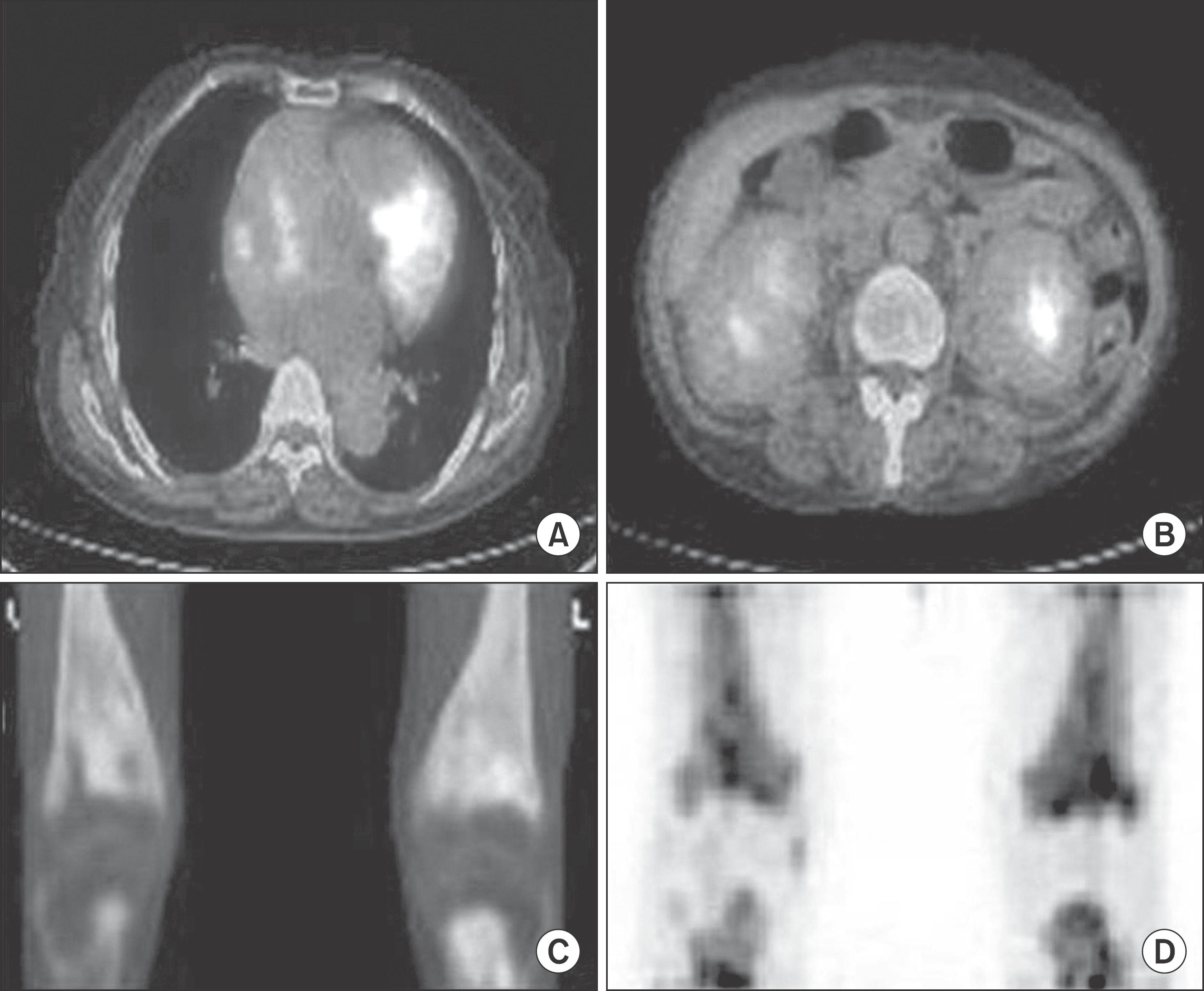
 | Figure 3.On PET CT, it showed high metabolic lesions of SUV 4.7 in heart (A), kidney (B), bilateral distal femoral, and proximal tibia (C, D). |


 PDF
PDF ePub
ePub Citation
 Print
Print


 XML Download