

 PDF
PDF ePub
ePub Citation
 Print
Print


Abstract
Purpose
Primary bone tumors of hindfoot are uncommon compared with other locations, and there have been few large-group studies. This study was designed to analyze the characteristics and the clinical results of the primary bone tumors of hindfoot.
Materials and Methods
Forty five cases in 44 patients who have been diagnosed from 1989 to 2011 were reviewed. The minimum follow-up period was 1 year. We retrospectively reviewed the medical records and images.
Results
Twenty six cases were male and 18 cases were female. Mean follow-up period was 33.1 months and mean age was 25.1 years. Forty four cases were benign and 1 case was malignant. Thirty six cases occurred in calcaneus and 9 cases were in talus. The most common benign bone tumor was simple bone cyst (20 cases), followed by intraosseous lipoma (12 cases), and chondroblastoma (4 cases). In calcaneus, there were 18 cases of simple bone cyst, and 12 cases of intrasosseous lipoma. In talus, there were 3 cases of chondroblastoma, 2 cases of simple bone cyst, and 2 cases of intraossesous ganglion. Many patients with hindfoot bone tumors presented with pain, but some were found accidentally. Patients received surgical procedures, such as curettage and bone graft, open reduction and internal fixation, tumor resection, and below knee amputation.
Go to : 
References
1. Walling AK, Gasser SI. Soft-tissue and bone tumors about the foot and ankle. Clin Sports Med. 1994; 13:909–38.

2. Murari TM, Callaghan JJ, Berrey BH Jr, Sweet DE. Primary benign and malignant osseous neoplasms of the foot. Foot Ankle. 1989; 10:68–80.
3. Steinberg MD, Steinberg LB, Calihman N. Tumors of the foot: benign and malignant. J Am Podiatry Assoc. 1979; 69:135–8.
4. Choi WJ, Shin KH, Lee JW, Han CW. Tumors in the foot and ankle (185 cases). J Korean Foot Ankle Soc. 2007; 11:1–7.
5. Buchner M, Bernd L, Zahlten-Hinguranage A, Sabo D. Bone and soft-tissue tumors of the foot and ankle. Chirurg. 2005; 76:391–7.
6. Ozdemir HM, Yildiz Y, Yilmaz C, Saglik Y. Tumors of the foot and ankle: analysis of 196 cases. J Foot Ankle Surg. 1997; 36:403–8.
7. Bakotic B, Huvos AG. Tumors of the bones of the feet: the clinicopathologic features of 150 cases. J Foot Ankle Surg. 2001; 40:277–86.
8. Young PS, Bell SW, MacDuff EM, Mahendra A. Primary osseous tumors of the hindfoot: why the delay in diagnosis and should we be concerned? Clin Orthop Relat Res. 2013; 471:871–7.
9. Shin DS, Park SH, Ahn JC. Tumors of the foot. J Korean Bone & Joint Tumor Soc. 2003; 9:69–76.
10. Dhillon MS, Singh B, Singh DP, Prabhu V, Nagi ON. Primary bone tumors of the talus. J Am Podiatr Med Assoc. 1994; 84:379–84.
11. Monroe MT, Manoli A 2nd. Osteoid osteoma of the lateral talar process presenting as a chronic sprained ankle. Foot Ankle Int. 1999; 20:461–3.
12. Killian JT, Wilkinson L, White S, Brassard M. Treatment of unicameral bone cyst with demineralized bone matrix. J Pediatr Orthop. 1998; 18:621–4.
13. Glaser DL, Dormans JP, Stanton RP, Davidson RS. Surgical management of calcaneal unicameral bone cysts. Clin Orthop Relat Res. 1999; 360:231–7.
14. Hollawell SM. Allograft cellular bone matrix as an alternative to autograft in hindfoot and ankle fusion procedures. J Foot Ankle Surg. 2012; 51:222–5.
15. Delawi D, Kruyt MC, Huipin Y, et al. Comparing autograft, allograft, and tricalcium phosphate ceramic in a goat instrumented posterolateral fusion model. Tissue Eng Part C Methods. 2013. [Epub ahead of print].
16. An HS, Lynch K, Toth J. Prospective comparison of autograft vs. allograft for adult posterolateral lumbar spine fusion: differences among freeze-dried, frozen, and mixed grafts. J Spinal Disord. 1995; 8:131–5.
17. Aumar DK, Dadjo YB, Chagar B. Intraosseous lipoma of the calcaneus: report of a case and review of the literature. J Foot Ankle Surg. 2013; 52:360–3.
18. Greenspan A, Raiszadeh K, Riley GM, Matthews D. Intraosseous lipoma of the calcaneus. Foot Ankle Int. 1997; 18:53–6.
19. Weinfeld GD, Yu GV, Good JJ. Intraosseous lipoma of the calcaneus: a review and report of four cases. J Foot Ankle Surg. 2002; 41:398–411.
20. Bloem JL, Mulder JD. Chondroblastoma: a clinical and radiological study of 104 cases. Skeletal Radiol. 1985; 14:1–9.
21. Edel G, Ueda Y, Nakanishi J, et al. Chondroblastoma of bone. A clinical, radiological, light and immunohistochemical study. Virchows Arch A Pathol Anat Histopathol. 1992; 421:355–66.
22. Lee YK, Han IK, Oh JH, Lee SH, Kim HS. Treatment and prognosis of chondroblastoma. J of Korean Bone & Joint Tumor Soc. 2007; 13:81–7.
23. Giudici MA, Moser RP Jr, Kransdorf MJ. Cartilaginous bone tumors. Radiol Clin North Am. 1993; 31:237–59.
24. Han CS, Park PY, Park JS, Park KH. Osteochondroma of the Os calcaneum – A case report. J of Korean Bone & Joint Tumor Soc. 2008; 14:152–6.
25. Karasick D, Schweitzer ME, Eschelman DJ. Symptomatic osteochondromas: imaging features. AJR Am J Roentgenol. 1997; 168:1507–12.
26. Blitz NM, Lopez KT. Giant solitary osteochondroma of the inferior medial calcaneal tubercle: a case report and review of the literature. J Foot Ankle Surg. 2008; 47:206–12.
27. Kennedy JG, Frelinghuysen P, Hoang BH. Ewing sarcoma: current concepts in diagnosis and treatment. Curr Opin Pediatr. 2003; 15:53–7.
28. Weber KL. Current concepts in the treatment of Ewing's sarcoma. Expert Rev Anticancer Ther. 2002; 2:687–94.
29. Weber KL, Sim FH. Ewing's sarcoma: presentation and management. J Orthop Sci. 2001; 6:366–71.
30. Leeson MC, Smith MJ. Ewing's sarcoma of the foot. Foot Ankle. 1989; 10:147–51.
Go to :
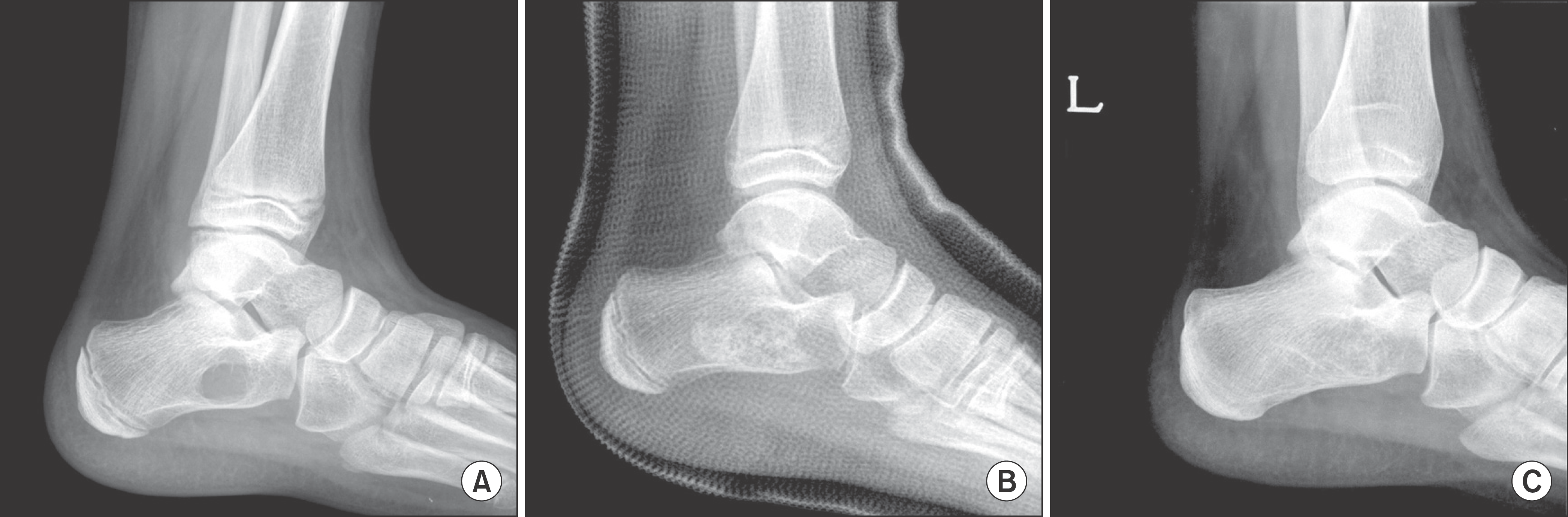 | Figure 1.(A) Cystic lesion with well-defined margin in calcaneus was found accidentally after ankle sprain. (B) Cyst was diagnosed as simple bone cyst, filled with allograft chip bone graft. (C) Radiograph of 5 year follow-up showed consolidation of grafted bone post-operatively. |
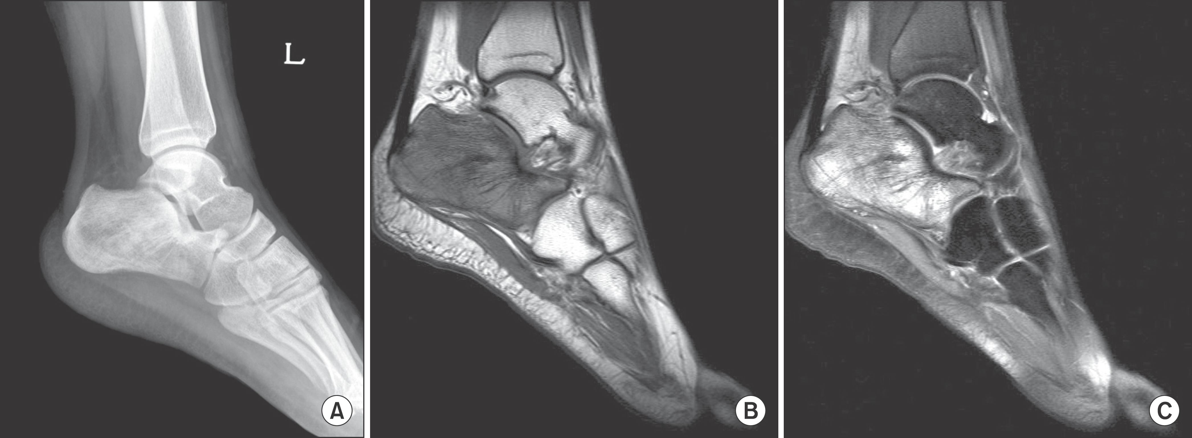 | Figure 2.(A) Irregular and sclerotic lesion in calcaneus was found after sprain. (B) T1-weighted MRI of the calcaneus demonstrating low intensity compared to the normal high intensity of bone marrow. (C) T2-weighted MRI of the calcaneus reveal heterogeneous high intensity and extent of marrow infiltration. |
 | Figure 3.(A) Cystic lesion with pathologic fracture was found in radiograph after fall-down. (B) CT scan showed comminuted fracture extending to large low density lesion. (C) Postoperative radiograph showed state of open reduction and internal fixation with allo chip bone graft. (D) Radiograph of 3 year follow-up showed consolidation of grafted bone. |
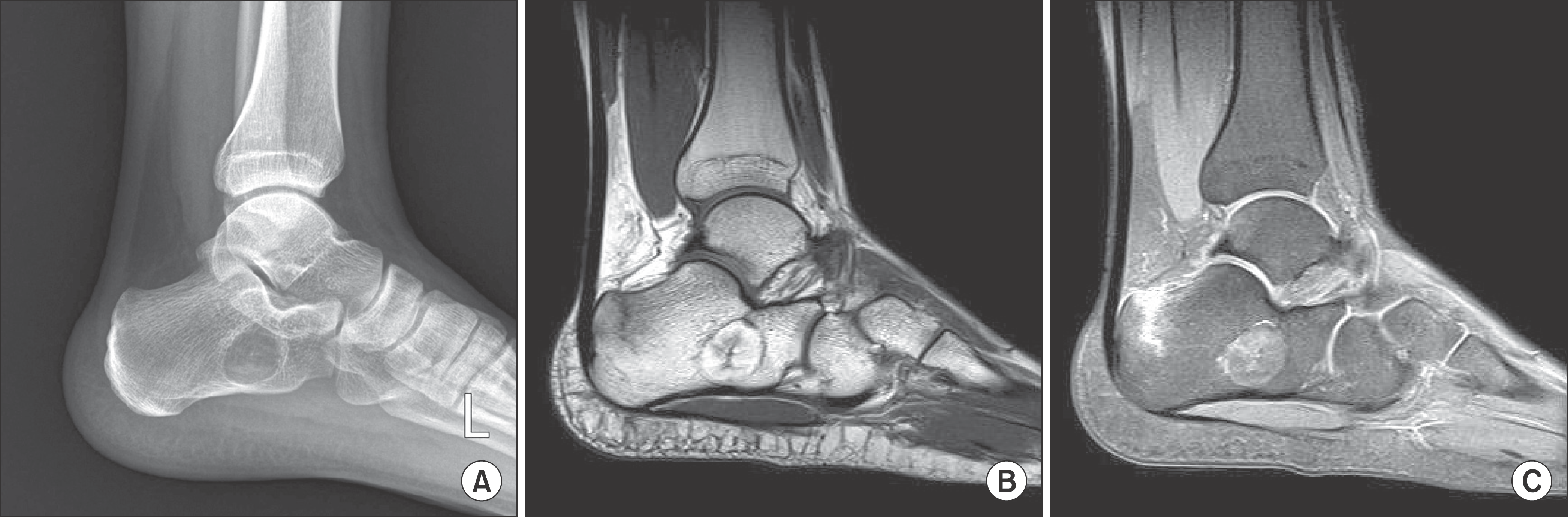 | Figure 4.(A) Radiolucent cystic lesion with well-definded margin in calcaneus found in radiograph after playing soccer. (B) T1-weighted MRI showed well-defined fat signal mass and vertical fracture line on posterosuperior aspect of calcaneus. (C) T2-weighted MRI showed well-defined mass and vertical fracture on calcaneus with perilesional bone contusion. |
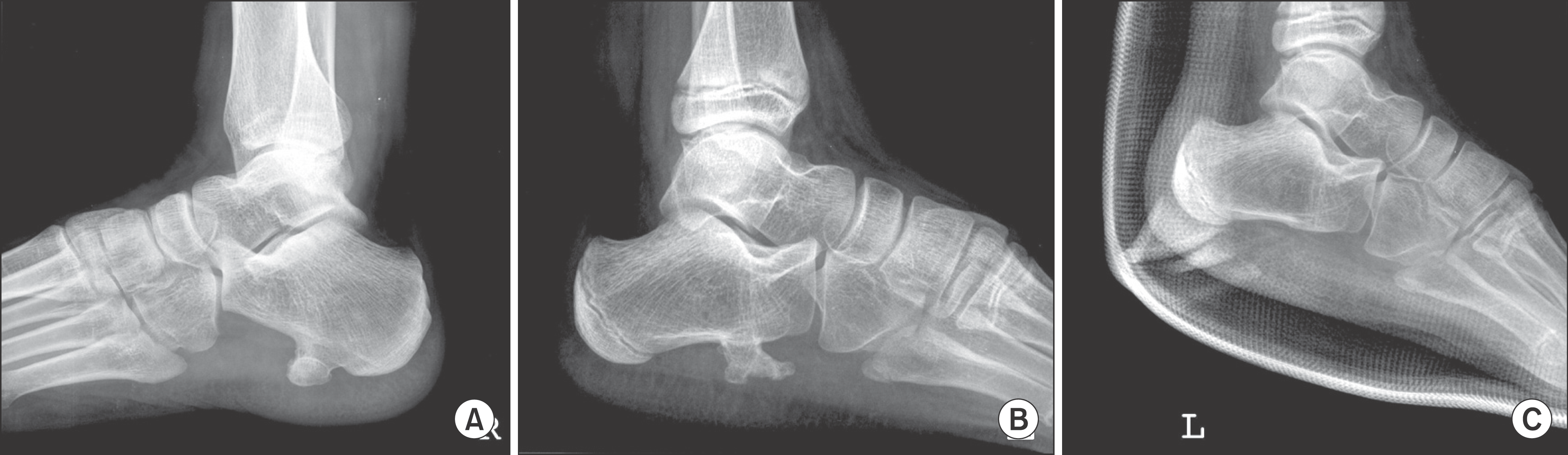 | Figure 5.(A) An exophytic osseous mass at plantar surface of calcaneus was found in radiograph. (B) A large exophytic osseous mass at plantar surface of calcaneus was found in plain-film radiograph. (C) Postoperative radiograph showed excised osteochondroma. |
Table 1.
Distributions of Hind Foot Tumors
 XML Download