

 PDF
PDF ePub
ePub Citation
 Print
Print


Abstract
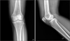
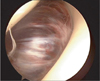
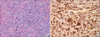
Benign fibrous hitiocytoma of the infrapatella fat pad is very rare. It has usually do not induced a pain or a symptom because it was located deep tissue. So it was very difficult to be diagnosed. We experienced a case of deep benign fibrous histiocytoma in a 53-year-old woman. It was diagnosed by MRI. Diagnostic arthroscopic procedure was performed and the lesion was completely resected by open excision. We report a rare case of benign fibrous hitiocytoma presenting as an intra-articular tumor in the joint causing pain and limitation of movement.
Figures and Tables
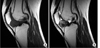 | Figure 2(A) MRI of her right knee. The ovoid mass like lesion in the infrapatellar fat pad area, shows isointense on sagittal SE T1-weighted image. (B) MRI SE T2-weighted image of her right knee. This mass is relatively well defined margin and homogenous signal intensity.
|
References
1. Calonje E, Mentzel T, Fletcher CD. Cellular benign fibrous histiocytoma. Clinicopathologic analysis of 74 cases of a distinctive variant of cutaneous fibrous histiocytoma with frequent recurrence. Am J Surg Pathol. 1994. 18:668–676.
2. Ha KI, Han SH, Jung MY, Yang BG, Ryu JH. Benign fibrous histiocytoma of the knee. J Korean Knee Soc. 1993. 5:222–225.
3. Ushijima M, Hashimoto H, Tsuneyoshi M, Enjoji M. Giant cell tumor of the tendon sheath (nodular tenosynovitis) A study of 207 cases to compare the large joint group with the common digit group. Cancer. 1986. 57:875–884.
4. Yi GJ, Kim YS, Oh CW. A case of giant cell tumor of tendon sheath. Korean J Dermatol. 1993. 31:416–420.
5. Lee HP, Park HJ, Park YH, Kim JW, Suh EJ. A case of giant cell tumor of the tendon sheath developing on the dorsum of the foot. Korean J Dermatol. 1995. 33:1168–1171.
6. Fletcher CD. Benign fibrous histiocytoma of subcutaneous and deep soft tissue: a clinicopathologic analysis of 21 cases. Am J Surg Pathol. 1990. 14:801–809.
7. Jun IS, Kim NI, Haw CR. A case of giant cell tumor of the tendon sheath. Korean J Dermatol. 1994. 32:939–943.
8. Pinar H, Ozkan M, Ozaksoy D, Pabuççuoğlu U, Akseki D, Karaoğlan O. Intraarticular fibroma of the tendon sheath of the knee. Arthroscopy. 1995. 11:608–611.
9. Azouz EM. Benign fibrous histiocytoma of the proximal tibial epiphysis in a 12-year-old girl. Skeletal Radiol. 1995. 24:375–378.
10. Lee KB, Park RS, Lee EJ, Lee JY, Song KW, Park IH. Benign fibrous histiocytoma of the patellar fat pad: a report of one case. J Korean Knee Soc. 1997. 9:224–228.
 XML Download