

 PDF
PDF ePub
ePub Citation
 Print
Print


Abstract
We reviewed the clinical features of 2 patients who underwent surgery for subungal exostosis, focusing on postoperative deformity of the nail. The lesion destroyed the nail bed and was excised with a direct approach. then thin split-thickness sterile matrix graft was done after excision of the tumor because the defect of the nail bed was large. Good postoperative appearance of the nail was obtained by thin split-thickness sterile matrix graft. The use of thin split-thickness sterile matrix graft for the replacement of a nail bed defect can regain a smooth, adherent, and normal-looking nail and avoid donor-site morbidity. Thin split-thickness toe-nail bed graft is a good choice for the prevention of postoperative deformity.
Figures and Tables
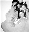
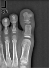
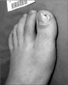 | Figure 1A 14 year old male patient had subungal exostosis of great toe with ulceration of medial border of the nail and elevation the nail plate.
|
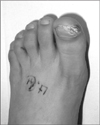
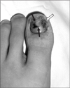 | Figure 4Thin split-thickness nail bed graft was done to the defect. The black arrows show donor site and the yellow arrow shows recipient site.
|
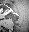
 | Figure 5Pathologic features of Case 1. (A) A cartilaginous cap overlies the underlying bone of the stalk (Hematoxylin & Eosin, ×40).
|
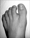
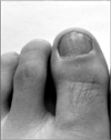 | Figure 6Seven months postoperatively, postoperative clinical photo showed improvement of nail deformity.
|
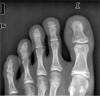
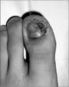
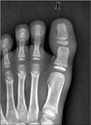
 | Figure 8A 10 year old male patient had subungal exostosis of great toe with severe nail bed destruction and onycholysis.
|
 | Figure 10Pathologic features of case 2. The maturing chondrocytes in osteochondroma simulate endochondral ossification in the growth plate at the junction between the cartilaginous cap and stalk (Hematoxylin & Eosin, ×100).
|
References
1. Hatoko M, Tanaka A, Kuwahara M, et al. Hard palate mucosal grafts for defects of the nail bed. Ann Plast Surg. 2002. 49:424–428.
2. Dumontier CA, Abimelec P. Nail unit enchondromas and osteochondromas: a surgical approach. Dermatol Surg. 2001. 27:274–279.
3. Lokiec F, Ezra E, Krasin E, Keret D, Wientroub S. A simple and efficient surgical technique for subungual exostosis. J Pediatr Orthop. 2001. 21:76–79.
4. Iizuka T, Kinoshita Y, Fukumoto K. Subungual exostosis of the finger. Ann Plast Surg. 1995. 35:330–332.
5. Ilyas W, Geskin L, Joseph AK, Seraly MP. Subungual exostosis of the third toe. J Am Acad Dermatol. 2001. 45:6 Suppl. S200–S201.
6. Dahlin DC. Bone tumor: general aspects and data on 8542 cases. 1986. 4th ed. Springerfield, Illinois: Charles C Thomas;19–30.
7. Landon GC, Johnson KA, Dahlin DC. Subungual exostoses. J Bone Joint Surg Am. 1979. 61:256–259.
8. de Palma L, Gigante A, Specchia N. Subungual exostosis of the foot. Foot Ankle Int. 1996. 17:758–763.
9. Moon MS, Lee IJ, Chung KH. Subungual exostoeis. J Korean Orthop Assoc. 1986. 21:502–506.
10. Koshima I, Soeda S, Takase T, Yamasaki M. Free vascularized nail grafts. J Hand Surg Am. 1988. 13:29–32.
 XML Download