

 PDF
PDF ePub
ePub Citation
 Print
Print


Introduction
Chemokines are proinflammatory cytokines that are produced locally in tissues and function as the directional cue to sort, direct, and fine-tune cell trafficking.1) Chemokines and their receptors are known to be involved in the development and progression of malignant tumors. It has been demonstrated that certain chemokines serve as tissue-specific attractant molecules for tumor cells, promoting tumor cell migration to particular sites in vivo.2) Recent evidence suggests that cancer cells use a chemokine receptor axis for metastasis formation at secondary sites.3)
CXCR4 is a seven-domain transmembrane chemokine receptor expressed predominantly on lymphocytes where it activates chemotaxis. It has been demonstrated that normal breast tissue expresses low levels of CXCR4, whereas neoplastic breast tissue expresses higher levels of CXCR4.4) Recently, CXCR4 and its corresponding ligand, stromal cell-derived factor 1 (SDF-1)/CXCL12 have been implicated in the pathogenesis and progression of various cancers, as a number of studies have reported that CXCR4 expression predicts poor prognosis in lung cancer, melanoma, esophageal cancer, and ovarian cancer.5-8) Data from breast carcinoma studies have suggested that the specific effects mediated via chemokines could be significantly different depending on the source of the ligand or receptor expression.9) In pediatric osteosarcoma, CXCR4 expression by tumor cells may participate in metastasis to tissues containing SDF-1.10)
In a recent retrospective osteosarcoma patient study, expressions of CXCR4, CCR7 and CCR10 receptor mRNA in osteosarcoma tissue was linked to a clinical outcome.11) Involvement of the CXCR4/SDF-1 pathway in the metastatic process in the lung has also been demonstrated in osteosarcoma cell lines.12) However, the expression and distribution of some receptors in osteosarcoma is still controversial and documentation of the expression of CXCR4 is scarce in clinical samples of malignant mesenchymal tumors including osteosarcoma. As osteosarcoma consists of tumor cells derived from mesenchymal origin and of infiltrating mononuclear cells, we attempted to define the source of chemokine receptor expression in patient samples. We hypothesized that there may be an association between CXCR4 expression and prognostic factors. Therefore, we examined the expression of CXCR4 in osteosarcomas and investigated the relationship between its expression and clinicopathological features.
Materials and Methods
1. Patients and samples
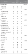
A total of 63 patients with osteosarcoma were included in the present study, including 38 and 25 cases of conventional high-grade and low-grade central osteosarcomas respectively. The medical records were reviewed and information about age, gender, tumor location, presence of metastasis, and follow-up data were retrieved. The clinical data for tumor size and AJCC stage were only available for 33 patients with high-grade conventional osteosarcomas, and information for neo- and adjuvant chemotherapy was available for only 25 patients. All specimens were primary osteosarcoma tissue and were obtained prior to any chemotherapy treatment. One of the conventional high-grade osteosarcomas included a pulmonary metastatic lesion. Two pathologists (H.R.P. and Y.K.P.) reviewed all sections from each case to confirm the diagnosis and determine the histologic subtype and grade. Tumors were subclassified as osteoblastic, chondroblastic, or fibroblastic according to the predominant histological element, and were graded using a two-tiered system of low-grade (1 and 2) and high-grade (3 and 4).13) This study was approved by the Institutional Review Board at Kyung Hee University Hospital.
2. Immunohistochemistry
The avidin-biotin complex (ABC) method was performed on 4 µm thick tissue sections for immunohistochemical analysis. Sections were deparaffinized with xylene for 15 min and pretreated in a microwave oven using 0.01 M citrate buffer (pH 6.0) for 30 min. Sections were incubated with mouse IgG monoclonal antibody against CXCR4 (1:60 dilution, Abnova Co., Taipei, Taiwan) for 30 min at room temperature. Both positive and negative controls were used in each experiment. Sections from breast carcinoma specimens which had resulted in lymph node metastasis were used as a positive control for CXCR4. The utility of all antibodies after decalcification was confirmed using a decalcified breast carcinoma specimen.
3. Evaluation
Consensus judgment was adopted to determine immunohistochemical cytoplasmic positivity of the tumors based on the distribution of positive cells: negative, focal (positive cells ≤30%), and diffuse (positive cells >30%). The intensities of CXCR4 expression were similar in the positive samples. Two independent pathologists (H.R.P. and Y.K.P.) judged the immunoreactivities.
4. Statistical analysis
All statistical analyses were carried out with the DBSTAT program (version 4.1, Seoul, Korea). Relationships between clinicopathologic variables and CXCR4 expression were determined using the Chi-square test. The clinicopathologic variables studied were histologic grade, age at diagnosis, gender, tumor location, histologic subtype, tumor size, AJCC stage, and presence of metastatic disease at any time during the progression of the disease. Overall survival was defined as the time from diagnosis until the date of the final follow-up or death. Overall survival rates were calculated using the Kaplan-Meier method, and differences in survival were compared using the log-rank test. When the p value was less than 0.05, the statistical difference was considered significant.
Results
1. Patient characteristics
The clinical and pathological features of 63 patients are shown in Table 1. In total, we analyzed 38 cases of conventional high-grade and 25 low-grade central osteosarcomas. For 63 patients with osteosarcomas, the age distribution ranged from 7 to 66 years. Of these cases, 35 were male and 28 were female. For the histologic subtypes of conventional high-grade osteosarcomas, twenty-one patients were osteoblastic, ten were chondroblastic, and five were fibroblastic. Each case of giant cell rich and telangiectatic type was included.
2. Expression of CXCR4 and its correlation with clinicopathologic characteristics (Table 1)
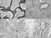
Immunohistochemistry showed that CXCR4 was present in the cytoplasm of osteosarcoma cells. In addition, we found some expression of CXCR4 in admixed inflammatory cells and in the lining of intratumoral vessels. Twenty-nine cases (76.3%) out of the 38 conventional high-grade osteosarcomas showed increased staining for CXCR4 (Fig. 1). Eleven cases (28.9%) were considered focal and 18 (47.4%) were diffuse. For one patient who died with pulmonary metastasis, paraffin blocks were available. They exhibited diffuse CXCR4 expression in both primary bone and pulmonary metastatic lesions (Fig. 1D). Twenty-five low-grade osteosarcoma samples were also analyzed for CXCR4 expression. The CXCR4 expression was 36% for the low-grade samples compared to 76.3% for the 38 high-grade osteosarcoma samples. Diffuse expression was noted in 47.4% of the high-grade osteosarcomas whereas all positive cases with low-grade were focally positive. CXCR4 expression showed a significant correlation with higher histologic grade (p<0.0001). In addition, younger patients (under 20 years of age) showed more CXCR4 expression compared to older patients (over 20 years of age) (p=0.0022). However, no statistically significant differences were found in CXCR4 expression with respect to gender (p=0.0649) or tumor location (p=0.3511).
Among the variable histological subtypes in high-grade osteosarcomas, no statistically significant differences were found in CXCR4 expression (p=0.4953). Among all 38 cases of high-grade conventional osteosarcoma, nine cases had a history of metastasis, seven of which showed cytoplasmic CXCR4 expression. However, this difference was not statistically significant (p=0.1426). In addition, no statistically significant differences were found in CXCR4 expression with respect to tumor size (p=0.8682) or AJCC stage (p=0.1065).
3. Overall survival data (Table 2)
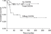
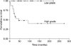
Follow-up data were available for 24 patients with high-grade osteosarcomas and all 25 patients with low-grade central osteosarcomas. The follow-up period ranged from one to 275 months. In the high-grade group, thirteen patients died while the other eleven patients survived. In contrast all patients with low-grade central osteosarcomas survived. CXCR4 expression (p=0.0058) (Fig. 2), histologic grade (p<0.0001) (Fig. 3), and age (p=0.0140) were found to be prognostically relevant in terms of overall survival by univariate analysis. However, overall survival rate was not statistically correlated with gender (p=0.3260), tumor size (p=0.2315), or AJCC stage (p=0.2996).
Discussion
The chemokine receptor CXCR4 and its corresponding ligand SDF-1 are thought to play a major role in the metastatic cascade and prognosis for cancer patients. Chemokines released from the endothelium in target tissues bind to their receptors on tumor cells in the primary lesion and result in both the release of matrix metalloproteinases (MMPs) and a chemoattractant gradient, leading to metastasis to the specific target tissue.14,15) The CXCR4/SDF-1 system has been found to correlate significantly with the presence of metastases in a number of tumors including prostate, melanoma, breast, and rhabdomyosarcoma.15-18)
In this study, we compared the immunohistochemical expressions of CXCR4 between conventional high-grade and low-grade central osteosarcomas and found that its expression was increased in the conventional high-grade osteosarcomas. Our results elucidated that CXCR4 is frequently expressed in osteosarcomas and that its level of expression correlates with the higher-grade tumor in osteosarcoma. In addition, increased CXCR4 expression correlated with reduced overall survival, suggesting that CXCR4 could have a prognostic implication. Recently, Bai et al.19) showed that CXCR4 expression levels were higher in high-grade chondrosarcoma cells than in low-grade specimens. This result provides more evidence for the importance of CXCR4 expression in tumor progression. Unfortunately, CXCR4 expression showed no significant association with the incidence of metastasis or the AJCC stage, in contrast to other previously reported human malignancies. One of the limitations of this study, however, is that the number of samples analyzed is small and insufficient for such comparison. Another limitation is the incomplete information about neo- and adjuvant chemotherapies for the high-grade osteosarcoma patients.
CXCR4 expression has been linked to osteosarcoma aggressiveness. In our study, one patient exhibited diffuse CXCR4 expression in both primary bone and pulmonary metastatic lesions. A recent report by Oda et al.2) suggested that CXCR4 was produced at high levels on tumor cells in the metastatic site as compared to the primary osteosarcoma tumor. Laverdiere et al.11) showed that increased CXCR4 mRNA expression in osteosarcoma tumor samples correlated with reduced overall survival and with the presence of metastases at diagnosis. These data suggest that CXCR4 could be useful as a prognostic factor and as a predictor of potential metastatic development in osteosarcoma. According to de Nigris et al.,20) cell invasiveness and metastasis implantation by wild-type SaOs-2 cells were associated with the upregulation of CXCR4 expression. CXCR4 seems to intervene in the pathogenesis of the malignant phenotype of osteosarcoma by acting on cell invasiveness and metastasis growth. In osteosarcoma cell lines, the release of matrix metalloproteinases following chemokine activation has been described.14) Another study also suggests an autocrine role for CXCR4 in angiogenic factors like vascular endothelial growth factor.21)
Perissinotto et al.12) showed that osteosarcoma cell lines produced CXCR4 proteins on flow cytometric analysis. Osteosarcoma tumors are usually rich with stromal cells like fibroblasts, macrophages, endothelial cells, and lymphocytes. It is important to remember that the CXCR4 receptor is expressed by virtually all leukocytes; thus the level of mRNA expression detected in primary or metastatic tumors will be due, at least in part, to the extent of mononuclear inflammatory cells infiltration. A recent review using metastatic models has indeed shown that interactions between primary tumor cells and stromal cells are critical for invasion into tissues.22) Furthermore, other studies have suggested that the metastatic potential correlates with the expression of chemokine receptors in non-tumoral stromal cells rather than in primary tumor cells.23) In our series, we found some expression of CXCR4 in admixed inflammatory cells, verifying this hypothesis in osteosarcoma. von Luettichau et al.24) also reported on their immunohistochemical analysis for chemokine receptors on a series of archival osteosarcoma samples. Importantly, their results show that infiltrating inflammatory cells often represent a significant, or even the only, source of chemokine receptor expression signal in osteosarcoma tissue. These results demonstrate the danger of using mRNA analysis alone when determining chemokine receptor expression in tumor samples.
Therapeutic strategies are classified mainly into two categories: the application of CXCR4-neutralizing antibodies and specific CXCR4 antagonists. In any case, both AMD3100- and T140-derived CXCR4 antagonists appear to have activity in animal tumor models, providing the rationale for future clinical trials of these agents in patients with various cancers.17,25) SDF-1, the ligand for CXCR4, is expressed at high levels in lung and lymph node tissue, which are the respective primary sites to which these tumors metastasize. These findings suggest that therapy aimed at disruption of this specific receptor/ligand complex may lead to a decrease in metastases. CTCE-9908, a small peptide CXCR4 antagonist, was utilized in two murine metastasis models to test this hypothesis; the results showed that inhibition of the CXCR4/SDF-1 pathway decreased metastatic disease in these models.26) Clinical experiments with larger samples and further study will be needed.
In conclusion, our results suggested that CXCR4 expression was involved in the higher-grade tumor and reduced overall survival in osteosarcomas. There is also the possibility that CXCR4 antagonists may actually prevent distant metastasis and tumor progression in cases of osteosarcoma. A larger cohort of osteosarcoma patient samples should allow for more specific conclusions and the inclusion of potential prognostic variables.
 XML Download