

 PDF
PDF ePub
ePub Citation
 Print
Print


Abstract
The development of different entities of soft tissue sarcoma in one patient is rare. It usually affects head and neck or abdominal region, whereas those affecting the extremities are much rarer. We describe a patient with double primary presentation of liposarcoma and Ewing's sarcoma in extremity. This case implies that sarcoma patients are at increased risk of a second malignancy, and this implies a need to search for occult tumors during follow up.
References
1. Grobmyer SR, Luther N, Antonescu CR, Singer S, Brennan MF. Multiple primary soft tissue sarcomas. Cancer. 2004; 101:2633–5.

2. Daigeler A, Lehnhardt M, Sebastian A, et al. Metachronous bilateral soft tissue sarcoma of the extremities. Langenbecks Arch Surg. 2008; 393:207–12.
3. Abramson DH, Melson MR, Dunkel IJ, Frank CM. Third (fourth and fifth) nonocular tumors in survivors of retinoblastoma. Ophthalmology. 2001; 108:1868–76.
4. Birch JM, Alston RD, McNally RJ, et al. Relative frequency and morphology of cancers in carriers of germline TP53 mutations. Oncogene. 2001; 20:4621–8.
5. Hope DG, Mulvihill JJ. Malignancy in neurofibromatosis. Adv Neurol. 1981; 29:33–56.
6. Li FP, Fraumeni JF Jr, Mulvihill JJ, et al. A cancer family syndrome in twenty-four kindreds. Cancer Res. 1988; 48:5358–62.
7. Fiore M, Grosso F, Lo Vullo S, et al. Myxoid/round cell and pleomorphic liposarcomas: prognostic factors and survival in a series of patients treated at a single institution. Cancer. 2007; 109:2522–31.
8. Antonescu CR, Tschernyavsky SJ, Decuseara R, et al. Prognostic impact of P53 status, TLS-CHOP fusion transcript structure, and histological grade in myxoid liposarcoma: a molecular and clinicopathologic study of 82 cases. Clin Cancer Res. 2001; 7:3977–87.
9. Cheng EY, Springfield DS, Mankin HJ. Frequent incidence of extrapulmonary sites of initial metastasis in patients with liposarcoma. Cancer. 1995; 75:1120–7.
10. Estourgie SH, Nielsen GP, Ott MJ. Metastatic patterns of extremity myxoid liposarcoma and their outcome. J Surg Oncol. 2002; 80:89–93.
Figure 1.
(A) Axial T2-weighted fat-saturated image, (B) Axial T1-weighted image, and (C) gadolinium enhancing image showing mass in proximal thigh.
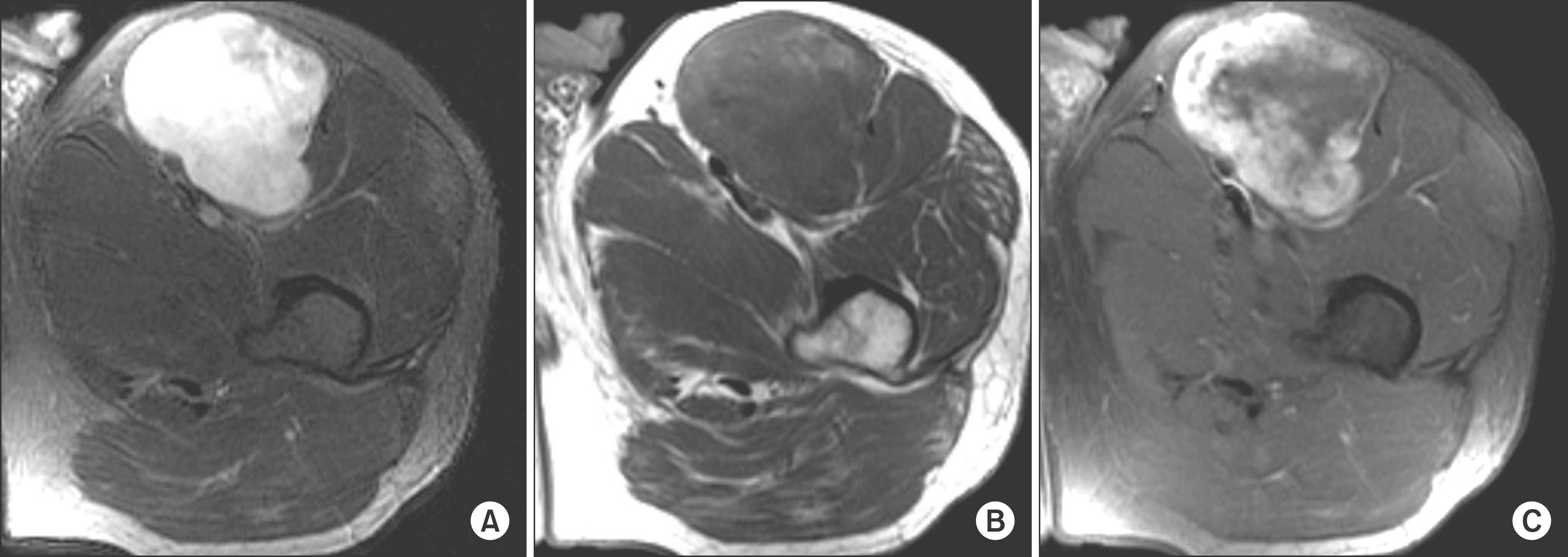
Figure 2.
Tc99-bone scans showing diffuse uptake left proximal thigh and no other site of disease. (A) Anteroposterior, and (B) posteroanterior bone scan view.
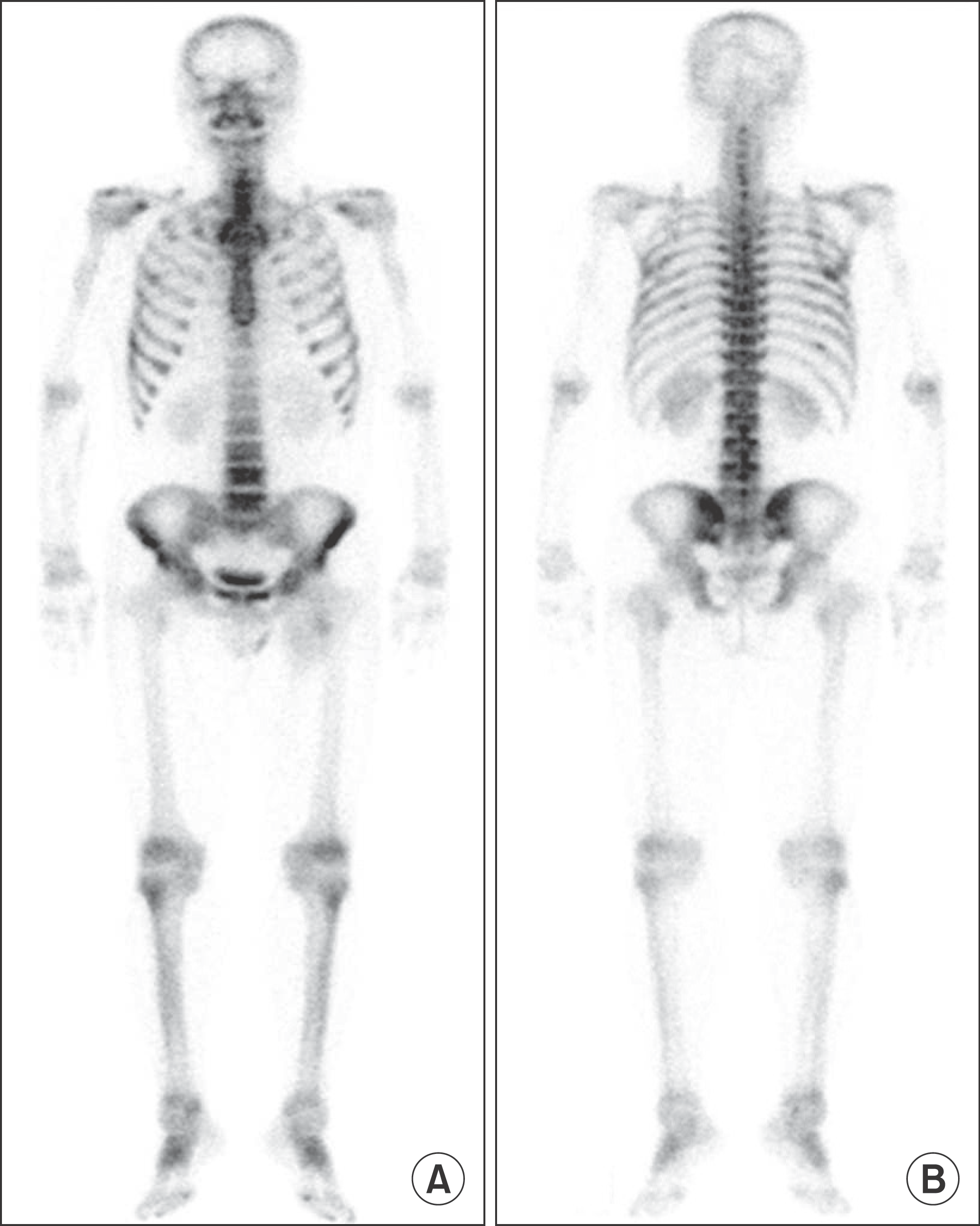
Figure 3.
(A) A high-power photomicrograph of the specimen shows findings compatible with liposarcoma (Stain, hematoxylin & eosin; magnification, ×200) and (B) positive S-100 immunohistochemical analysis stain.

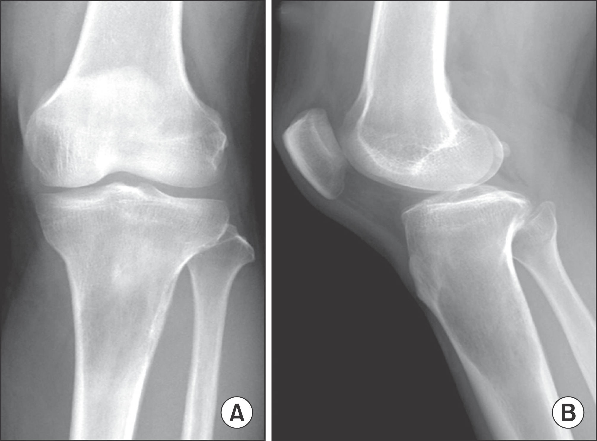
Figure 4.
2 years and 6 months follow up radiographs showing a new osteolytic lesion in proximal tibia. Anteroposterior (A) and lateral view (B).

 XML Download