

 PDF
PDF ePub
ePub Citation
 Print
Print


Abstract
Bizarre parosteal osteochondromatous proliferation (BPOP) otherwise known as Nora's lesion, is a benign surface tumor of the small tubular bone of the hands and feet with high probability of local recurrence. The report of BPOP in the foot is very rare in our country. We report a case of BPOP of proximal phalanx of right 3rd toe in 44-year-old female, successfully managed by en-bloc marginal excision with a review of the literatures.
References
1. Nora FE, Dahlin DC, Beabout JW. Bizarre parosteal osteochondromatous proliferation of hands and feet. Am J Surg Pathol. 1983; 7:245–50.
2. Meneses MF, Unni KK, Swee RG. Bizarre parosteal osteochondromatous proliferation of bone (Nora's lesion). Am J Surg Pathol. 1993; 17:691–7.

3. Chung DW, Lee JH, Bae SC. Bizarre parosteal osteochondromatous proliferation (Nora's lesion): a case report. J Korean Soc Surg Hand. 2002; 7:101–4.
4. Kang HJ, Cho NH, Park JH, Ha JW. Nora's lesion in the foot. J Korean Foot Ankle Soc. 1998; 2:48–51.
5. Kim KT, Lee S, Kim JH, Ji MK, Park JS, Park KY. Bizarre parosteal osteochondromatous proliferation (Nora's-lesion) which affects humeral shaft: a case report. J Korean Bone & Joint Tumor Soc. 2004; 10:142–6.
6. Shin BK, Cho HD, Yum BW, Choi JS, Kim CH. Bizarre parosteal osteochondromatous proliferation of the femur (Nora's lesion): a case report. J Korean Bone & Joint Tumor Soc. 1999; 5:178–81.
7. Spjut HJ, Dorfman HD. Florid reactive periositis of the tubular bones of the hands and feet. A benign lesion which may simulate osteosarcoma. Am J Surg Pathol. 1981; 5:423–33.
8. Breidahl WH, Wylie EJ. Bizarre parosteal osteochondromatous proliferation of the hands and feet. Australas Radiol. 1995; 39:401–4.
9. Twiston Davies CW. Bizarre parosteal osteochondromatous proliferation in the hand. A case report. J Bone Joint Surg Am. 1985; 67:648–50.
Figure 1.
Preoperative photograph shows a bony mass located on the medial-plantar aspect of proximal phalanx of the 3rd toe.
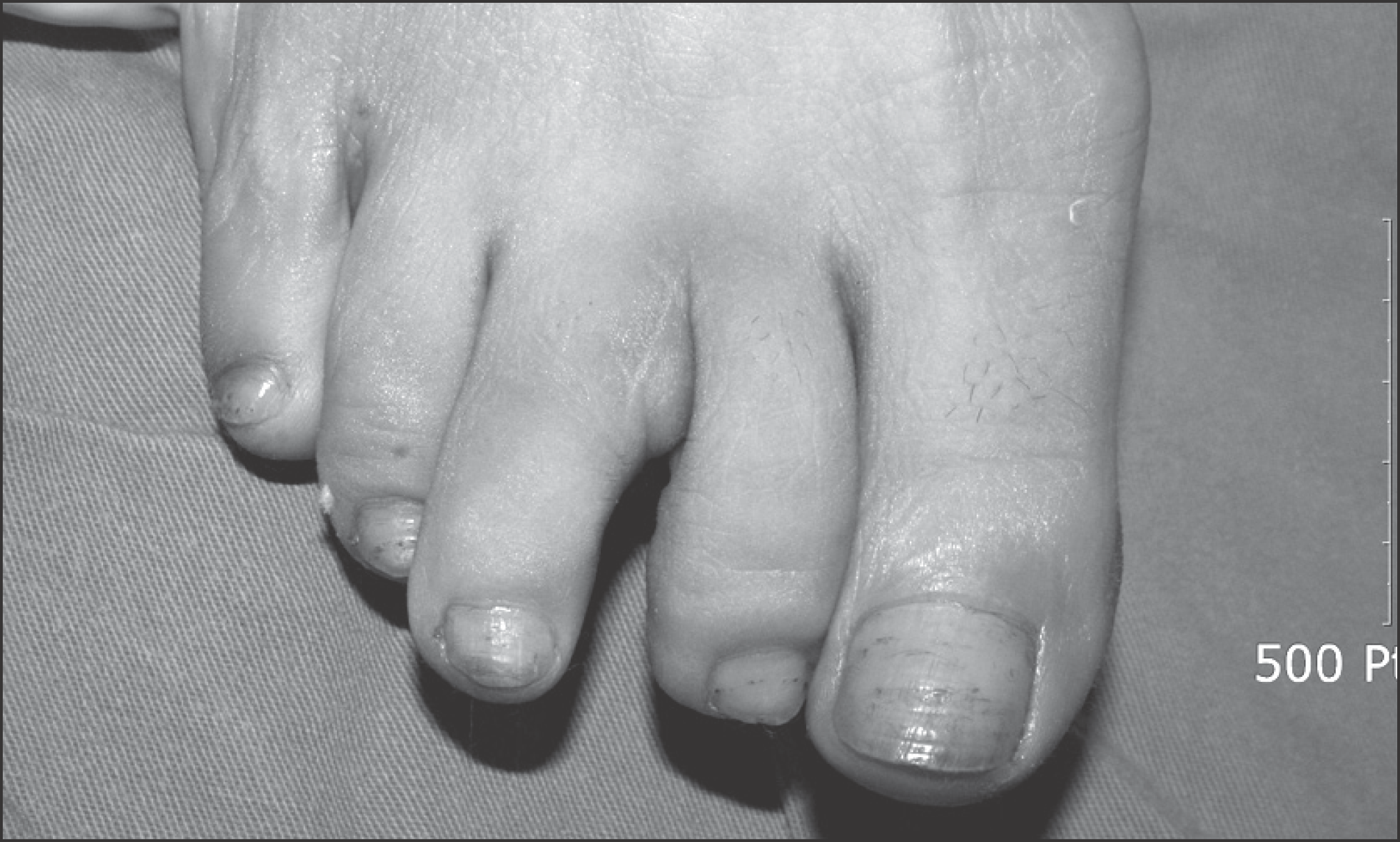
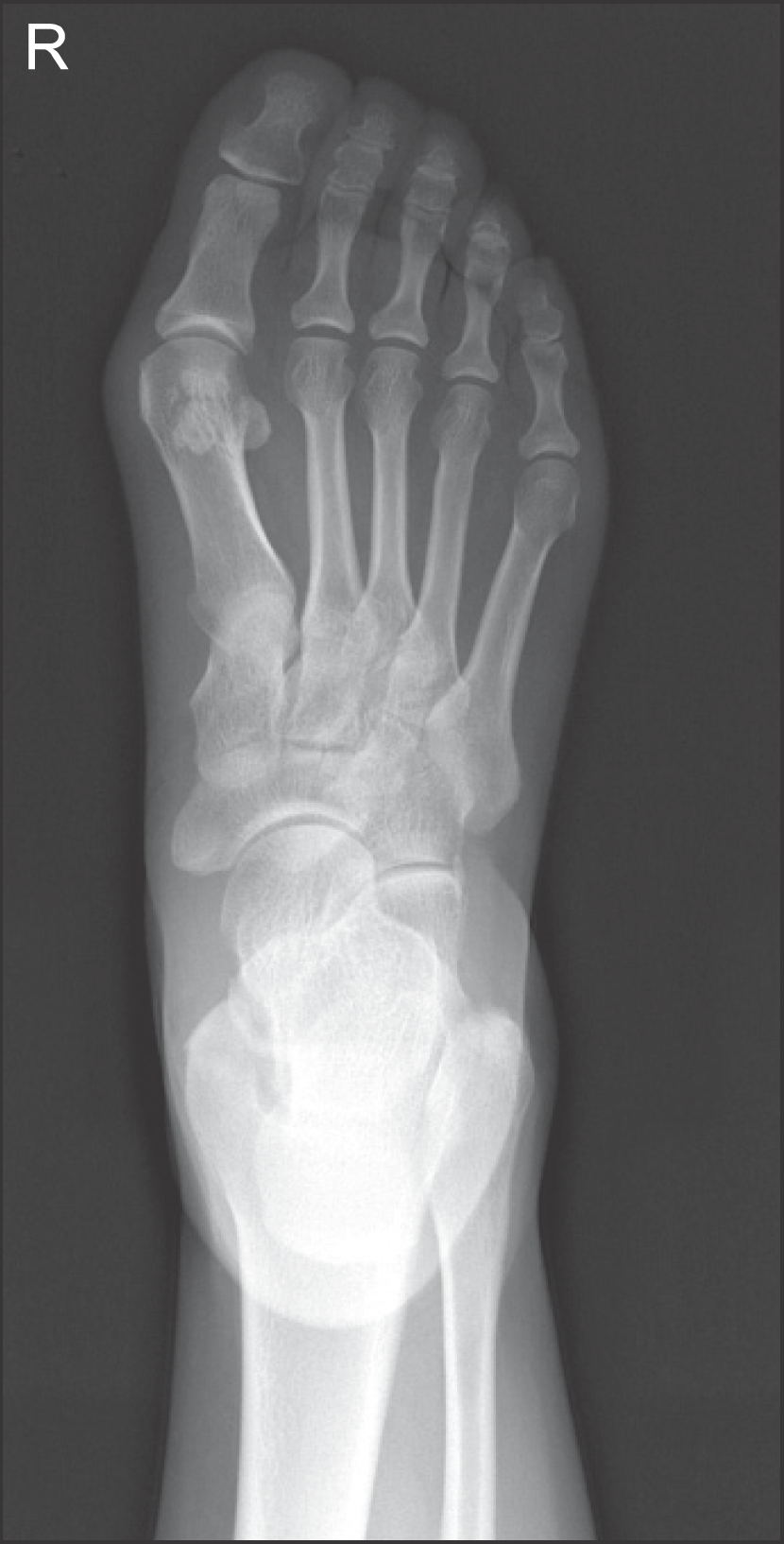
Figure 2.
Preoperative radiograph shows a 21×13 mm sized exostotic mass on the medial-plantar aspect of proximal phalanx of the 3rd toe (white arrow). The protruding bony mass does not connect with the medullary cavity of proximal phalanx.
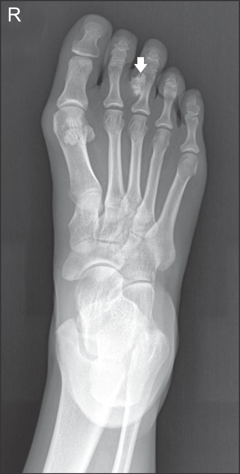
Figure 3.
Preoperative computed tomography scans show an irregular and calcified mass of the proximal phalanx.
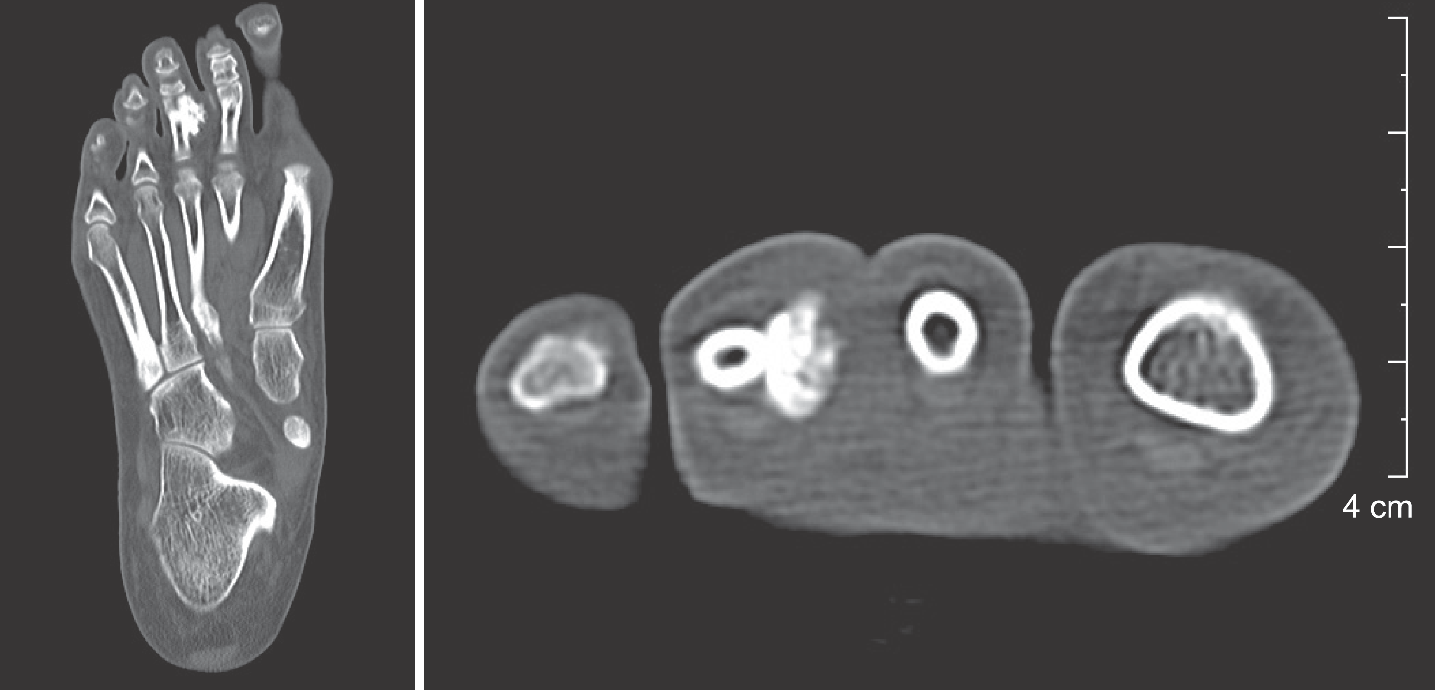
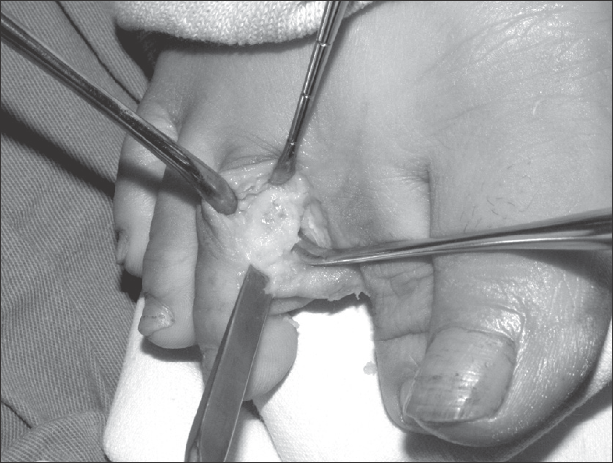
Figure 4.
Intraoperative photograph shows a relatively well margined whitish irregular calcified mass.
 XML Download