Abstract
Purpose
Modular tumor prosthesis is the most popular recontructive modality after resection of malignat tumor in extremity. Complications and survival of tumor prosthesis reconstruction are well-known. however, reports on the long-term outcome of tumor prosthesis in osteosarcoma patientss are scarece.
Materials and Methods
In 158 cases as diagnosed as osteosarcoma from feburary 1989 to December 2006 in a single cancer center. We retrospectively reviewd 48 osteosarcoma patients who under went tumor prosthetic reconstruction. Mean follow up preiod was 75.6 months (range; 60 to 179 months). There were 28 males, 20 females and mean age was 22.4 years (range; 11–71). Pathologic subtypes were conventional central osteosarcoma in 46 cases and periosteal in 2 cases. The location of the tumor was proximal tibia (26 cases), distal femor (20 cases), femor diaphysis (1 case), and tibia diaphysis (1 case). In 41 cases built-up-type tumor prosthesis have been used and 7 cases expansion-type tumor prosthesis have been used. We used Musculoskeletal tumor society (MSTS) grading system to asses post operation function, and we analyzed survival rate of patient and tumor prosthesis and complication.
Results
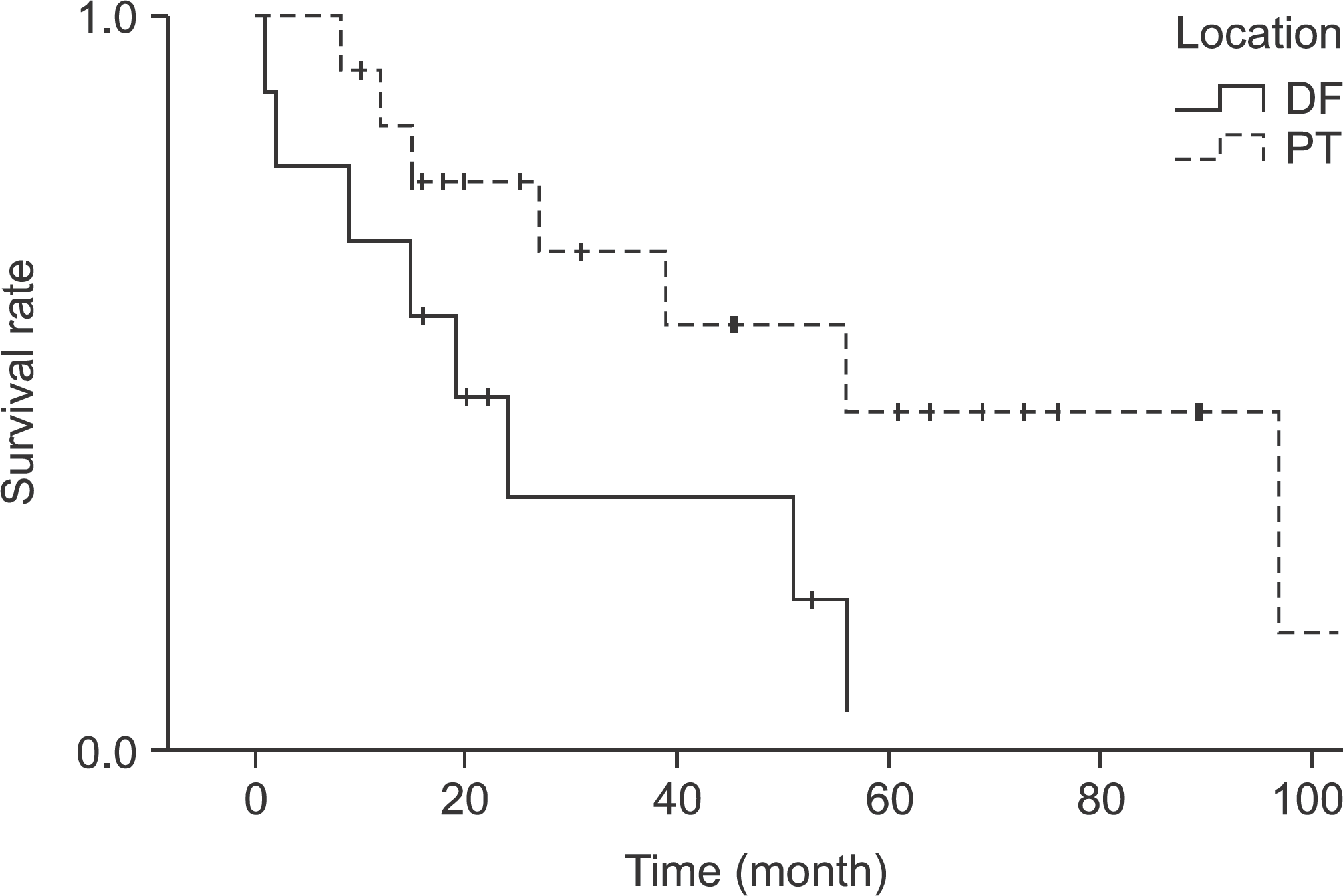
The overall survival rate was 77.7% and disease free survival rate was 68.9%. The survival rate of tumor prosthesis was 73%, in last follow up tumor prosthesis has been removed in 12 cases. All of them, 17 complications occurred, which included infection in 16 cases, Periprosthetic Fracture and Loosening of tumor prosthesis in 4 cases, articular instability in 4 cases. MSTS functional score was 74.1% in post operation.
References
1. Abudu A, Carter SR, Grimer RJ. The outcome and functional results of diaphyseal endoprosheses after tumor excision. J Bone and Joint Surg. 1996; 78B:652–7.
2. Rougraff BT, Simmon MA, Kneisl JS, Greenberg DB, Mankin HJ. Limb salvge compaired with amputation for osteosarcoma of the distal end of the femur: a longterm oncological, functional, and quality of life study. J Bone and Joint Surg. 1994; 76:649.
3. Grimmer RJ, Carter SR, Tillman RM, et al. Endoprosthetic replacement of the proximal tibia. J Bone Joint Surg. 1999; 81B:488–94.
4. Kabukcuoglu Y, Grimmer RJ, Tillmann RM, Carter SR. Endoprosthetic replacement for primary maligant tumors of the proximal femur. Clin Orthop. 1999; 358:8–14.
5. Kawai A, Muschler GF, Lane JM, Otis JC, Healey JH. Prosthetic knee replacement after resection of maligant tumor of the distal part of the femur. J Bone Joint Surg Am. 1998; 80:636–47.
6. Lee SH, Oh JH, Yoo KH, et al. Evaluation of Prosthetic Reconstruction in Lower Extremity. J Korean Bone Joint Tumor Soc. 2002; 8:46–9.
7. Chung SH, Cho Y, Kim JD. Recycling total joint autotransplantation in osteosarcoma around knee joint. J Korean Bone Joint Tumor Soc. 2007; 13:31–6.
8. Dick HM, Malinin TI, Mnaymn WA. Massive allograft implantations following radical resection of high-grade tumors requiring adjuvant chemotherapy treatment. Clin Orthop Relat Res. 1985; 197:88–95.
9. Gosheger G, Gebert C, Ahrens H, Streitbuerger A, Winkelmann W, Hardes J. Endoprosthetic reconstruction in 250 patients with osteosarcoma. Clin Orthop Relat Res. 2006; 450:164–71.
10. Hardes J, Gebert C, Schwappach A, et al. Characteristic and outcome of infections associated with tumor endoprostheses. Arch Orthop Trauma Surg. 2006; 126:289–96.
11. Hillmann A, Hoffmann C, Gosheger G, Krakau H, Winkelmann W. Malignant tumor of the distal part of the femur or the proximal part of the tibia: endoprosthetic replacement or rotationplasty. Functional outcome and quality-of life measurements. J Bone Joint Surg Am. 1999; 81:462–8.
12. Lindner NJ, RAMM O, Hillmann A, et al. Limb salvage and outcome of osteosarcoma. The University of Muenster experience. Clin Orthop. 1999; 358:83–9.
13. Quill G, Gitelis S, Morton T, Piasecki P. Complications associated with limb salvage for extremity sarcomas and their management. Clin Orthop. 1990; 260:242–50.

14. Sanjayy BK, Moreau PG. Limb salvage surgery in bone tumor with modular endoprosthesis. Int Orthop. 1999; 23:41–6.
15. Eckardt JJ, Kabo MK, Kelly CM, et al. Expandible endoprosthesis reconstruction in skeletally immature patients with tumors. Clin Orthop. 2000; 373:51–61.
16. Hornicek FJ, Gebhardt MC, Sorger JI, Mankin HJ. Tumor reconstruction. Orthop Clin N AM. 1999; 30:673–84.

17. Kneisl JS, Finn HA, Simon MA. Mobile knee reconstruction after resection of malignant tumors of the distal femur. Orthop Clin N Am. 1991; 22:105–19.
18. Enneking WF, Mindell ER. Observations on massive retrieved human allografts. J Bone Joint Surg. 1991; 73:1123–42.

19. Jensen KL, Johnston JO. Proximal humeral reconstruction after excision of a primary sarcoma. Clin Orthop Relat Res. 1995; 311:164–75.
20. Kohler P, Kreicberg A. Chondrosarcoma treated by reimplantation of resected bone aft er autoclaving and supplement with allogenic bone matrix. Clin Orthop Relat Res. 1993; 294:281–4.
21. Unwin PS, Cobb JP, Walker PS. Distal femoral arthroplsty using custom-made prostheses. The first 218 cases. J Arthroplasty. 1993; 8:259–68.
22. Eckardt JJ, Paebody TD. Complications of Prostehtic Reconstructions. Surgery for Bone and Soft tissue Tumors. 1998. 467–79.
23. Tsuchiya H, Tomita K. Prognosis of osteosarcoma treated by limb-salvage surgey: the ten-year intergroup study in Japan Jpn J Clin Oncol. 1992; 22:347–53.
24. Schiller C, Windhager R, Fellinger EJ, Salzer-Kuntschik M, Kalder A, Kotz R. Extendable tumour endoprostheses for the leg in children. J Bone Joint Surg Br. 1995; 77:608–14.

25. Malawer MM, Chou LB. Prosthetic survival and clinical results with use of large-segment replacements in the treatment of high-grade bone sarcomas. J Bone Joint Surg. 1995; 77:1154–65.

26. Enneking WF, Dunham W, Gebhardt MC, Malawar M, Pritchard DJ. A system for the functional evaluation of reconstructive procedures after surgical treatment of tumors of the musculoskeletal system. Clin Orthop Relat Res. 1993; 286:241–6.

27. Robert P, Chan D, Grimmer RJ, Sneath RS, Scales JT. Prosthetic replacement of the distal femur for primary bone tumors. J Bone Joint Surg. 1991; 73:762–9.
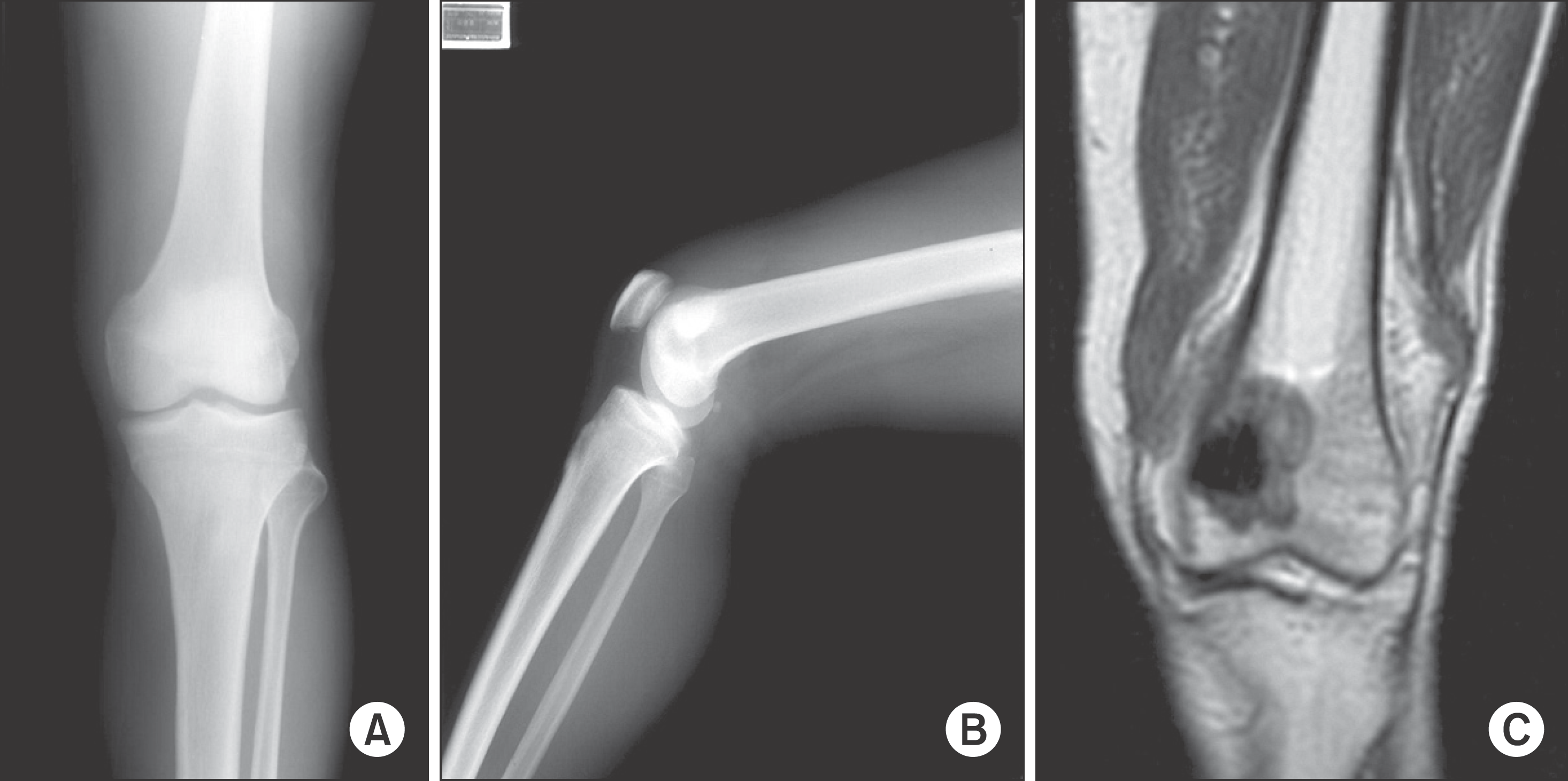
Figure 2.
19 years-old male patient. (A, B) Preoperative radiograph show radiolucent bony lesion in left distal femur and (C) inhomogenous low signal intensity of mass and surrounging muscles. The result of insional bouposy was grade 2B osteosarcoma.

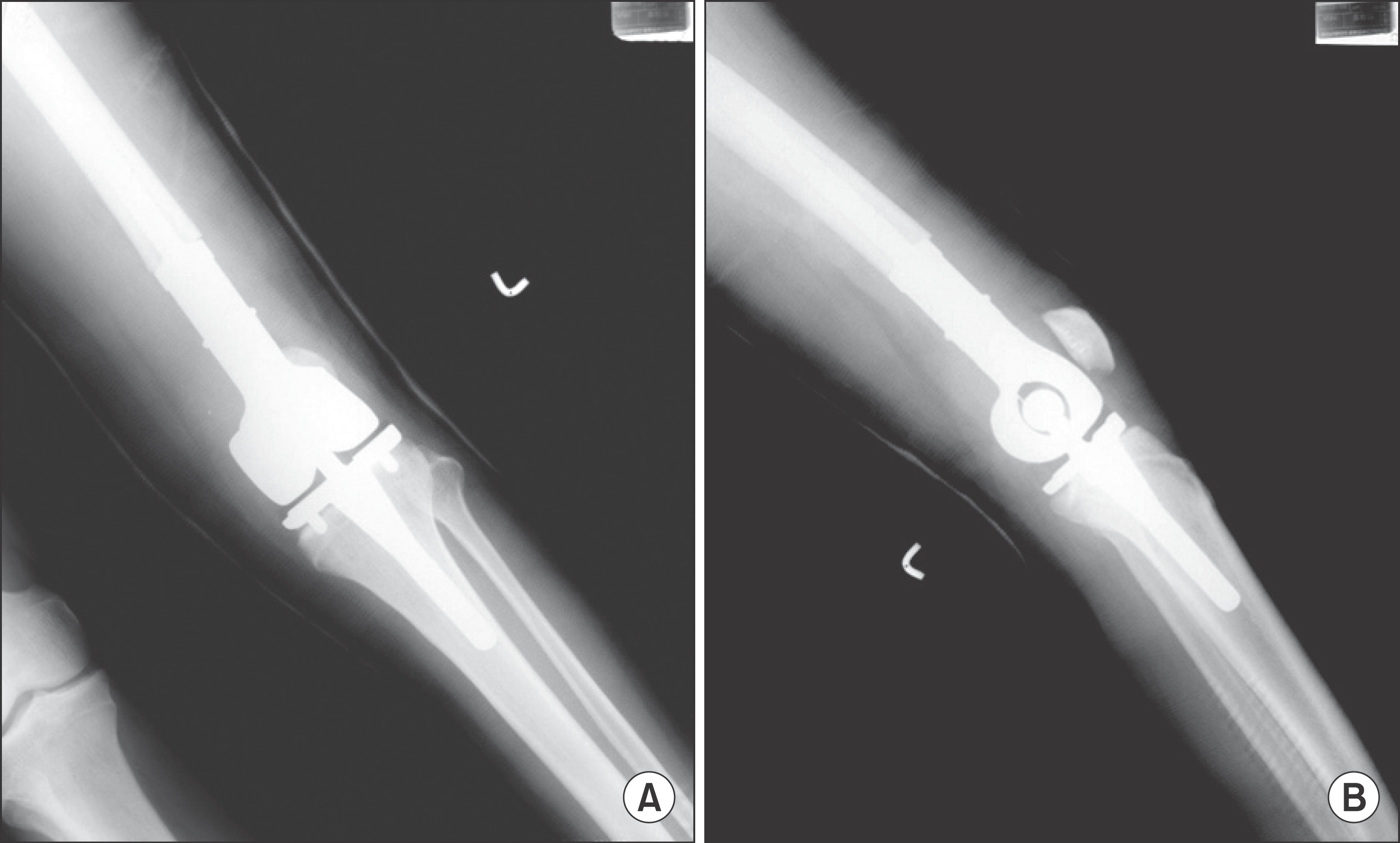
Figure 3.
(A, B) After Wide excision, resconstruction with Mutars Prosthesis. 15 months follow up infection was occurred.

Table 1.
Summary of Cases
Table 2.
Prevalence of Complication
| Site of tumor | No. of patients | No. of infection | No. of loosening | No. of periprosthetic Fx. | c No. of LLD |
|---|---|---|---|---|---|
| Dist. Femur | 20 | 6 | 2 | 1 | 0 |
| Prox. Tibia | 28 | 4 | 3 | 1 | 3 |
| Total | 48 | 10 | 5 | 2 | 3 |
Table 3.
MSTS Score according to Surgical Site
| Location | N | Average | T-score |
|---|---|---|---|
| PT | 28 | 73.6 | |
| DF | 20 | 73.8 | p=0.979 |
Table 4.
Published Literature Review about Reconstruction Using Tumor Prosthesis


 PDF
PDF ePub
ePub Citation
 Print
Print


 XML Download