Abstract
Purpose
This study was designed to evaluate the treatment results of malignant melanoma and to analyze the factors influencing prognosis.
Materials and Methods
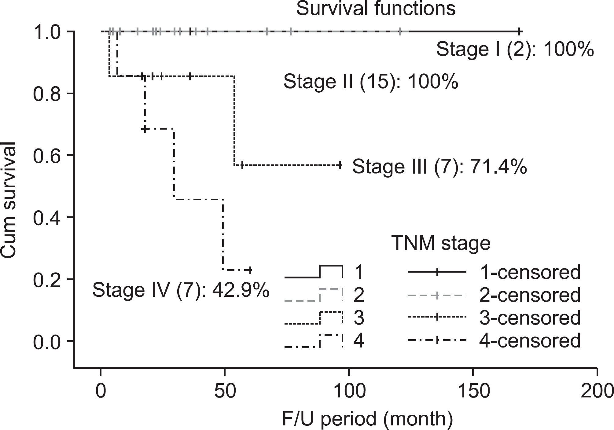
Thirty one cases of malignant melanoma were included in this study. They were treated in our hospital surgically, medically and immunologically from January 1996 to December 2005, and were followed more than 5 years. We compared 5 year survival rate (5YSR) according to the age, gender, anatomical site, depth of tumor, TNM stage, involvement of lymph node and immuno-chemotherapy.
Results
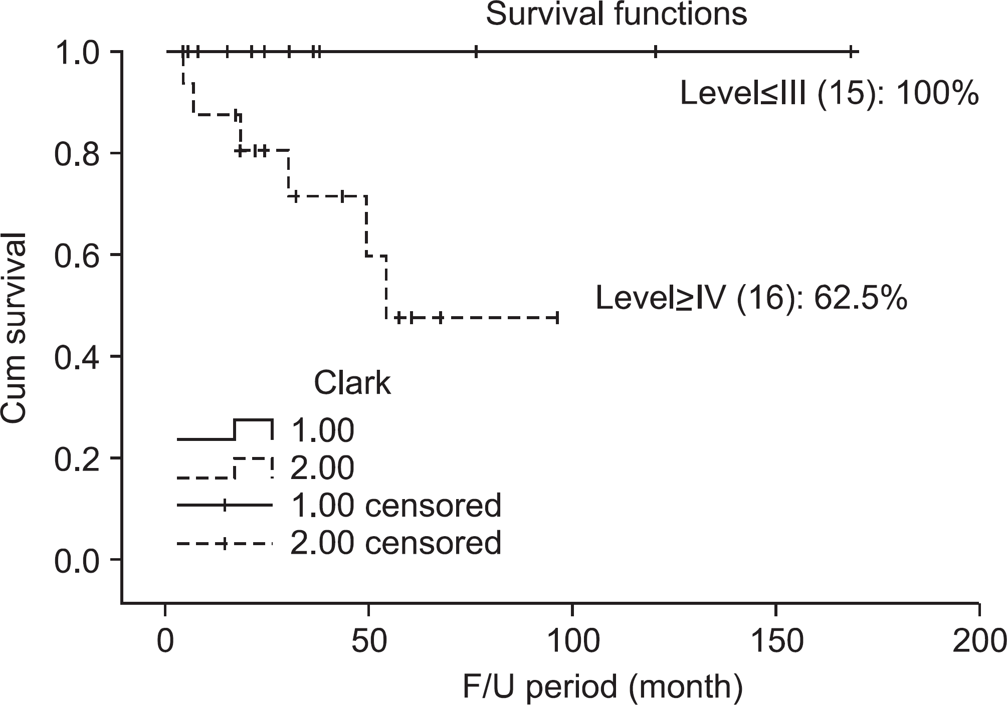
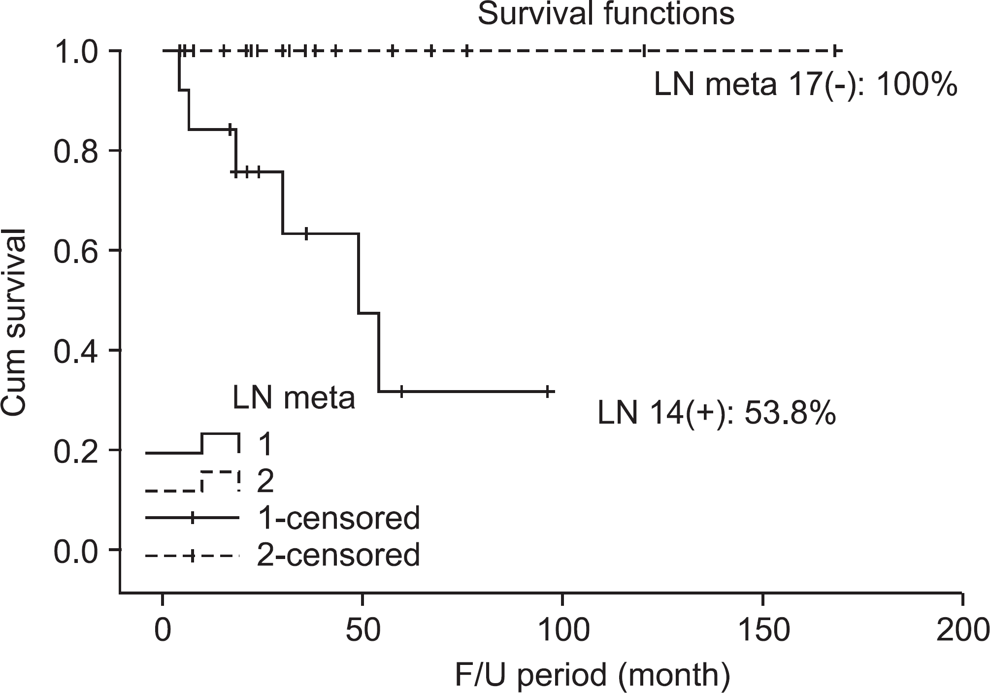
Overall 5YSR was 80.6%. 5YSR of the age group below 65 years was 89.7% and 66.7% for the age group over 65 (p=0.033). 5YSR for men was 75% and 90.9% for women. 5YSR according to the site of occurrence showed 66.7% in upper extremities, 89.5% in lower extremities, and 66.7% in other site. 5YSR was 100% for the Clark level below III and 62.5% for the level above IV (p=0.032). 5YSR was 53.8% for lymph node metastasis group and 100% for non-lymph node metastasis group (p=0.021).
REFERENCES
1. Rigel DS, Carucci JA. Malignant melanoma: prevention, early detection, and treatment in the 21st century. CA Cancer J Clin. 2000; 50:215–36.
2. Park JH, Lee HS, Ham DH, Kim JR. An analysis of the prognostic factors of malignant melanoma. J Korean Bone & Joint Tumor Soc. 2009; 15:122–9.
3. Cummins DL, Cummins JM, Pantle H, Silverman MA, Leonard AL, Chanmugam A. Cutaneous malignant melanoma. Mayo Clin Proc. 2006; 81:500–7.

4. Clark WH. A classification of malignant melanoma in man correlated with histiogenesis and biologic behavior. Adv Biol Skin. 1976; 8:621–47.
5. Balch CM, Buzaid AC, Soong SJ, et al. Final version of the American Joint Committee on Cancer staging system for cutaneous melanoma. J Clin Oncol. 2001; 19:3635–48.

6. Breslow A. Thickness, cross-sectional areas and depth of invasion in the prognosis of cutaneous melanoma. Ann Surg. 1790; 172:902–8.

7. Verma S, McCready D, Bak K, Charette M, Iscoe N. Systematic review of systemic adjuvant therapy for patients at high risk for recurrent melanoma. Cancer. 2006; 106:1431–42.

8. Sekulic A, Haluska P, Miller AJ, et al. Malignant melanoma in the 21st century: the emerging mole cular landscape. Mayo Clin Proc. 2008; 83:825–46.
9. Su WP. Malignant melanoma: basic approach to clinicopathologic correlation. Mayo Clin Proc. 1997; 72:267–72.
10. Rhee SK, Kang YK, Park WJ, Chung YG, Lee HJ. Malignant melanoma. J Korean Bone & Joint Tumor Soc. 2001; 7:12–9.
11. Nahabedian MY. Malignant melanoma. Clin Plast Surg. 2005; 32:249–59.
Figure 1.
48 year-old women patient with malignant melanoma on right hand. Wide excision and split skin graft is done over the hand.
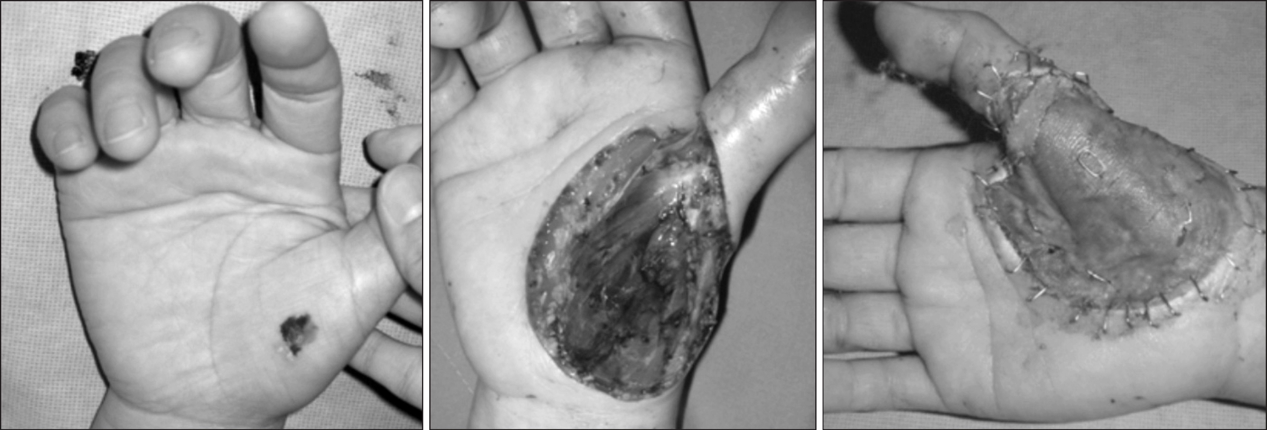
Figure 2.
67 year-old man patient with malignant melanoma on left heel. Wide excision and anterolateral thigh free flap is done over the heel.
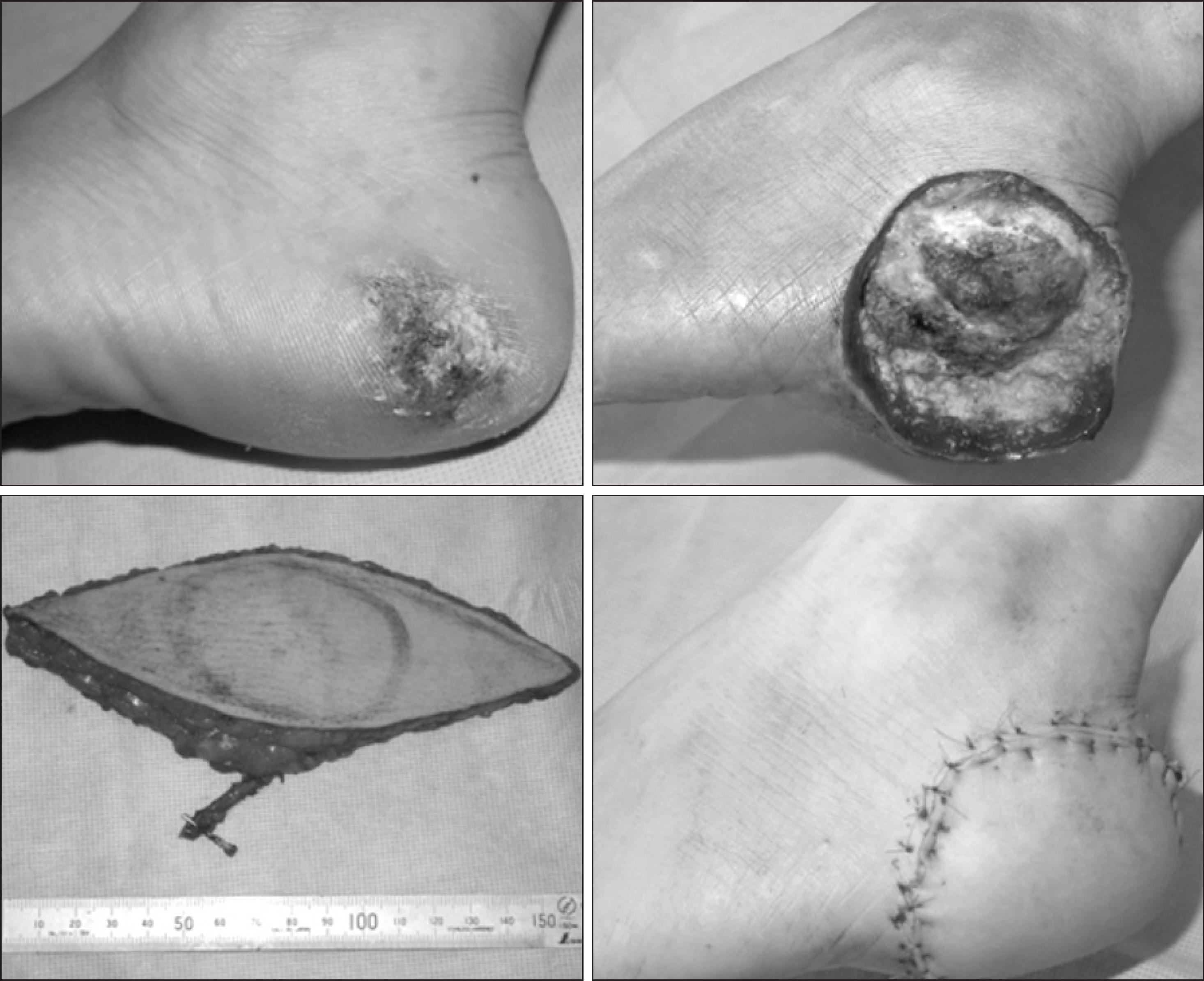
Figure 3.
50 year-old man patient with malignant melanoma on right 2nd finger. Finger amputation is done.
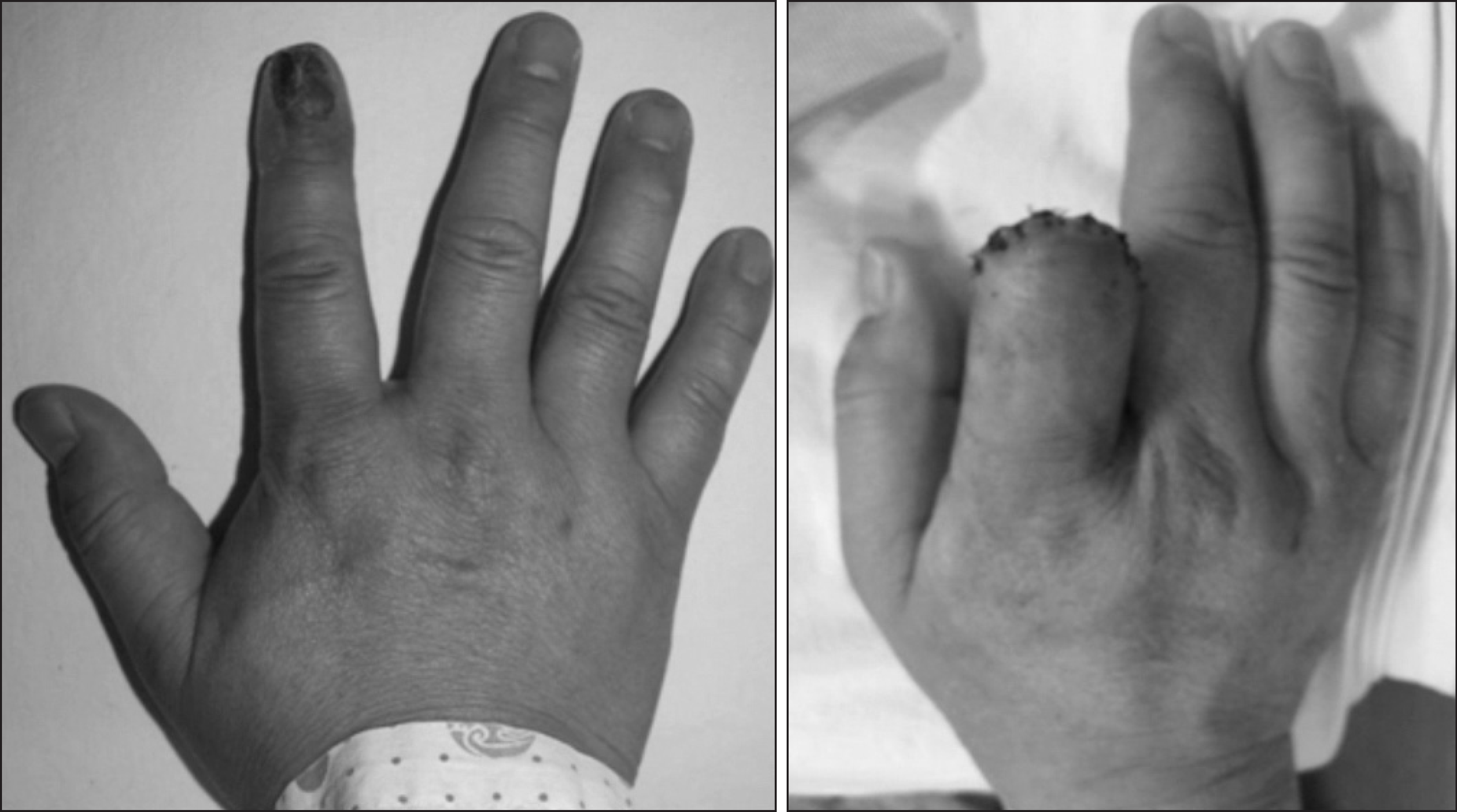
Figure 4.
Overall cumulative survival curves according to age. Figure 5. Overall cumulative survival curves according to Clark's level.
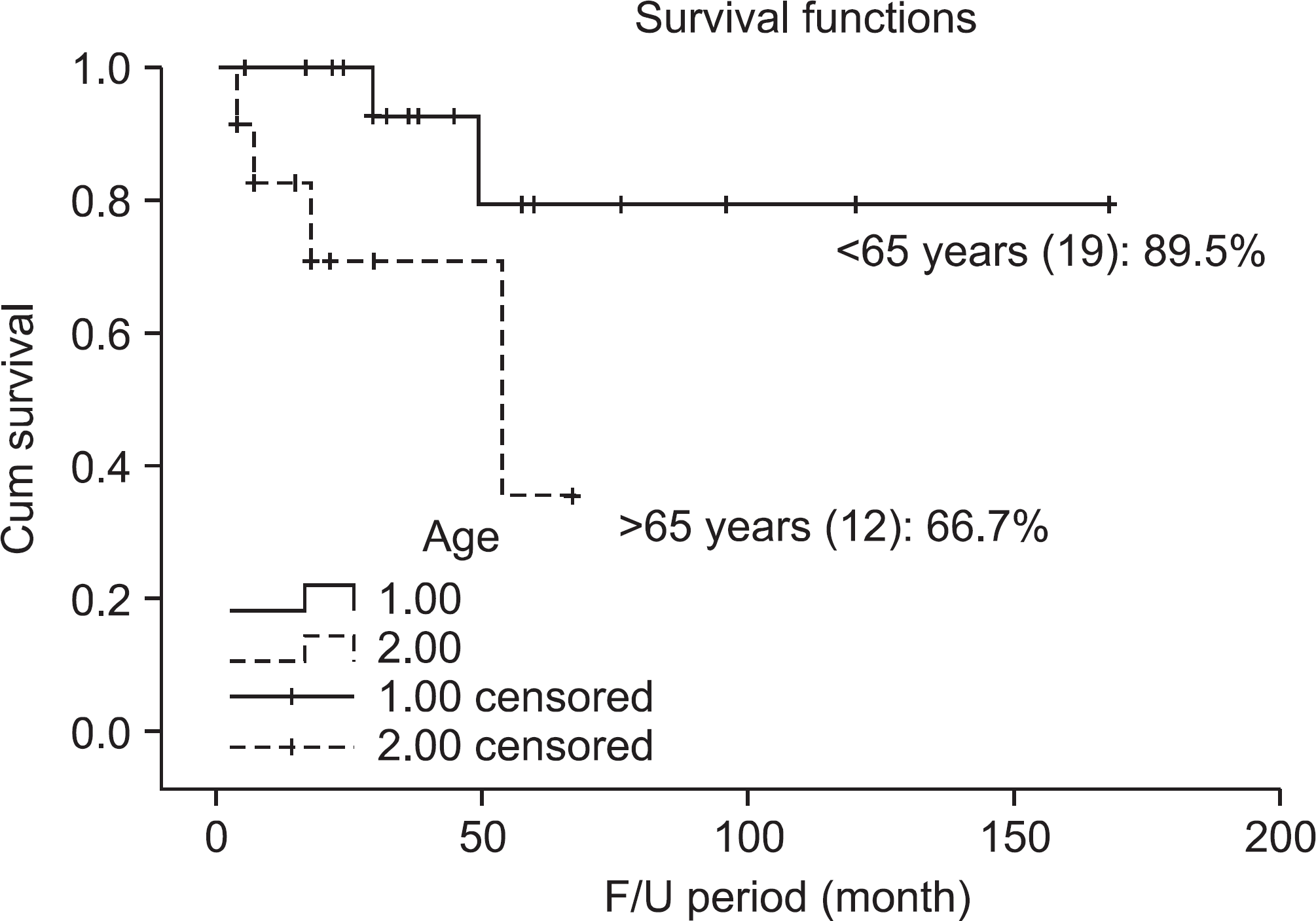
Table 1.
Patients’ Data
| Mean age | 56.9 |
|---|---|
| Sex (M:F) | 20/11 |
| LN meta. | 13:+/18:- |
| Site | LE (19), UE (6), Others (6) |
| Mean F/U (mo.) | 46.5 |
Table 2.
Clark Level, TNM Stage


 PDF
PDF ePub
ePub Citation
 Print
Print


 XML Download