

 PDF
PDF ePub
ePub Citation
 Print
Print


Abstract
Purpose
This study was aimed to analyze the incidence and the anatomical distributions of HME (Hereditary Multiple Exostoses) on upper limbs and its related change in alignment of the upper limbs in HME patients.
Materials and Methods
Thirty eight patients who had been diagnosed HME between 2001 and 2009, were categorized into two groups; (1) group A (1-2 involvements); (2) group B (≥3 involvements). We checked the carrying angle, VAS (Visual Analogue Scale), limitations in daily activities, cosmetic satisfaction according to the number of exostoses invasion.
Results
Among the 38 patients, 23 patients (43 cases) had exostoses in the upper limbs. The locations of exostoses in the upper limbs were proximal humerus in 33 cases (30%), distal ulna in 31 cases (28.2%), and distal radius in 24 cases (21.8%). The carrying angle of group A and B was 10.7o, 13.8o, VAS was 1.3, 3.5, and the limitations in daily activities was 7.3, 6.6 of 8 points. The cosmetic satisfactory cases were 13 and 10 cases, respectively.
REFERENCES
1. Boyer A. Trait des malaidies chirurgicals et des operation quileur conviennent. Paris: ve Migneret. 1814; 3:594.
2. Beals R. Metachondromatosis. Clin Orthop Relat Res. 1982; 169:167–70.

3. Jaffe H. Hereditary multiple exostoses. Arch Pathol. 1943; 36:335–57.
4. Alvarez C, De Vera M, Heslip T, Casey B. Evaluation of the anatomic burden of patients with hereditary multiple exostoses. Clin Orthop Relat Res. 2007; 462:73–9.
5. Burrows H. An operation for the correction of Madelung's deformity and similar conditions. Proc Roy Soc Med. 1937; 30:565–72.
6. Shin E, Jones N, Lawrence J. Treatment of multiple hereditary osteochondromas of the forearm in children: a study of surgical procedures. J Bone Joint Surg Br. 2006; 88:255–60.
7. Hennekam R. Hereditary multiple exostoses. J Med Genet. 1991; 28:262–6.
8. Krooth R, Macklin M, Hilbish T. Diaphysealaclasis (multiple exostosis) on Guam. Am J Hum Genet. 1961; 13:340–7.
9. Peterson H. Deformities and problems of the forearm in children with multiple hereditary osteochondromata. J Peiatr Orthop. 1994; 14:92–100.
10. Solomon L. Hereditary multiple exostosis. J Bone Joint Surg Br. 1963; 43:292–304.
11. Black B, Dooley J, Pyper A, Reed M. Multiple hereditary exostoses. An epidemiologic study of an isolated community in Manitoba. Clin Orthop Relat Res. 1993; 287:212–7.
12. Matsubara H, Tsuchiya H, Sakurakichi K, Yamashiro T, Watanabe K, Tomita K. Correction and lengthening for deformities of the forearm in multiple cartilaginous exostoses. J Orthop Sci. 2006; 11:459–66.
13. Schmale G, Conrad E, Raskind W. The natural history of hereditary multiple exostoses. J Bone Joint Surg Am. 1994; 76:986–92.
14. Oppenheim W, Clader T, Smith C, Bayer M. Supracondylar humeral osteotomy for traumatic childhood cubitus varus deformity. Clin Orthop Relat Res. 1984; 188:34–9.
15. Masada K, Tsuyuguchi Y, Kawai H, Kawabata H, Noguchi K, Ono K. Operations for forearm deformity caused by multiple osteochondromatosis. J Bone Joint Surg Br. 1989; 71:24–9.
16. Nawata K, Teshima R, Minamizaki T, Yamamoto K. Knee deformities in multiple hereditary exostoses: a longitudinal radiographic study. Clin Orthop Relat Res. 1995; 313:194–9.
17. Fogel G, McElfresh E, Peterson H, Wocklund P. Management of deformities of the forearm in multiple hereditary osteochondromatosis. J Bone Joint Surg Am. 1980; 66:670–80.
18. Akita S, Murase T, Yonenobu K, Shimada K, Masada K, Yoshikawa H. Long-term results of surgery for forearm deformities in patients with multiple cartilaginous exostoses. J Bone Joint Surg Am. 2007; 89:1993–9.
19. Fogel G, McElfresh E, Peterson H, Wicklund P. Management of deformities of the forearm in multiple hereditary osteochondromas. J Bone Joint Surg Am. 1984; 66:670–80.
Figure 1.
A radiograph shows whole upper extremity teleradiograph. Carrying angle is measured with two drawing axes of the arm and forearm. Line A-B passes through the lateral border of the acromion to the midpoint of the lateral and medial epicondyles of the humerus. Line C-D passes through the midpoint of the lateral and medial epicondyles of the humerus to the midpoint of the distal radial and ulnar styloid process.
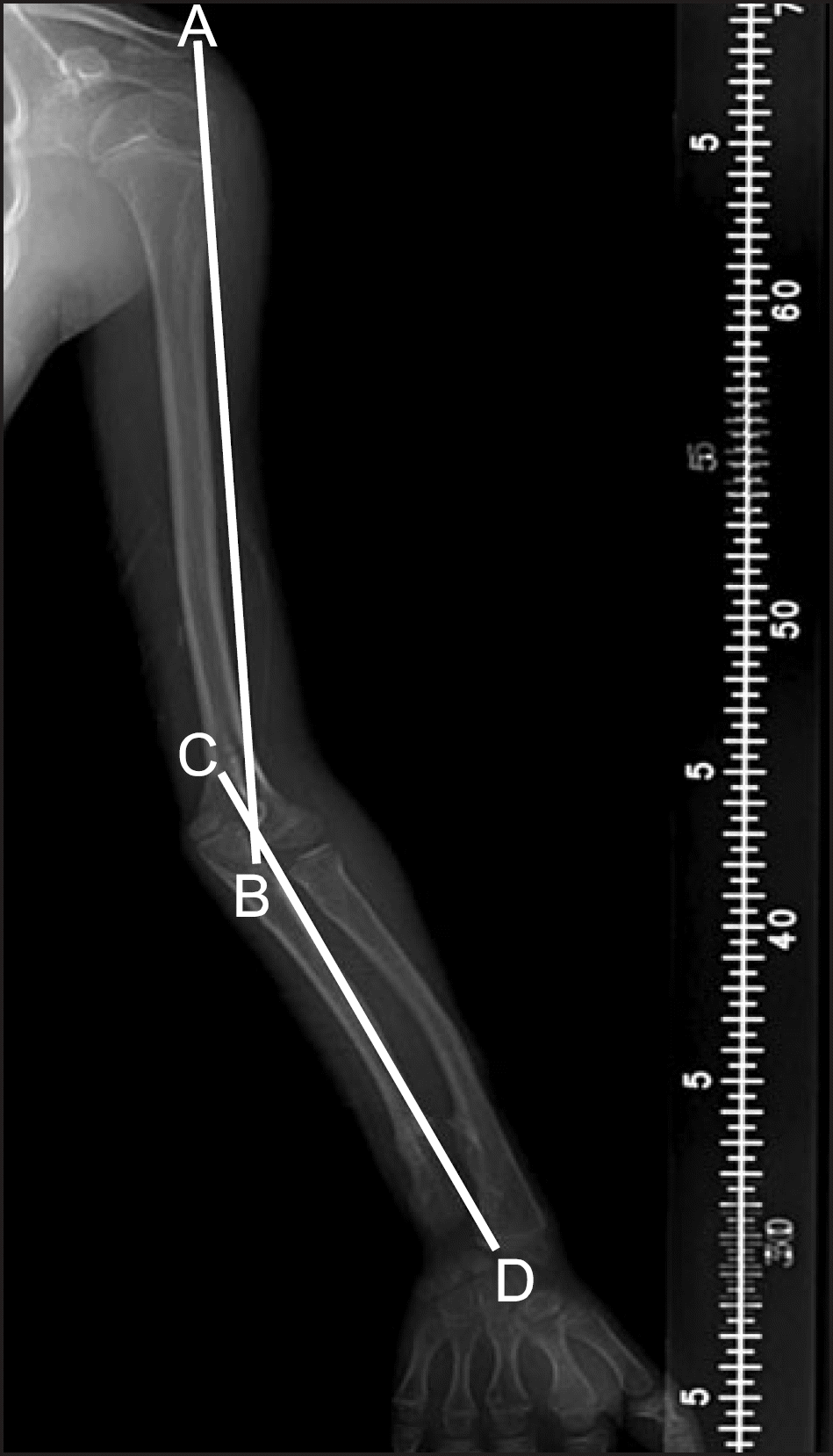
Figure 2.
Masada classification(Masada et al. 1989) (A) Type I: Primary exostosis formation is in the distal portion of the ulna. (B) Type IIa: In addition to ulnar shortening, the radial head is dislocated. (C) Type IIb: The radial head is dislocated without a proximal radial exostosis. (D) Type III: Primary exostosis formation is in the metaphysis of the distal radius.
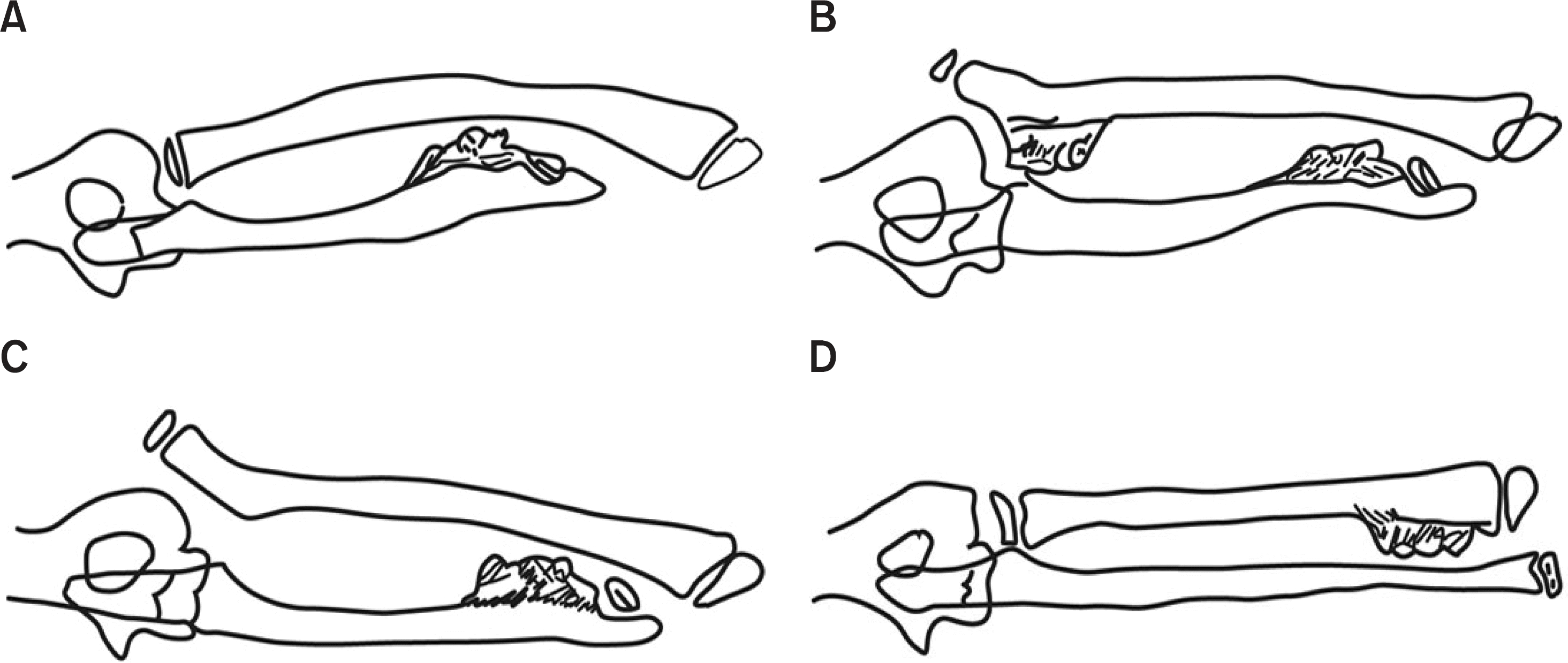
Figure 3.
(A) Radial articular angle: Angle between two constructed lines - One along the articular surface of the radius and the other perpendicular to a line that bisects the head of the radius and passes through the radial edge of the distal radial epiphysis. (B) Carpal slip: Percentage of the attachment of a lunate with the radius, determined by an axial line drawn from the center of the olecranon through the ulnar edge of the radius. (C) Ulnar shortening: Distance between a line that is perpendicular to the longitudinal axis of the forearm and a line that is constructed at the distal end of the ulna.
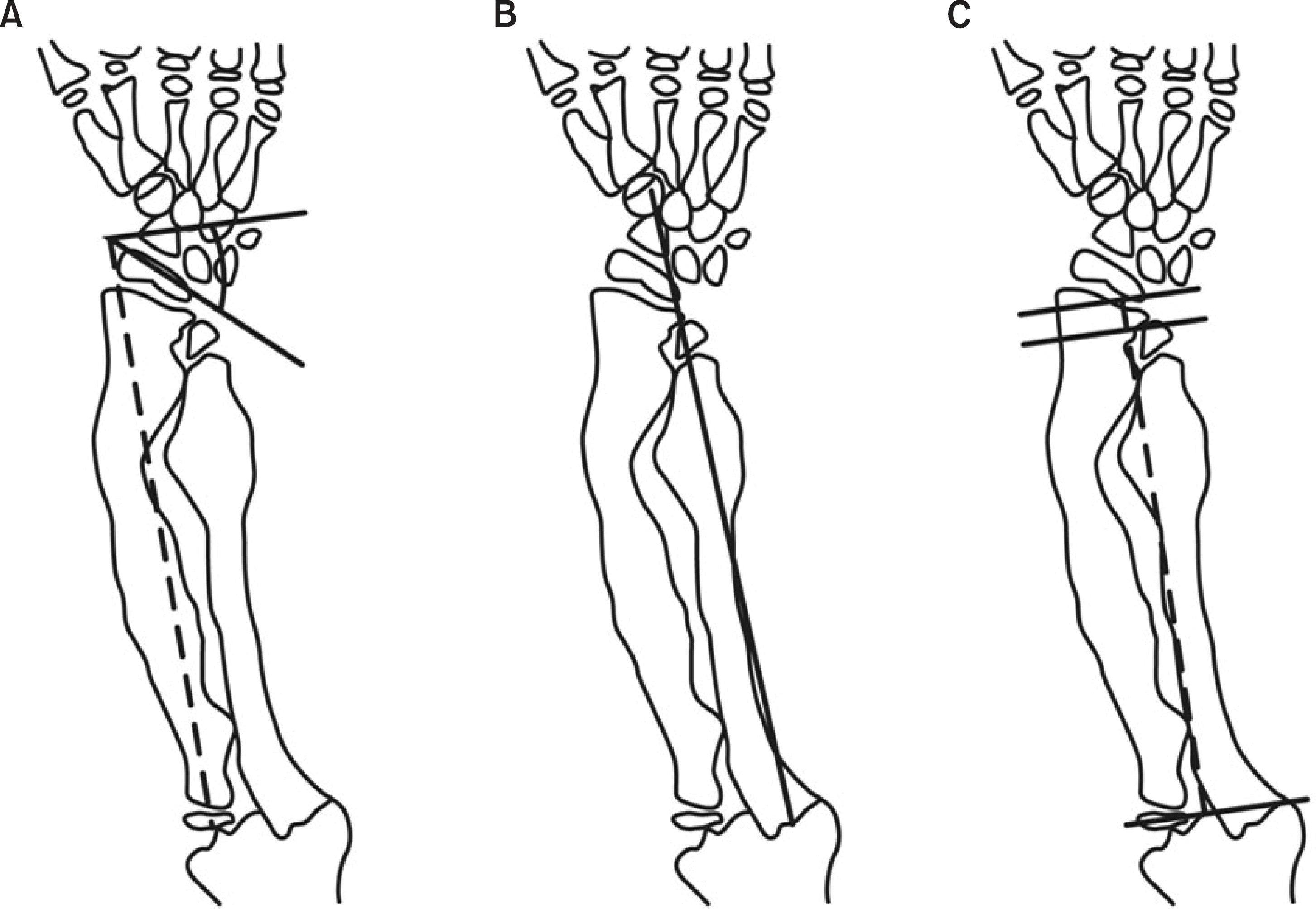
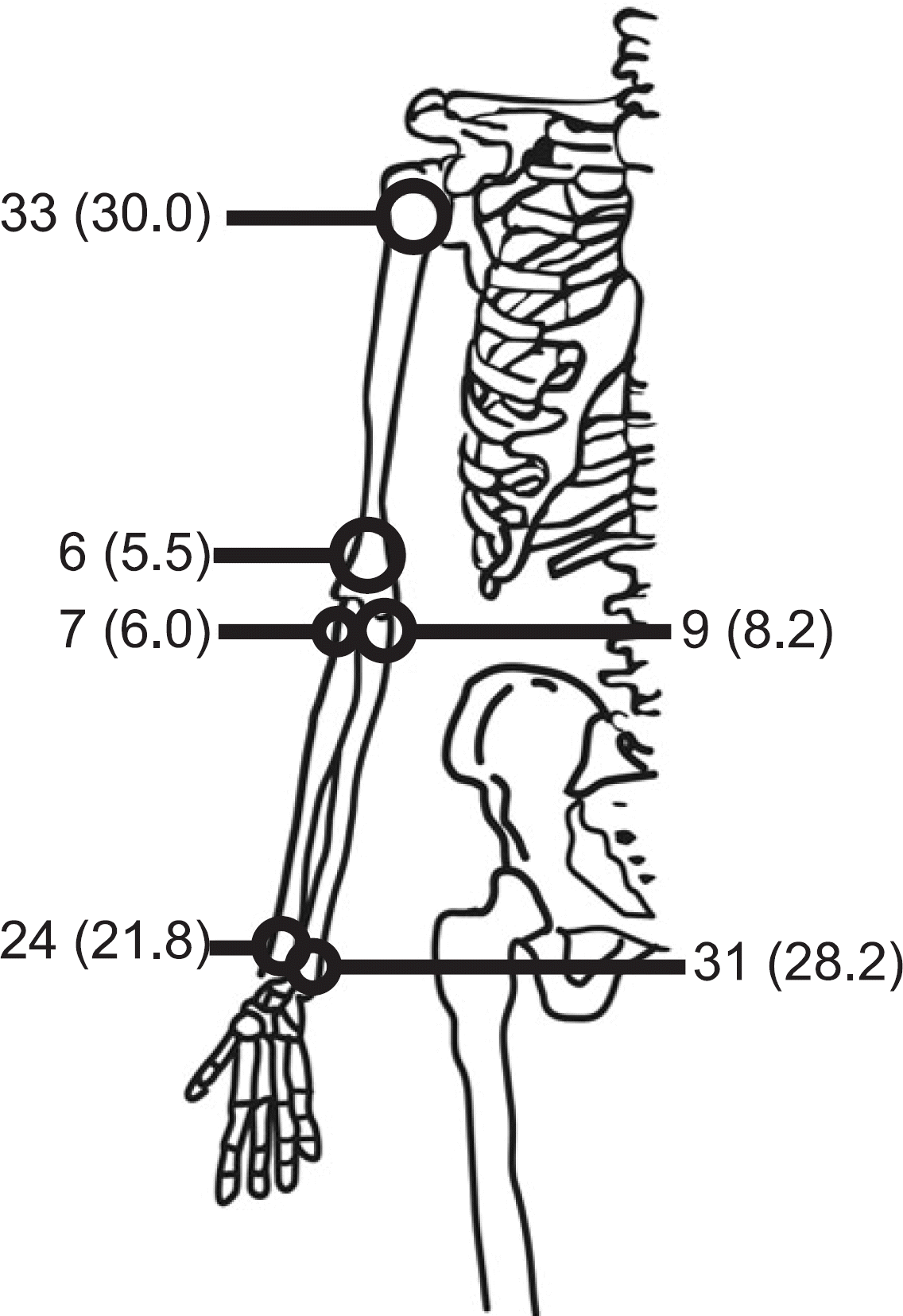
Table 1.
Demographic Data
| A | B | Total | |
|---|---|---|---|
| Number of cases | 18 | 25 | 43 |
| Age (range)∗ 15 | 5.5 (5.7-25.2) | 9.5 (2.9-17.1) | 12.2 (2.9-25.2) |
| Sex | |||
| Male | 6 | 6 | 12 |
| Female | 4 | 7 | 11 |
Table 2.
Clinical Parameters
| A | B | p-value | |
|---|---|---|---|
| VAS∗ | 1.3 (0-3.0) | 3.5 (2.0-5.0) | 0.003 |
| ADL† | 7.3 (5.0-8.0) | 6.6 (3.0-8.0) | 0.001 |
| Satisfactory to cosmetic outcome | 13 | 10 |
Table 3.
Forearm Deformity Classified as Masada Classification
| Type | No∗ of cases | Radial articular angle (degree) | Carpal slip (%) | Ulnar shortening (mm) | Carrying angle (degree) |
|---|---|---|---|---|---|
| I | 25 | 41.9 (20.7-50.5) | 44.3 (10.0-100.0) | -10.9† (3.4-22.9) | 13.4 (4.6-23.6) |
| II | 1 | 47.5 | 80 | -3.4 | 20.2 |
| III | 4 | 17.9 (14.2-32.2) | 27.5 (10.0-70.0) | -3.5 (1.8-10.2) | 15.6 (6.8-25.4) |
| Total | 30 | 42.2 (14.2-50.5) | 49.7 (10.0-100.0) | -9.1 (3.4-22.9) | 12.6 (4.6-25.4) |
 XML Download