Abstract
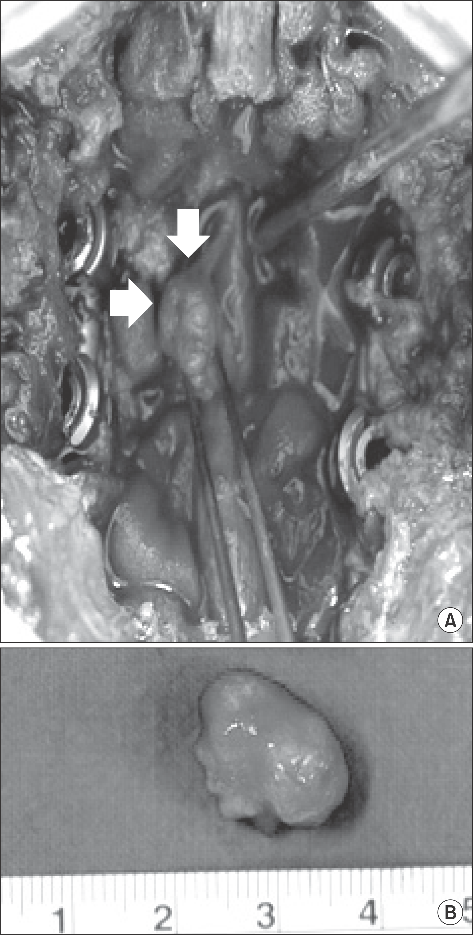
In the patient who has intradural mass associated with spinal stenosis, if the operation for spinal stenosis is performed alone, the symptom may remain. We report with literature review that we achieved the successful outcome after simultaneous decompression of spinal stenosis and space occupying mass removal in the case of intradural and extradural compression. A 71-year-old female patient suffering from low back pain and radiating pain of both lower extremities admitted. In magnetic resonance imaging, spinal stenosis on L4–5 and spondylolisthesis on L5-S1 compressed dural sac and intradural space occupying mass on L4 level compressed. By posterior approach, decompression and interbody fusion were carried out. Then mass was removed with median durotomy. Pathologic diagnosis was schwannoma and the symptom was improved remarkably.
References
2. De Verdelhan O, Haegelen C, Carsin-Nicol B, et al. MR imaging features of spinal schwannomas and meningiomas. J Neuroradiol. 2005; 32:42–9.

4. Jung HW, Lim SB, Kim DW, Shin BJ, Kim YI. Pre-sacral giant schwannoma: removal by a combined anterior and posterior approach: a case report. J Korean Soc Spine Surg. 2001; 8:259–63.
5. Shin BJ, Lee JC, Yoon TK, et al. Surgical treatments of intradural extramedullary tumor. J Korean Soc Spine Surg. 2002; 9:230–7.

6. Broager B. Spinal neurinoma; a clinical study comprising 44 cases with a discussion of histiological origin and with special reference to differential diagnosis against spinal glioma and meningioma. Acta Psychiatr Neurol Scand Suppl. 1953; 85:1–241.
7. Cervoni L, Celli P, Scarpinati M, Cantore G. Neurinomas of the cauda equina clinical analysis of 40 cases. Acta Neurochir (Wien). 1994; 127:199–202.
8. Schneider RC, Kahn EA, Crosby ECC. Spine and spinal cord tumors. Mcgauley JL, editor. Correlative Neurosurgery. 3rd ed.Springfield: Charles C Thomas;1982. 975.
9. Wilkins RH, Rengachary SS. Spinal intradural tumors. Stein BM, editor. Neurosurgery. New York: McGraw-Hill;1985. 1048.
Figure 1.
Preoperative plain radiographs. (A, B) Anteroposterior (A) and lateral (B) radiographs show degenerative spondylosis in L4–5-S1 level. (C, D) Flexion (C) and extension (D) lateral radiographs show spondylolisthesis on L5-S1 level.
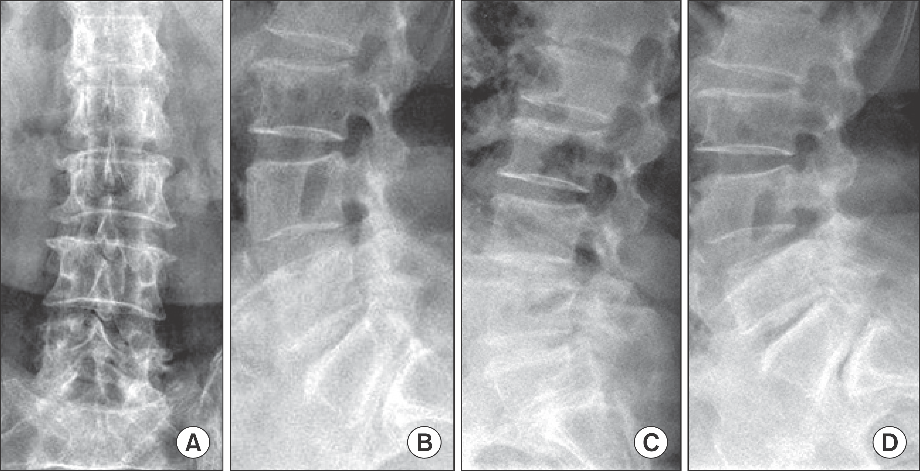
Figure 2.
T2-weighted sagittal and axial MR images. (A-C) Serial sagittal MR images show foraminal stenosis on L4–5 level and spondylolisthesis on L5-S1 level. (D) Axial MR image shows spinal stenosis on L4–5 level. (E) Axial MR image shows spondylolisthesis on L5-S1 level.
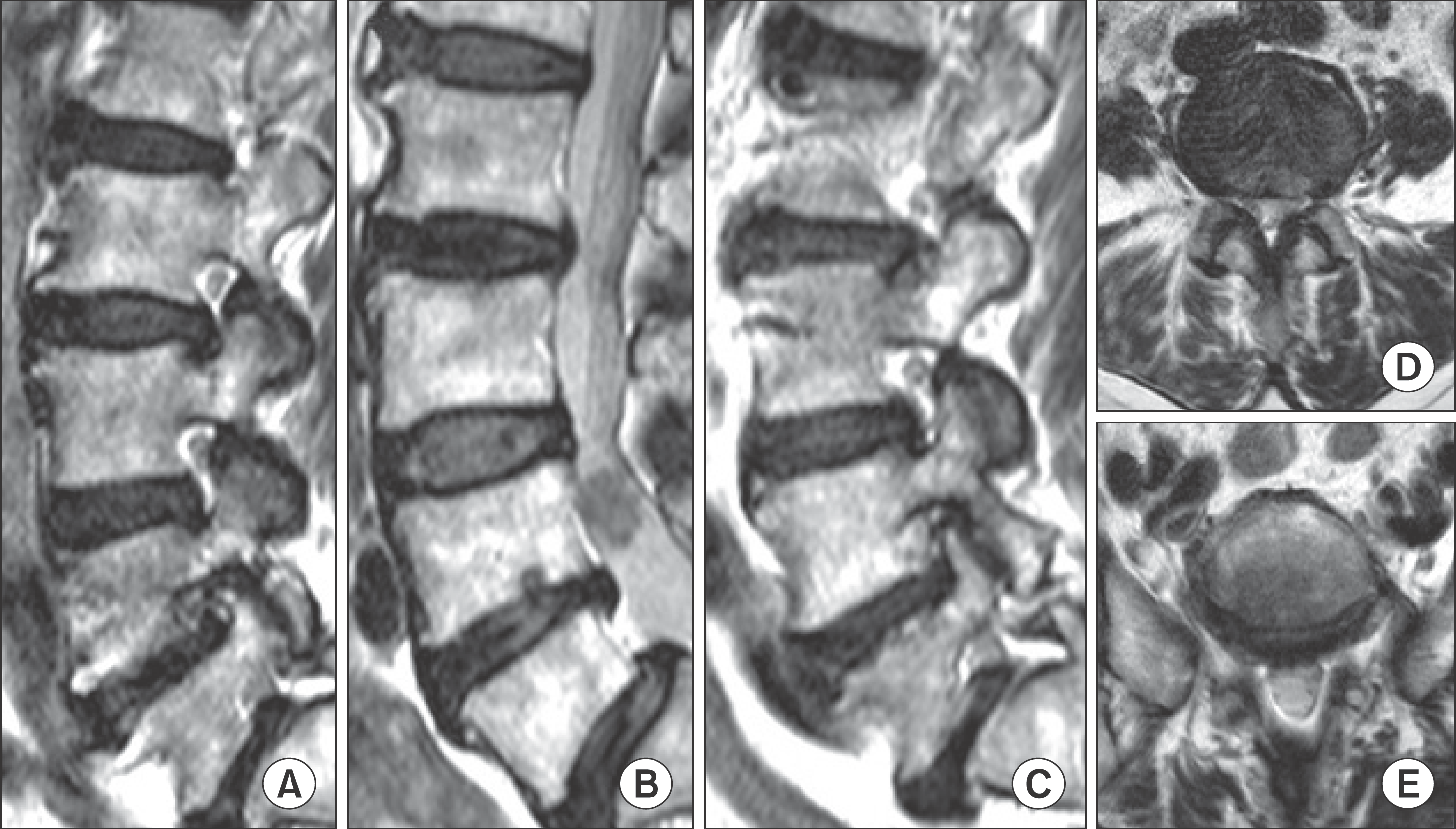
Figure 3.
MR images, myelogram and CT myelogram show space occupying mass on L4 level. (A) T1-weighted sagittal and axial MR image shows low-signal intensity lesion. (B) T2-weighted sagittal and axial image shows iso-signal intensity lesion. (C) Gadolinium enhanced sagittal and axial MR image shows high-signal intensity lesion. (D) Myelogram and CT myelogram show space occupying lesion.
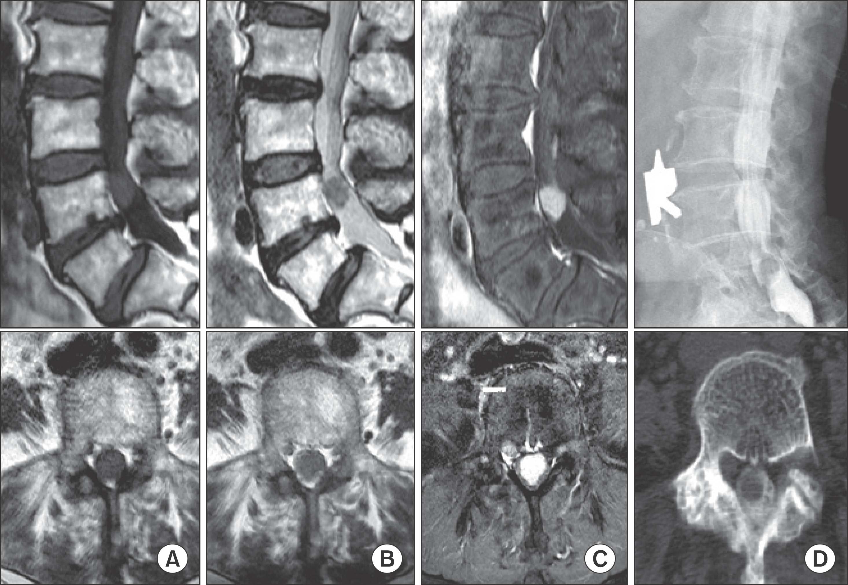
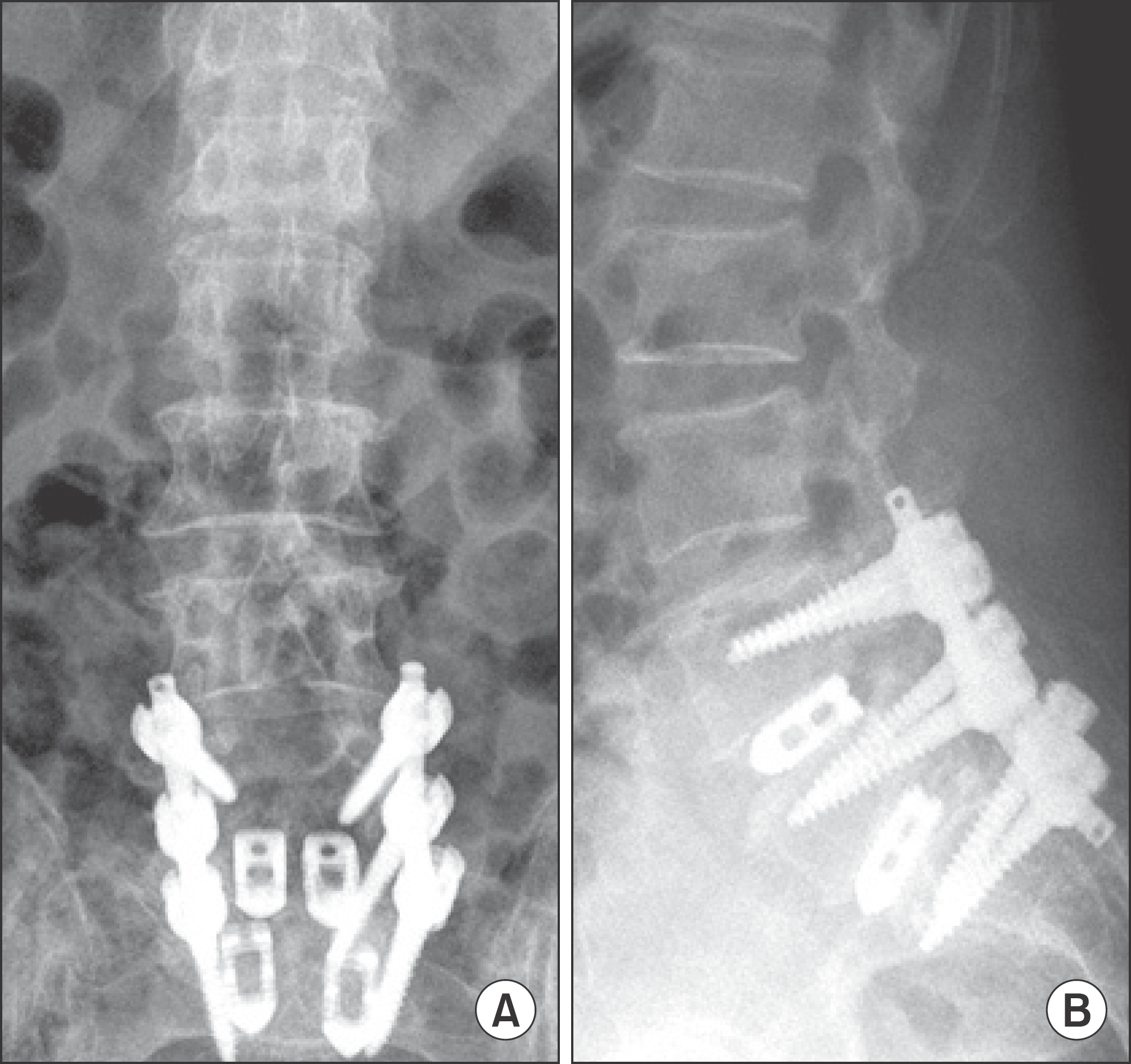
Figure 4.
Postoperative plain radiographs. (A, B) Anteroposterior (A) and lateral (B) radiographs show posterior decompression and interbody fusion using pedicle screws and cages on L4–5-S1 level.


 PDF
PDF ePub
ePub Citation
 Print
Print



 XML Download